
11-05-2026 12:32
 Bernard CLESSE
Pourriez-vous m'aider à identifier cette héloti
13-05-2026 15:26
 François Freléchoux
Bonjour,Voici une récolte faite il y a quelques j
12-05-2026 15:41
 Nicolas VAN VOOREN
Dear Ascolovers, especially interested in Pezizale
13-05-2026 12:05
 Thierry Blondelle
Bonjour à tous,J'aimerais avoir confirmation de c
10-05-2026 23:17
 Andreas Gminder
Hello,today we found in a moist steep decidous for
28-04-2026 20:07
 Lothar Krieglsteiner
... on twig in the air at standing Ceratonia siliq
27-04-2026 20:52
Lothar Krieglsteiner
Found on hanging tiwg of Olea europaea in dried-ou
11-05-2026 20:22
Lothar Krieglsteiner
on attached twig of standing Ficus caricaquite uns
29-04-2026 10:44
Lothar Krieglsteiner
growing at moist, drying-out soil at the side of a
 Hi,
Hi,I (totally inexperienced) would kindly ask for some help. Forgive me eventually stupid ideas.
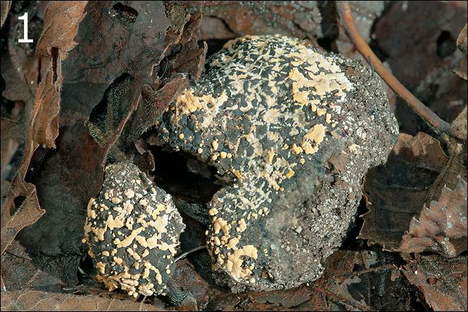
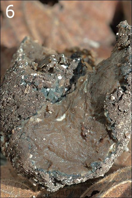
I found this tuber (hope so) with what I believe is a parasitic fungus on it. The fruitbody seemed to be slightly over-matured. Originally it was a single body, but it broke into two pieces during handling (Fig.1).
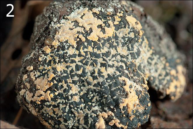
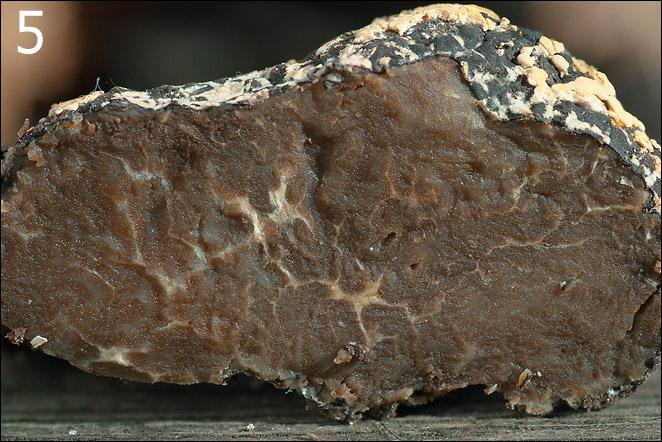
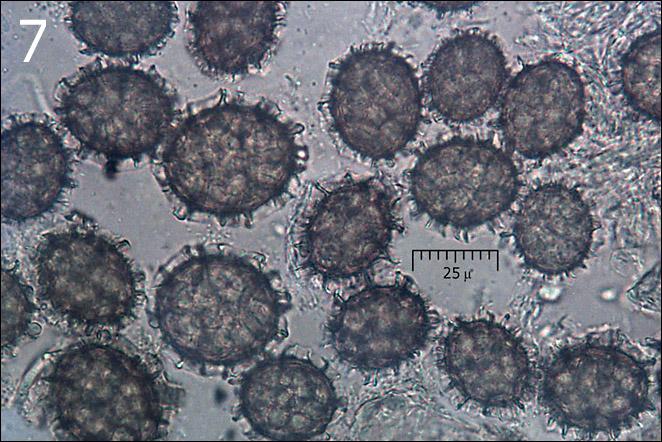
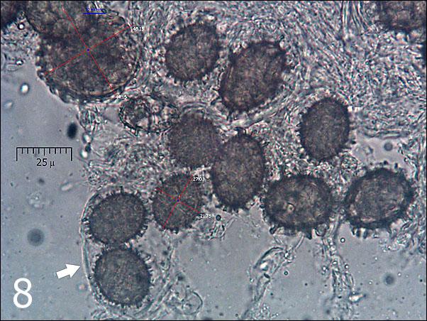
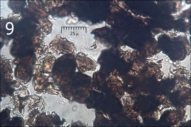
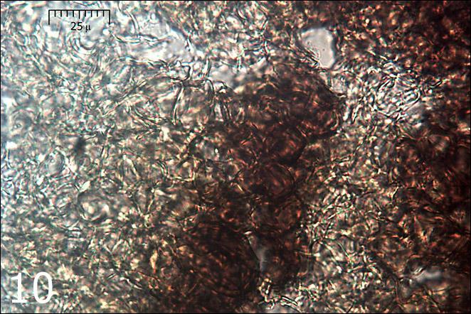
Based on spore dimensions (Me = 26 x 20.8 micr., n= 51) (Fig.7,8), their coarsely reticulated surface and tuber's cortex it could eventually be Tuber aestivum/uncinatum (Figs. 1-6). But the date of the find is very late, on November 3, and cross-section doesn't fit to what I can find in literature and internet (over-mature tuber?). It also had very faint smell. Also, it was not really buried in the ground. Gleba was almost 100% spore mass. I found only a few places where I believed I saw 'remnants' of asci (Fig.8, arrow). Variation in spore dimensions was enormous (Fig.8, from 21 to 45 micr. diameter). Tissue below cortex (Fig. 9, 10) was interesting, like a spatial 'hexangular' net (Fig.10) with no spores there. Can this really be Tuber aestivum?
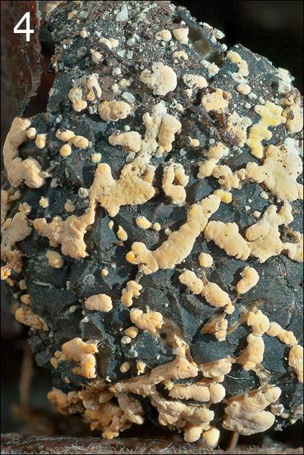
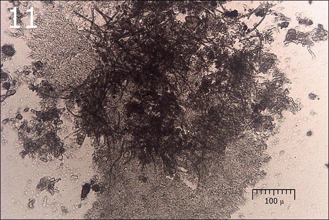
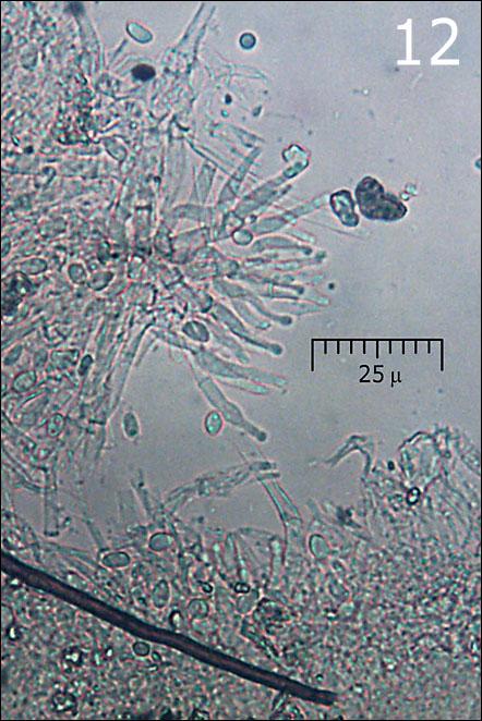
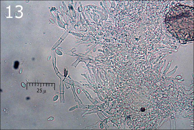
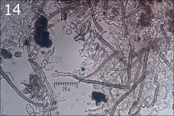
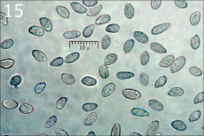
Orange blobs on tuber are probably a parasitic fungus, eventually an anamorph (Figs. 11-14) (Sympodiophora / Hypomyces ??). I've found neither asci nor basidia. Hypha is of two kinds, thin walled (Fig. 13) and yellow-brown thick walled (Fig.14). No clamps seen. Squashes of the blobs were full of small, smooth spores/conidia (Me = 5.7 x 3.4 micr., n = 40) shown on Fig.15. The surface of the blobs show a kind of flask shaped 'cystidia' with globose apex (Fig.12) - possibly conidiophores?
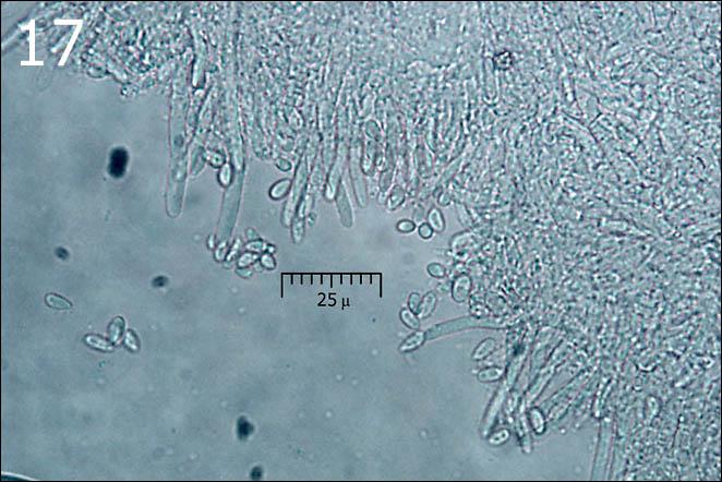
On the lower surface of the tuber I also noticed a few small (fi 1-2 mm) circular, cottony tufts of snow white hypha which seems to be a kind of mold (old tuber?). Under microscope I found only hypha without any other structure (Fig. 17) in them.
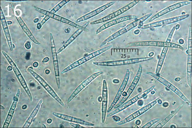
A third type of spores/conidia was also observed (Fig. 16) - very numerous, long and narrow, spindle shaped and septated (Me = 49.3 x 5.3 micr, n = 31; 3 - 7 septa, avg = 4.9, n = 35). They were everywhere, but seem to be associated mostly with the tuber's gleba (not sure). Where they actually belong I don't know.
All three types of spores/conidia were very numerous, so I exclude the possibility of an external contamination of the find.
I will be grateful to anybody for shedding some light on this interesting puzzle (for me).
Regards
Amadej
______________________
Habitat: mountain slope; Fagus sylvatica forest with some Picea abies; 590 m asl; in shade; colluvial, skeletal, calcareous ground; average temperature 7-9 deg C, average precipitations about 3.000 mm/year; alpine phytogeographical region.
Substratum: apparently on soil (not in!), but almost completely buried with ground humus, mostly rotten leaves of Fagus sylvatica.
Place: Trenta valley, East Julian Alps, Slovenia.
______________________


Best regards Walter
Warmest regards
Amadej
Please send it air-dry and not air-tight closed, because I shall be absent for 3 weeks.