
11-05-2026 12:32
 Bernard CLESSE
Pourriez-vous m'aider à identifier cette héloti
13-05-2026 15:26
 François Freléchoux
Bonjour,Voici une récolte faite il y a quelques j
12-05-2026 15:41
 Nicolas VAN VOOREN
Dear Ascolovers, especially interested in Pezizale
13-05-2026 12:05
 Thierry Blondelle
Bonjour à tous,J'aimerais avoir confirmation de c
10-05-2026 23:17
 Andreas Gminder
Hello,today we found in a moist steep decidous for
28-04-2026 20:07
 Lothar Krieglsteiner
... on twig in the air at standing Ceratonia siliq
27-04-2026 20:52
Lothar Krieglsteiner
Found on hanging tiwg of Olea europaea in dried-ou
11-05-2026 20:22
Lothar Krieglsteiner
on attached twig of standing Ficus caricaquite uns
29-04-2026 10:44
Lothar Krieglsteiner
growing at moist, drying-out soil at the side of a
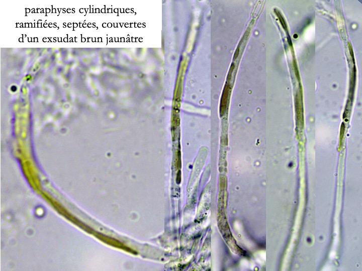
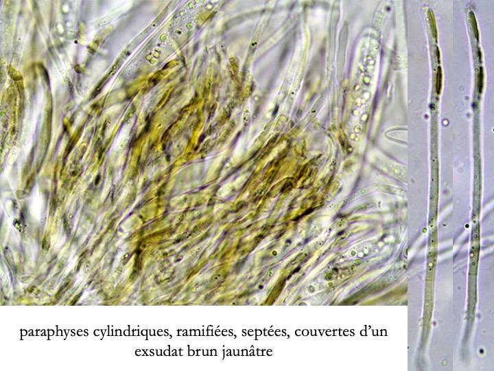
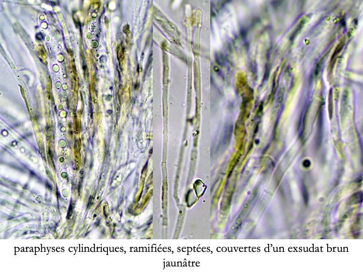
Hélotiale à paraphyses à exsudat brun jaunâtre
Bernard CLESSE,
11-05-2026 12:32
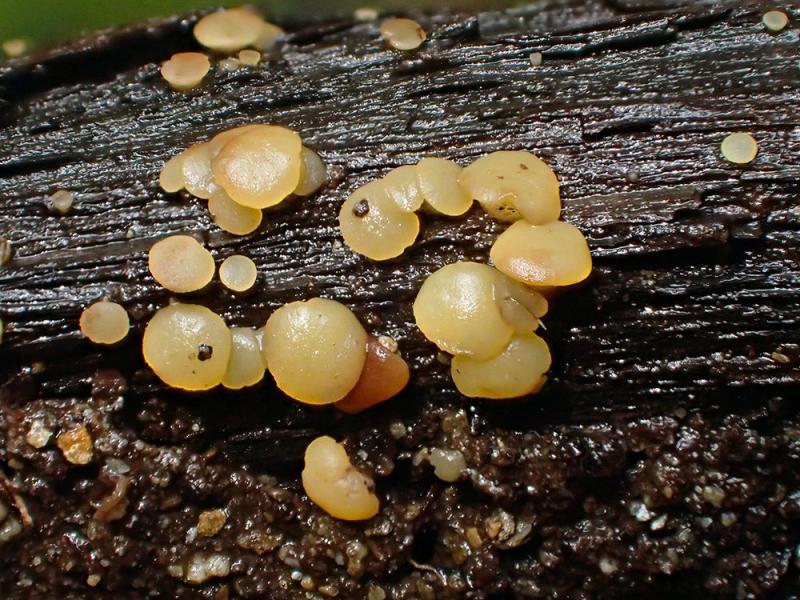
Pourriez-vous m'aider à identifier cette hélotiale dont les paraphyses sont couvertes d'un exsudat brun jaunâtre ? Récolte sur grosse branche pourrie, trempant dans un ruisseau.Merci d'avance !
Bernard
Andreas Gminder,
11-05-2026 13:56
Re : Hélotiale à paraphyses à exsudat brun jaunâtre
Bonjour,
je pense a une espèce du group Phaeohelotium imberbe, notament H. kathiae, meis assez vielli deja.
Je pense que ce n'est pas une couverture sur les paraühyses, mais des vacuoles internes, comprimé et detorié par l'age.
amicalement,
Andreas
je pense a une espèce du group Phaeohelotium imberbe, notament H. kathiae, meis assez vielli deja.
Je pense que ce n'est pas une couverture sur les paraühyses, mais des vacuoles internes, comprimé et detorié par l'age.
amicalement,
Andreas
Bernard CLESSE,
11-05-2026 14:59
Re : Hélotiale à paraphyses à exsudat brun jaunâtre
Grand merci pour ton avis, Andreas !
Cordialement,
Bernard
Cordialement,
Bernard
Camille Mertens,
11-05-2026 20:40
Re : Hélotiale à paraphyses à exsudat brun jaunâtre
Bonsoir Bernard.
Je suis d'avis d'Andreas.
Voici une fiche d' Hymenoscyphus Kathiae.
Camille
Je suis d'avis d'Andreas.
Voici une fiche d' Hymenoscyphus Kathiae.
Camille
 Sans-nom-1-0001.pdf
Bernard CLESSE,
11-05-2026 20:44
Re : Hélotiale à paraphyses à exsudat brun jaunâtre
Grand merci pour ton avis, Camille !
Amitiés,
Bernard
Amitiés,
Bernard
Patrice TANCHAUD,
13-05-2026 09:05
Re : Hélotiale à paraphyses à exsudat brun jaunâtre
Bonjour,
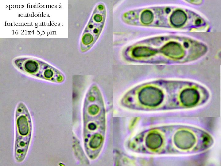
je reviens sur cette récolte de Bernard qui annonce des spores de 16-21 de long, et le nom d'Hymenoscyphus kathiae semble faire l'unanimité. Dans le DVD de Zotto, aussi bien dans la clé que sur ses fiches, les spores mesurent 9-15,5 de long.
Il y a quelque chose qui m'échappe...
Patrice
Andreas Gminder,
13-05-2026 09:18
Re : Hélotiale à paraphyses à exsudat brun jaunâtre
Bonjour,
ah, je n'ai pas vu des mesurements des spores ....
Cet mesurements, ils sont correct? Si oui, ce n'est pas H. kathiae, et même pas une espèce du imberbe group peut-être. Hymenoscyphus laetus?
amicalement,
Andreas
ah, je n'ai pas vu des mesurements des spores ....
Cet mesurements, ils sont correct? Si oui, ce n'est pas H. kathiae, et même pas une espèce du imberbe group peut-être. Hymenoscyphus laetus?
amicalement,
Andreas
Bernard CLESSE,
13-05-2026 09:39
Re : Hélotiale à paraphyses à exsudat brun jaunâtre
Bonjour Andreas et Patrice,
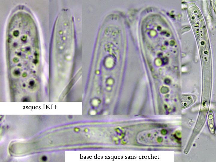
Oui, mes mesures de spores sont correctes. Mais un autre problème se pose, je n'ai pas trouvé de crochet à la base des asques or Patrice en voit (cf. sa fiche : https://www.mycocharentes.fr/pdf1/2227.pdf)
Ou alors, j'ai mal cherché…… :o)
Bernard
Oui, mes mesures de spores sont correctes. Mais un autre problème se pose, je n'ai pas trouvé de crochet à la base des asques or Patrice en voit (cf. sa fiche : https://www.mycocharentes.fr/pdf1/2227.pdf)
Ou alors, j'ai mal cherché…… :o)
Bernard
Bernard CLESSE,
13-05-2026 10:16
Re : Hélotiale à paraphyses à exsudat brun jaunâtre
Je viens de revérifier sur une vingtaine d'asques : aucun crochet visible à leur base.
Bernard
Bernard
Bernard CLESSE,
13-05-2026 21:00
Re : Hélotiale à paraphyses à exsudat brun jaunâtre
Que pensez-vous de cette idée : Phaeohelotium fulvidulum pour ma récolte ?
La forme, la taille et les guttules des spores conviendraient bien de même que l'absence de crochet à la base des asques ansi que l'écologie et la macro ?
Merci d'avance pour vos avis !
Bernard
La forme, la taille et les guttules des spores conviendraient bien de même que l'absence de crochet à la base des asques ansi que l'écologie et la macro ?
Merci d'avance pour vos avis !
Bernard
Patrice TANCHAUD,
13-05-2026 21:55
Re : Hélotiale à paraphyses à exsudat brun jaunâtre
Tout semblerait convenir, mais j'ai toujours vu les paraphyses garnies de guttules réfringentes chez cette espèce.
Patrice