
12-04-2026 17:56
 Hardware Tony
Found on dead stems in February earlier this year
12-04-2026 15:52
Gernot FriebesHi,I'm looking for help with this anamorph collect
12-04-2026 12:22
 William Slosse
In a dune grassland in Oostduinkerke (Belgium), on
11-04-2026 15:45
 Zuzana Sochorová (Egertová)
Please, could anyone send me this paper?Moyne G.,
11-04-2026 13:34
Artem PtukhaHello, I am seeking assistance with the identific

11-04-2026 10:19
 Michel Hairaud
Chers amis d'Ascofrance , voici une très bonne no
11-04-2026 10:10
Michel Hairaud
Dear Ascofrance members, here is some very good ne
10-04-2026 23:22
Gernot FriebesHi,ascospores are 1- to 3-septate, approximately
10-04-2026 15:51
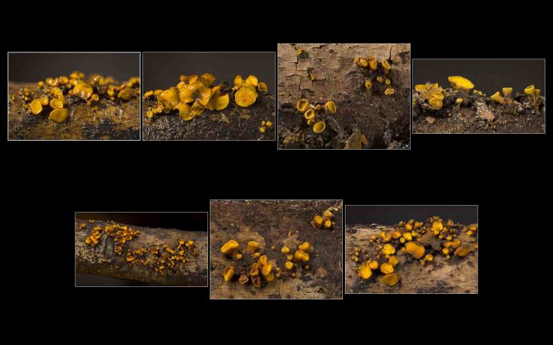
William Slosse
Hello everyone, On 08/04/26, I found a growth sit

Loc: Plantation Trail, Jean Lafitte National Historical Park and Preserve, Louisiana, USA
Coll: D. Newman, R. Cronce & K. Thorstad
Substrate: on dead, downed, corticate, firm/undecomposed, hardwood stick
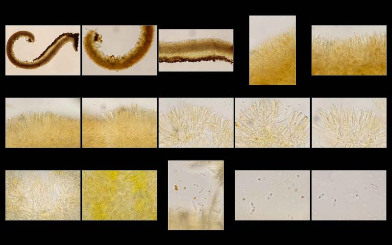
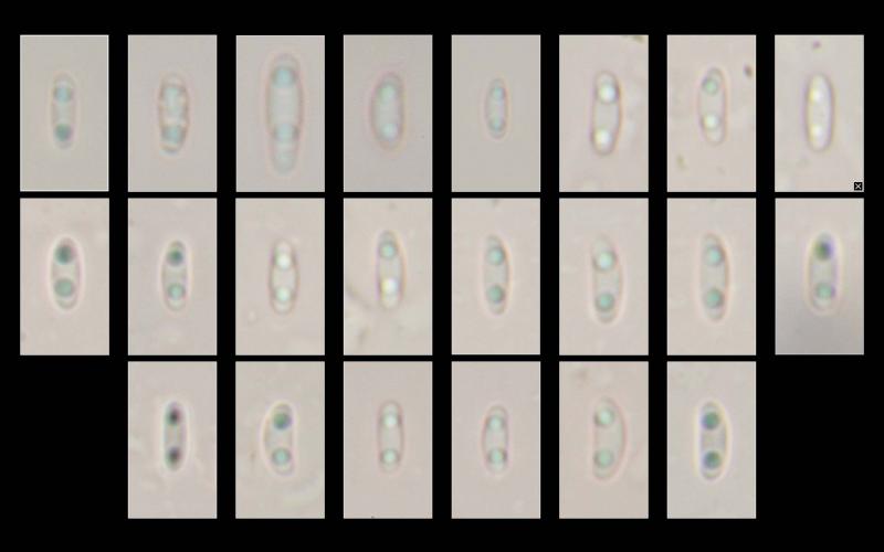
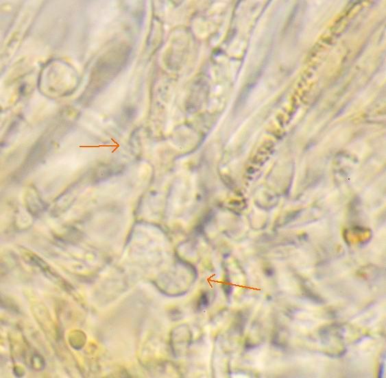
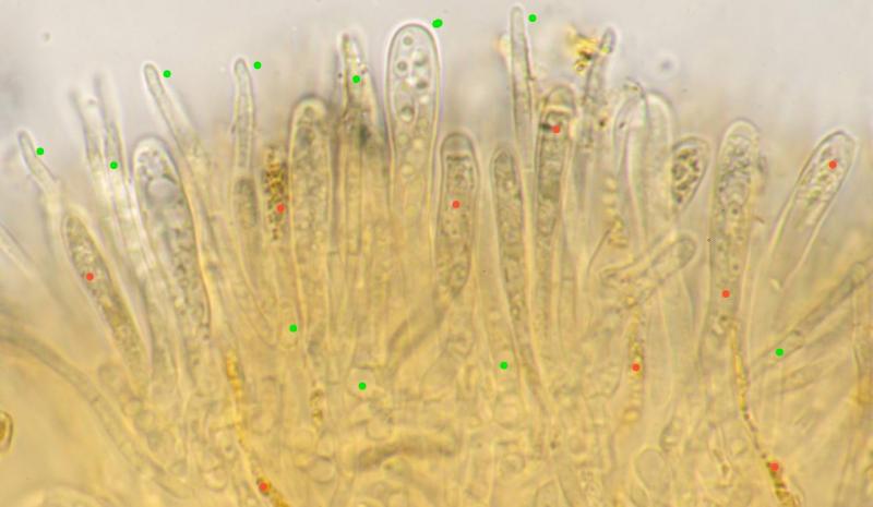
all mounts made in KOH. excipulum composed of irregularly inflated/swollen chains of cells (asexual propagules as in I. irregularis?); context composed of long, slender, even hyphae, possibly embedded in a gelatinous matrix; paraphyses filiform and short, only slightly exceeding asci; both hymenium and abhymenium appear to contain ionomidotic granules/contents; asci biseriate; spores bi-guttulate and (probably) aseptate.
Spores:
(5.1) 5.3 - 6.5 (6.8) × (2.1) 2.2 - 2.6 (2.7) µm
Q = (2) 2.2 - 2.9 (3) ; N = 20
Me = 5.9 × 2.4 µm ; Qe = 2.5
GIF of ionomidotic reaction available at https://inaturalist-open-data.s3.amazonaws.com/photos/347983283/original.gif
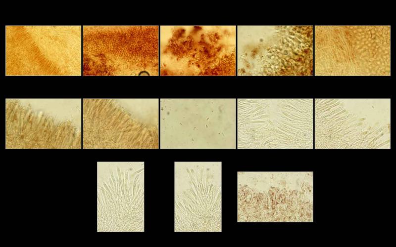

Two new things I've noticed in these recent mounts: 1: the paraphyses are multi-septate, and 2: many contain bright yellow, refractive contents. I had observed it rather vaguely in the hymenium before, but not inside the paraphyses.
Thank you for sharing these high-value images. I would like to sequence this specimen, it would help to clear the mysterious taxonomy in this group...
If you like to send a piece of specimen to our lab, please, use this address:
Kadri Pärtel
Chair of Mycology
Department of Botany
Institute of Ecology and Earth Sciences
University of Tartu
Oecologicum
J. Liivi St. 2
50409 Tartu
Estonia
kadri.partel@ut.ee
Phone +372 5226179
THANK YOU FOR COOPERATION!

https://inaturalist.org/observations?place_id=any&q=Ionomidotis%20cf.%20fulvotingens&search_on=tags&user_id=ikhom&iconic_taxa=Fungi
I kept the first two speciments.
I wonder about I. fulvotingens that grows on Pinus strobus. Has it been ever sequenced? I found a specimen in Feb 2020 and I kept it. (https://inaturalist.org/observations/38581250)
Thank you for sharing your found.
I checked my data: all specimens we have studied and sequenced are on hardwood. It would be intresting to add one from a conifer.
Best regards,
Kadri
(5.1) 5.3 - 6.5 (6.8) × (2.1) 2.2 - 2.6 (2.7) µm
Q = (2) 2.2 - 2.9 (3) ; N = 20
Me = 5.9 × 2.4 µm ; Qe = 2.5