
11-05-2026 12:32
 Bernard CLESSE
Pourriez-vous m'aider à identifier cette héloti
13-05-2026 15:26
 François Freléchoux
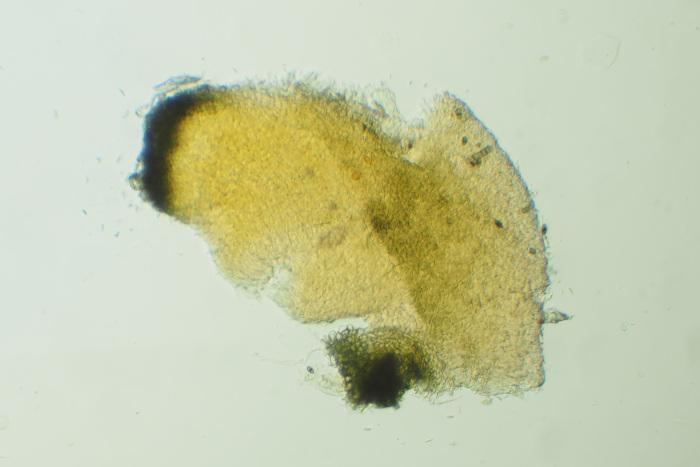
Bonjour,Voici une récolte faite il y a quelques j
12-05-2026 15:41
 Nicolas VAN VOOREN
Dear Ascolovers, especially interested in Pezizale
13-05-2026 12:05
 Thierry Blondelle
Bonjour à tous,J'aimerais avoir confirmation de c
10-05-2026 23:17
 Andreas Gminder
Hello,today we found in a moist steep decidous for
28-04-2026 20:07
 Lothar Krieglsteiner
... on twig in the air at standing Ceratonia siliq
27-04-2026 20:52
Lothar Krieglsteiner
Found on hanging tiwg of Olea europaea in dried-ou
11-05-2026 20:22
Lothar Krieglsteiner
on attached twig of standing Ficus caricaquite uns
29-04-2026 10:44
Lothar Krieglsteiner
growing at moist, drying-out soil at the side of a
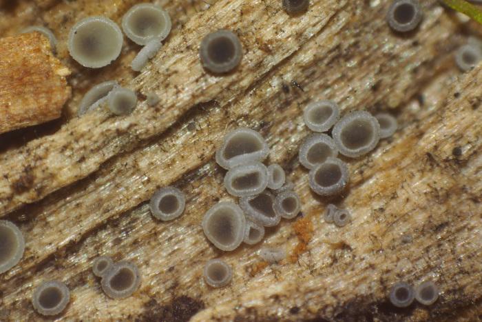
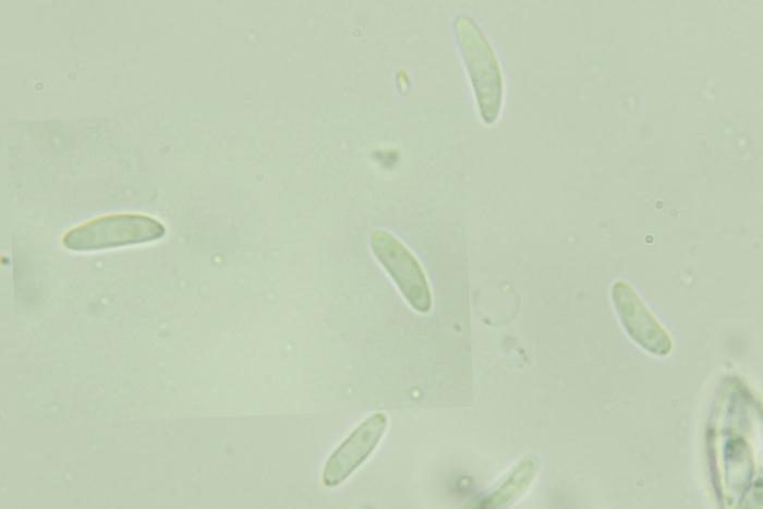
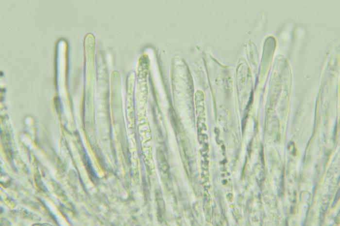
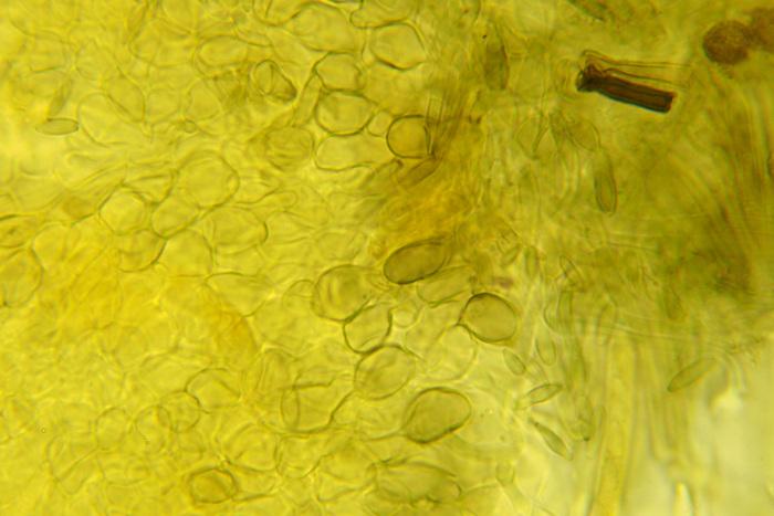
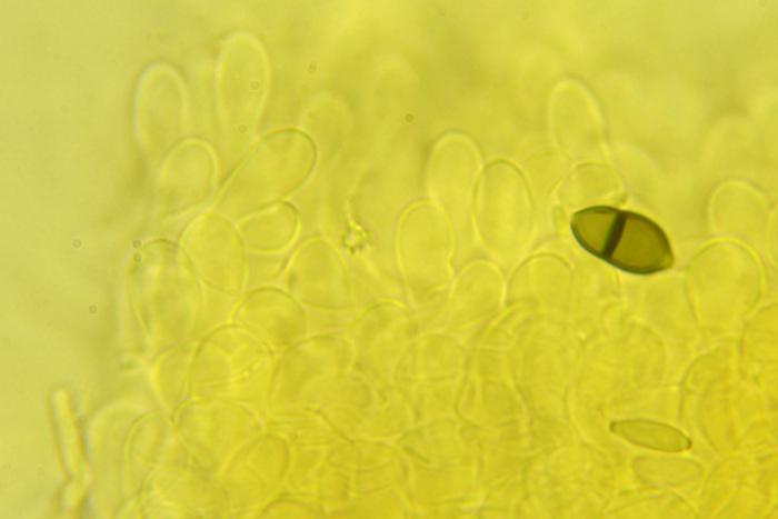
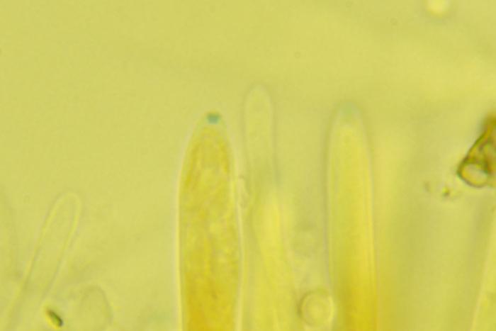
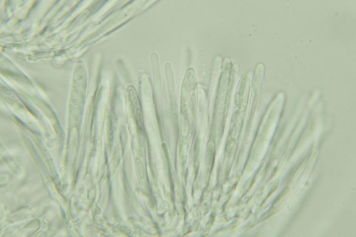
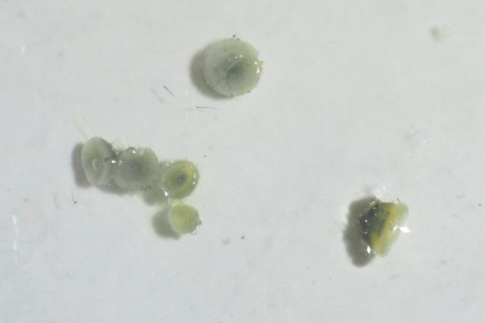
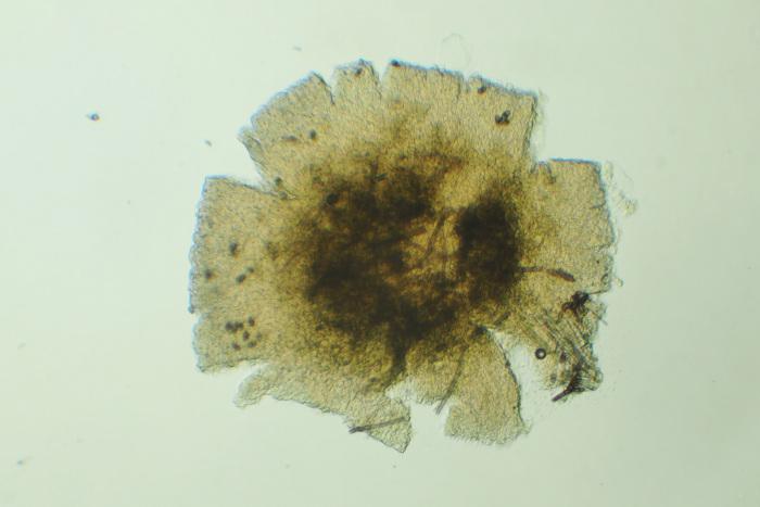
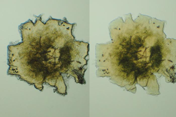
These grew on a fallen, decorticated hardwood branch. The size of the apos is much smaller than of the typical gray ones that I find often on moist wood. There was a faint yellow reaction with 40% KOH, but under the microscope in 3% KOH I didn't notice anything.
Spore measurements: (7.9) 8.9 - 10.7 (10.8) × (2.9) 2.93 - 3.1 (3.2) µm, N = 8.

This is not a good idea.
Mollisia is difficult but among them you can find a lot of interesting species.
Do not cover the preparation with a slide when checking the reaction to KOH. Put a drop of KOH on the slide slide and then dip a piece of ascocarp in it. If the fruiting body reacts to KOH, you will see a yellowish coating around it in a few seconds.
The second way is to apply a drop of KOH to the fruiting body hymenium. If the reaction is positive, the hymenium will change to +/- yellow.
The reaction is visible to the naked eye or after applying a magnifier.
To be sure, it is worth using both methods at the same time
Good luck
Mirek