
22-04-2026 01:06
 Richard VALERI
Bonjour à tous.Je vous présente cette Nectria s.
21-04-2026 22:14
Margot en Geert VullingsThis cup fungus was found on April 10, 2026, on lo
21-04-2026 13:36
Gernot FriebesHi,I am out of ideas for this one. I collected Sal
21-04-2026 13:19
Gernot FriebesHi,this Lophodermium on Typha has ascospores measu
21-04-2026 13:05
Gernot FriebesHi,this hyphomycete feels familiar but I was not a
20-04-2026 22:00
 Malcolm Greaves
These pale yellow, hairy ascos were growing on cul
19-04-2026 21:23
Steve ClementsBonjour, I found this anamorphic fungus on old pl
19-04-2026 20:46
Steve Clements1 mm diameter approx spherical conidiophores on pl
12-04-2026 17:56
 Hardware Tony
Found on dead stems in February earlier this year

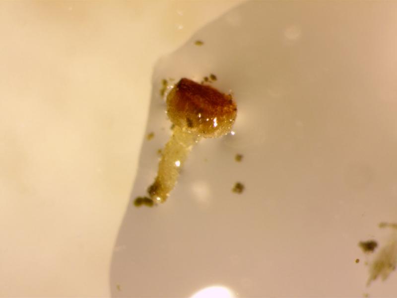
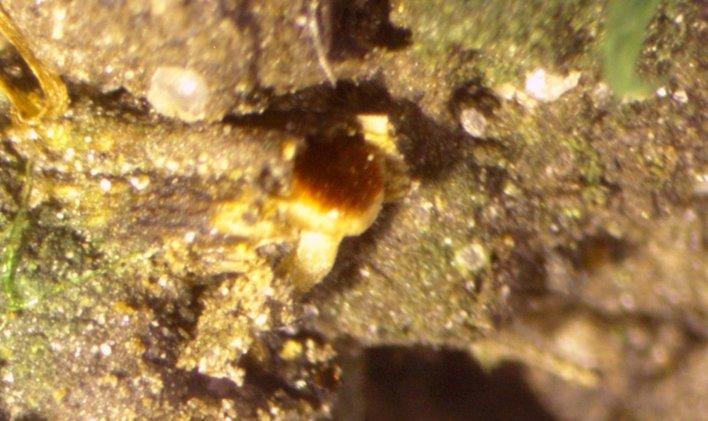
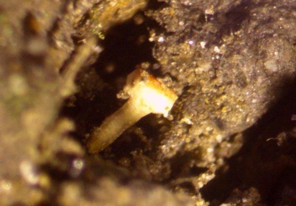
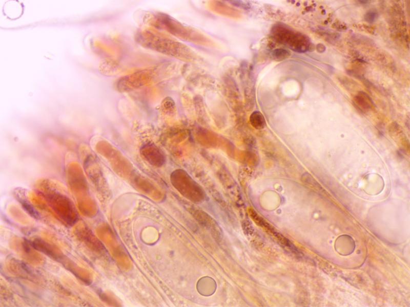
Hi All,
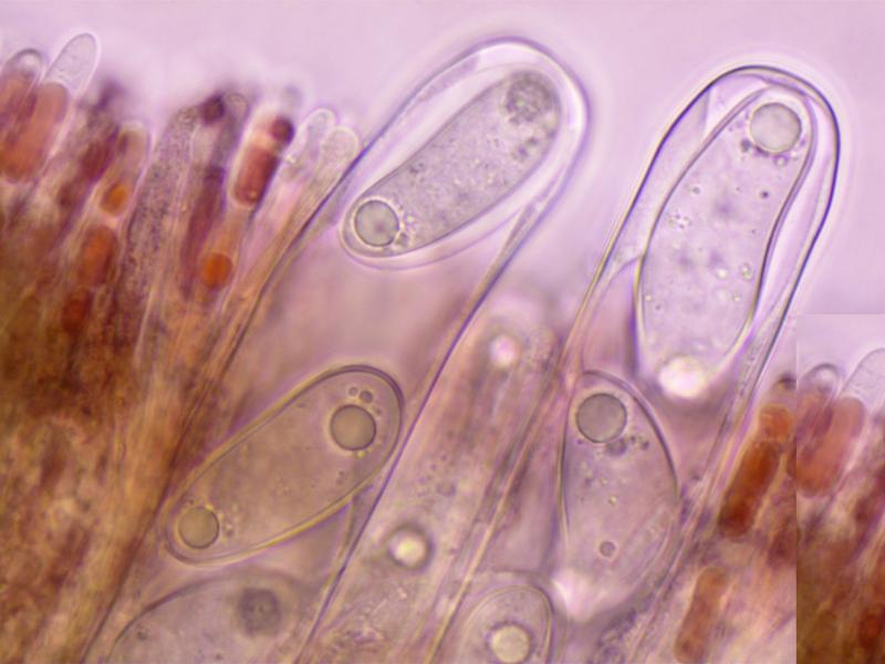
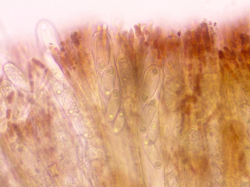
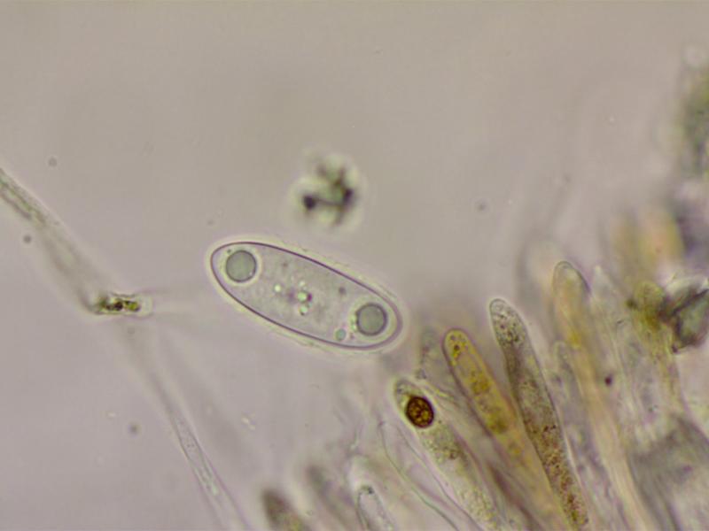
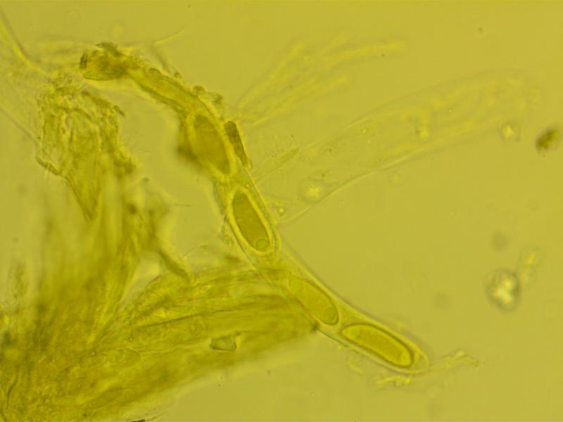
I have found a strange and distinctive Ascomycete growing on damp soil close to a brook. Only a single apothecium thus far.
Apothecium: c1x0.5mm. Hymenium convex, bright red, not clearly demarced from the excipulum.
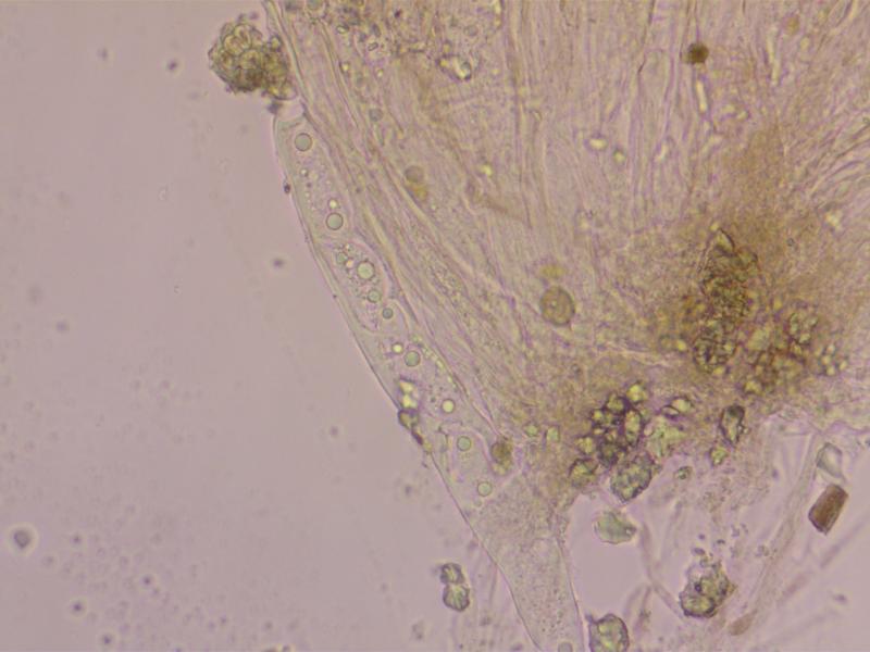
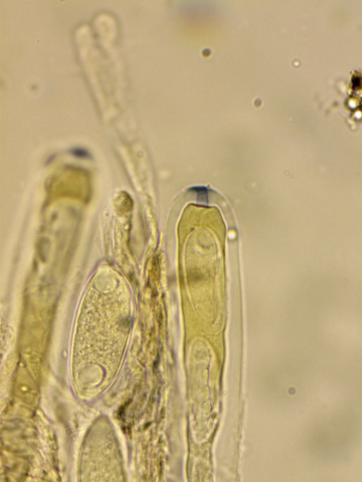
Asci: Cylindric, 250-325x18-25. 4-5 spored, uniseriate, with differetial maturation of the ascospores. Ascus pore blueing in Melzer' after treatment with KOH.
Ascospores:44-50x15-19. Ellipsoid-cylindric, some curved and some with strangely attenuated apices. With polar guttules. With a gelatinous coat.
Paraphyses with red pigment, swollen to 6-7.
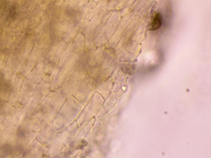
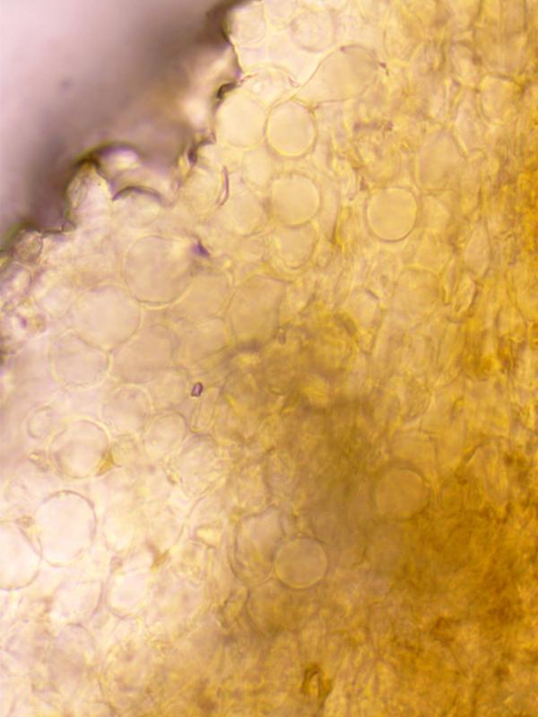
Excipulum: Textura Globosa.
I would be very interested in any thoughts on this. Thanks in advance.
Charles.
 rsz-red-stipitate-disco-asci-7-rhiwlas-pentraeth-7420-0001.jpg
Hi Zotto,
Yes, the t. prismatica is from the stipe. Have not tested with Lugol and very little material remains (on a couple of slides) Will definitely look out for more material-not far from where I live in Pentraeth, Anglesey.
Hi Zotto,
Its the ascus width that I had got wrong-it should be 250-325x18-25, so really quite large. Being Sclerotiniaceous I imagine the fungus would have been associated with plant material perhaps with a sclerotium but, unfortunately it quickly became detached from the substrate. There was some Mnium hornum fairly close but I don't think the fungus was associated with it. The site was in broad leaved woodland with ash etc.