

05-05-2026 22:40
Gernot FriebesHi,I believe this is a Plagiostoma growing on a Sa
06-05-2026 11:25
 Castillo Joseba
Me mandan el material seco de Galicia (España) re
06-05-2026 17:23
Thomas Læssøehttps://svampe.databasen.org/observations/10594257
28-04-2026 20:07
 Lothar Krieglsteiner
... on twig in the air at standing Ceratonia siliq
04-05-2026 18:13
 Stephen Martin Mifsud
ID request for what seems to be a true aquatic fun
04-05-2026 16:39
Stephen Martin Mifsud
ID request: This specimen was collected in Malta o
28-07-2011 18:31
 Alex Akulov
Dear FriendsToday I made the pdf file of Velenovsk
04-05-2026 09:50
Castillo Joseba
Me mandan el material seco de Galicia,(España) re
 Bonsoir à tous,
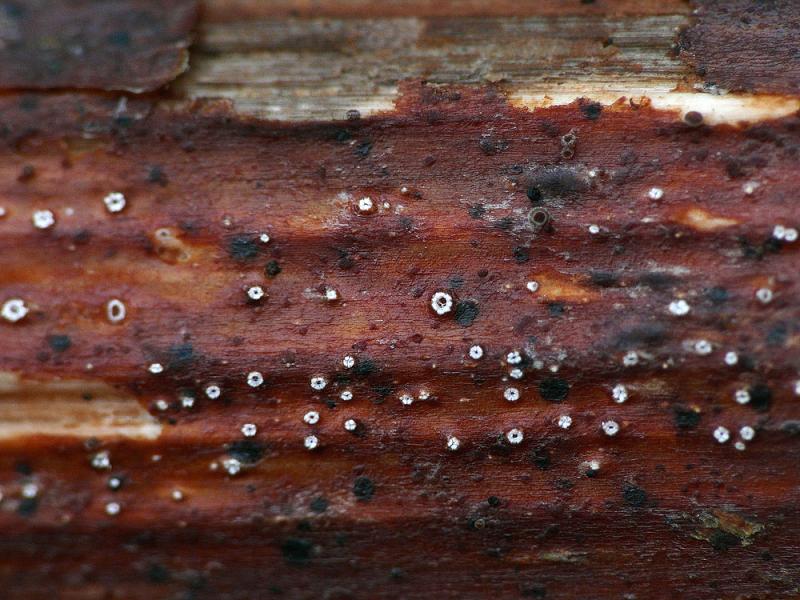
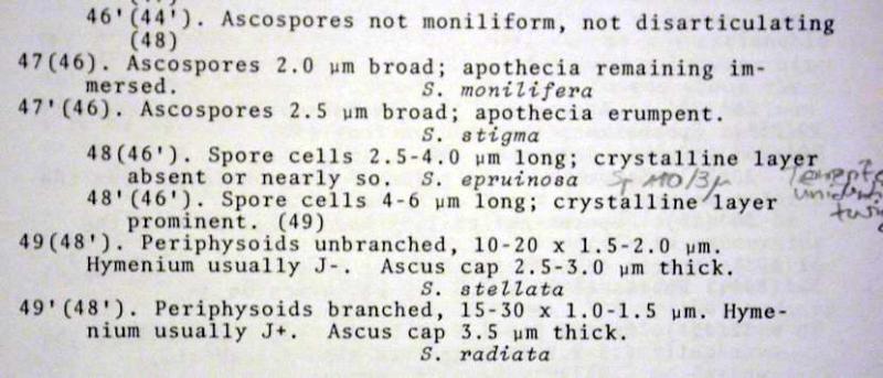
Suite et bientôt fin de mes récoltes sur renouée du Japon. Ici je pense à Stictis radiata, d'abord parce que les apothécies sont nettement plus petites (max. 0,3 mm de diamètre) que chez Stictis stellata (diamètre parfois dépassant le mm) montré tout récemment sur Fallopia japonica toujours et que d'autre part, les spores sont ici larges de 2-2,5 mu.
Qu'en pensez-vous ?
Bernard

Thanks for your message !
I will try to make a cut of apothecium but I admit that I do not know what are the periphyses. Would you possibly a drawing or a microscopic picture so that I know what I have to observe? I will try to redo pictures spores alive.
Regarding the iodine reaction, I just noticed a very slight blue color and diffuses into the ascus (especially upper half) with lugol but not a color "deep blue" suitable for this species.
Bernard
What is meant with periphysoids (not periphyses, sorry) you can see here.
Sherwoods distinction is perhaps a bit weird, and her opinion about amyloidity may be wrong as she appears not to have understood the influence of KOH and Melzer's.
But I must admit I have no clear concept of thse two species.
Thank you for your explanations !
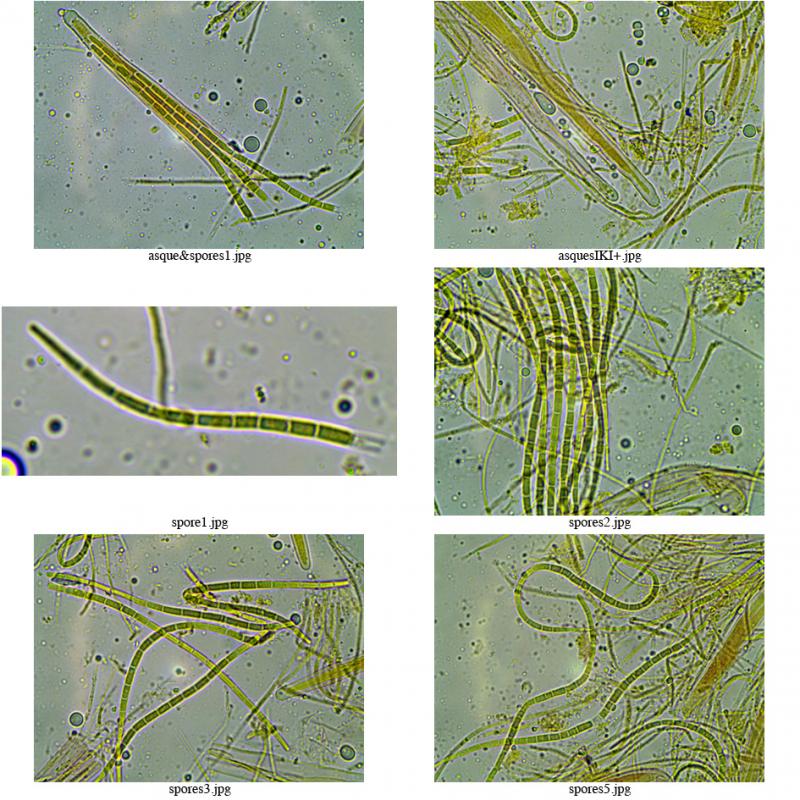
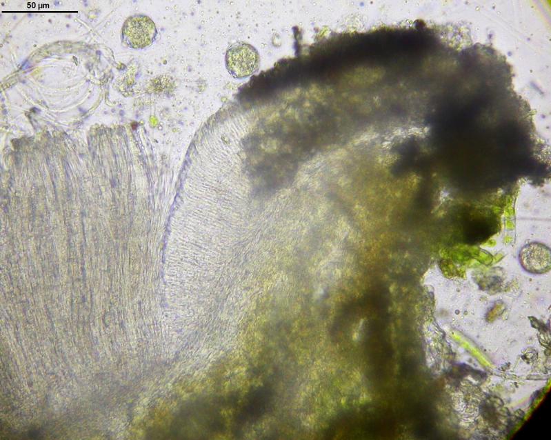
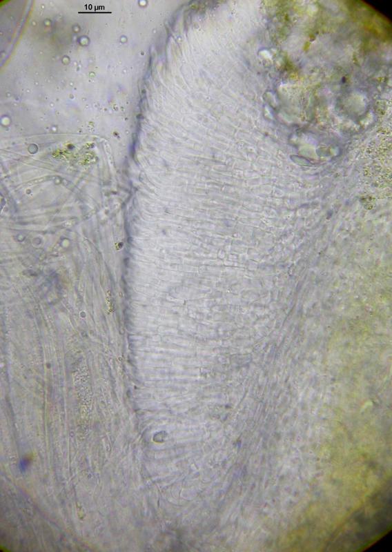
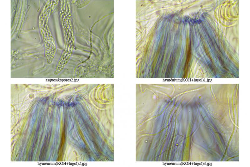
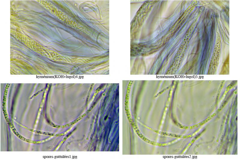
Here are some new pictures :
a) hymenium+KOH+lugol : well blue !
b) ascus+lugol : dextrinoid; ascus cap : 3,5 mu thick
c) spores guttulated
What do you think about that ?
Bernard
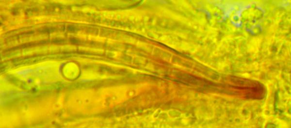
A red reaction inside the ascus could be due to glycogen and would then be called dextriniod, but then it should be seen also after KOH, or in Melzer without KOH. What reacts blue after KOH (I think the ascus wall surface) must be red in IKI without KOH (hemiamyloid). See my homepage:
http://invivoveritas.de/articles/iodine-reaction-in-ascomycetes-why-is-lugols-solution-superior-to-melzers-reagent/
Fig. 2 and 5.