

19-05-2026 10:27
Patrice TANCHAUDBonjour, récolte récente sur terre retournée i
04-06-2026 18:39
Gernot FriebesHi,I collected this species in two different locat
22-05-2026 13:29
Gernot FriebesHi,I am curious to hear your opinion on this mater
04-06-2026 10:50
 François Freléchoux
Bonjour, J'ai trouvé hier un petit asco observé
04-06-2026 07:02
François Freléchoux
Bonjour, Voici la description d'une espèce qui p
04-06-2026 13:34
Gernot FriebesHi,I am interested to hear your opinion on this Le
04-06-2026 11:36
Gernot FriebesHi,found on Vaccinium myrtillus.Asci: IKI –, 8-s
I thought with this special substrate it may be easy. But No...
Any help welcome!
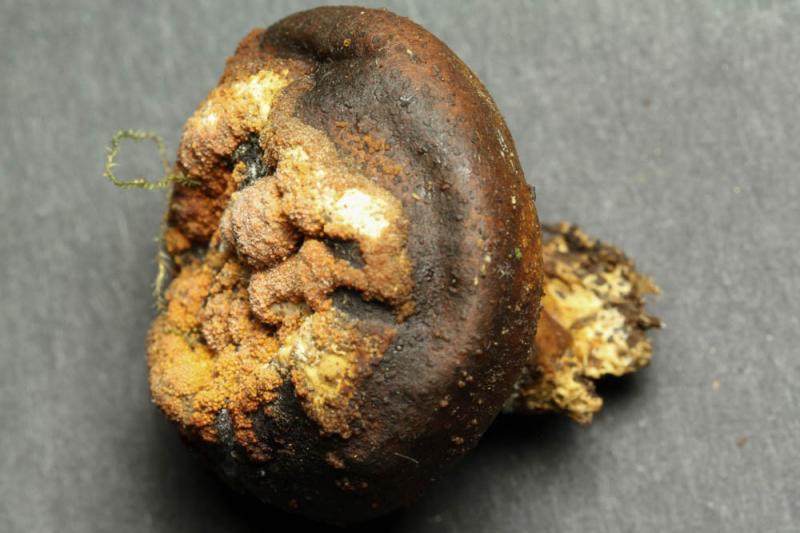
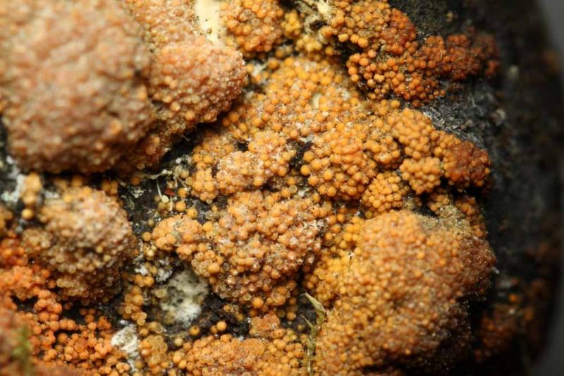
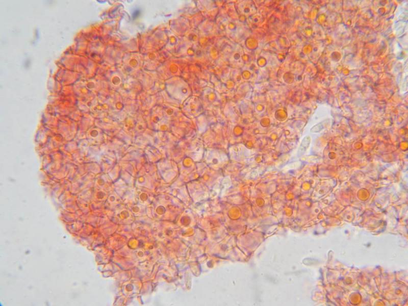
Substrate: The Basidiomycete Scleroderma sp.
Description:
Perithezia 0.2-0.25 mm Ø, bright orange, superficial, densely aggregated but not immersed in a stroma.
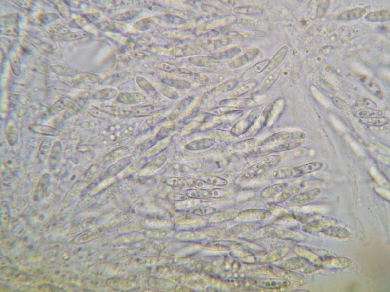
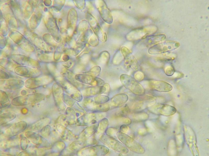
Spores 9-11 x 3-4 µm, 2-celled, hyaline, finely and indistinctly verrucose. Perithecial wall consist of round to angular cells of 5-10 µm diameter, containing large orange guttules.
Many thanks for inputs,
Stefan

Hi Stefan,
Have you test KOH ? It could be more one Bionectriaceae than Nectriaceae.
Alain
There was no reaction in KOH. So yes, likely Bionectriaceae...
Stefan

with orange oily droplet in the wall and hymenium, it is the genus Bionectria.
Your specimen looks like Bionectria solani but the ascomata should be seated on a basal, pseudoparenchymatous stroma, that seems the case in your images. Could you check this characteristic?
Christian
That is the case, yes.
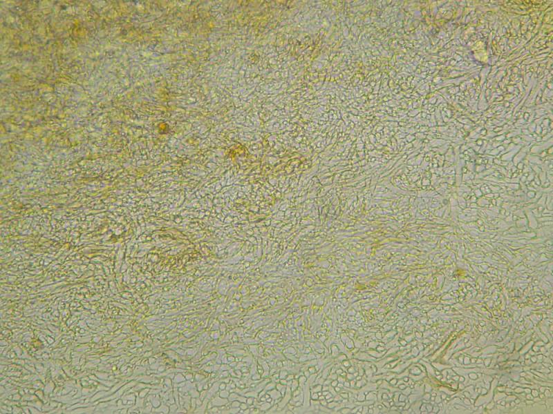
Here is a picture of the stromatic tissue beneath the perithecia.
Bionectria solani seems to have a very wide ecology. So I guess it ist possible on Scleroderma. I will have a close look at the species. Do you by any chance have a full description of B. solani.
Thank you very much
Stefan
Hi Stefan,
You have all informations in SIM 46, on line.
Alain
Merci bien, Alain et Christian!
Stefan