
30-05-2026 21:12
Philippe PELLICIERSur branche de mélèze (Larix) près de la neige,
31-05-2026 10:35
Hulda Caroline HolteHello,I collected this species growing on a rather
25-05-2026 16:35
 Bernard CLESSE
Bonjour à toutes et tous,J'ai trouvé récemment,
29-05-2026 15:35
daniel FERREBonjour à tous,Je voudrais votre aide pour cette
28-05-2026 16:15
James MitchellHello,Does anyone have the original publication of
28-05-2026 11:06
Thomas Læssøehttps://svampe.databasen.org/observations/10596750
23-05-2026 11:44
 Charles Grapinet
Hello, I am having trouble identifying this copro
25-05-2026 16:44
François BartholomeeusenHi forum members,During an excursion organised by
26-05-2026 21:25
Dirk GerstnerHello everyone, I'm completely stumped by this li
26-05-2026 22:44
Ethan CrensonHi all, I think I have Incrucipulum capitatum her
 Hello,
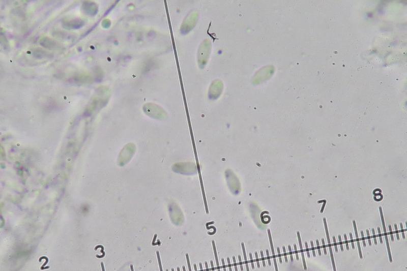
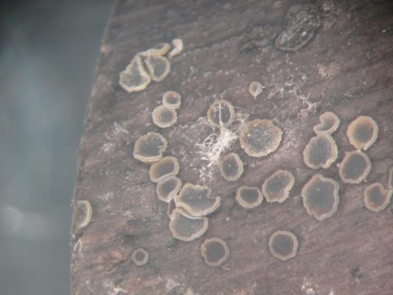
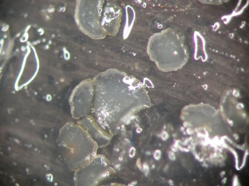
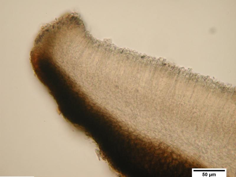
I found these small discs (max 0,8µm) between another unknown species on wood of populus. Asci 38/50x6/7 and small spores 4/6x2. J+
Anybody an idea?
regards,
Ralph

Andy

Zotto
Andy
Thx for your answer. Fits indeed perfect.
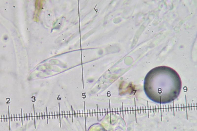
PS the large drop is indeed very good visible (this is not mentioned in E&E...)
regards,
Ralph
Put KOH to the water mount and the drop disappears. Likewise when killing by squashing.

Just to cast a little bit of doubt into the identification...
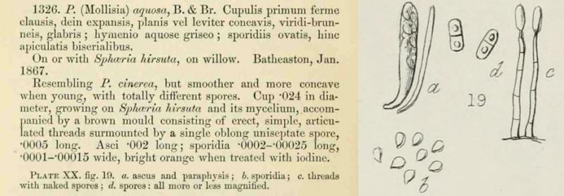
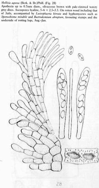
The original description of Peziza (Mollisia) aquosa describes egg-shaped ascospores with a single basal point (if my Latin translation is correct), as also shown in the illustrated plates (attached), and here:
http://biodiversitylibrary.org/item/78387#page/448/mode/1up
http://biodiversitylibrary.org/item/78387#page/509/mode/1up
So while Ralph's collection does fit the concept of Mollisia aquosa in the sense of Ellis & Ellis, I don't think this is the same species that was originally described under that name.
I have a collection of Mollisia dextrinospora (identical ITS sequence to the type, but morphologically a Pyrenopeziza) that looked very similar to Ralph's collection when fresh, although the ascospores in my collection are perhaps a little bit wider, and polar oil droplets a little bit more visible. The following AscoFrance threads also have what appears to be Mollisia dextrinospora.
http://www.ascofrance.com/forum/11799/difficult-inoperculate-discomycete
http://www.ascofrance.com/forum/23320/mollisia
So, I think that Ralph's collection is either Mollisia dextrinospora, or a closely related sibling species. I also suspect that the original Mollisia aquosa and Pyrenopeziza "ulicis" belong to the same group of Pyrenopeziza species due to similar morphology and lignicolous habitat - there is quite a few candidate groups based on DNA sequences.
I hope that helps.
Cheers,
Brian
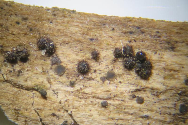
thank you very much for your interesting ideas. This is a frequent case: you think you know a species quite well, and you apply a name with a certain amount of hesitation because the original description is insufficent and a redescription of the type has not been made, as I suppose here. I agree that the spore drawing in the protologue does not match the present species. Nevertheless, I often experienced strong differences between type description and type material, which raises in the worst case the question whether a mixtum occurred in the type material or other confusion happened.
I fear that the description by Ellis & Ellis (see below) might perhaps be another species, possibly P. "ulicis". The two drops remote from the poles would point to this. But the available data are insufficient, for instance, are the asci amyloid, do they have croziers?
I know the description of Mollisia dextrinospora but never understood what it was. What I take from the description is that the asci are said to be inamyloid (pretreated with KOH), and arise from simple septa. Both features would exclude my P. aquosa. A dextrinoid reaction of the spores I never saw, but maybe this is only shown when using MLZ.
Now when you have a collection that shares the genetics with the type, could you please show us the morphology by photos or drawings? Did you obtain the dextrinoid reaction? I see three ITS sequences in GenBank, and I suppose that none of them concerns the type? So did you sequence the type yourself?
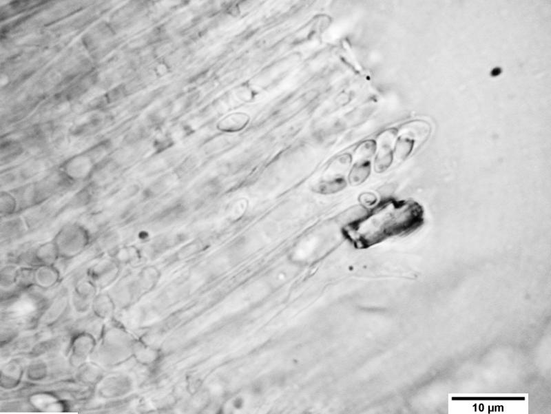
The two links you gave obviously refer to this P. "ulicis" (the link by Miguel I was unaware). Ralph drew my attention to the fact that the large drop below the spores is absent from those photos. This is true and a problem, because the drop is actually not constant. I did not see it, e.g., in my HB 6563 as well. Also in Psilachnum, where this drop is common, it is not always there. But you will never see it in a typical Mollisia I want to say.
Yes, it is a pity that so many species still remain to be sequenced. Perhaps Andreas Gminder has unpublished data on them too.
Zotto
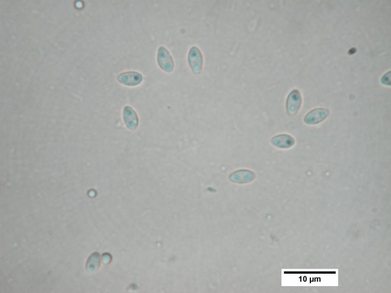
Mollisia dextrinospora, when fresh, has a blue ascus pore reaction in IKI, rather than the red reaction described in the protologue (in link).
http://www.librifungorum.org/Image.asp?Nav=yes&FirstPage=46056&LastPage=46319&NextPage=46252
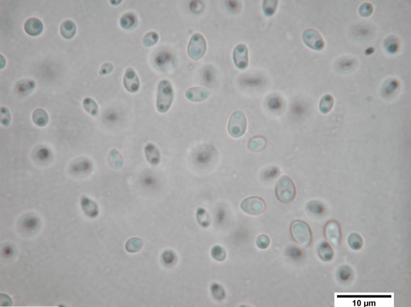
The living ascospores are also almost exactly like P. "ulicis", although without any visible warts that I could see; they always possess two distinct droplets at the poles, and appear to be consistently dextrinoid when exposed to IKI. I suppose it is possible that Mollisia dextrinospora and Pyrenopeziza "ulicis" could be the same species, but P. "ulicis" would really need to be sequenced to properly support this claim. Regardless, it is morphologically and phylogenetically a Pyrenopeziza, although in a basal clade to other Pyrenopezizas found on more diverse substrates.
Unlike the protologue , the ascospores are not visibly 1-septate, but could become visibly septate with age or dessication. They may have some sort of non-visible septum present when fresh, which could explain why the droplets in the ascospores are confined to the poles. As far as I can see, the difference in the morphology of my fresh collection and the protologue can only be due to dessication or KOH pretreatment.
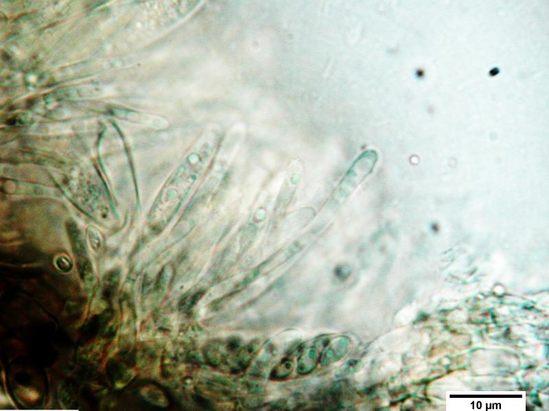
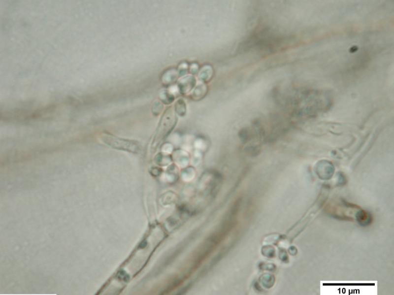
Mollisia dextrinospora also produces a Cadophora fastigiatia-like phialidic anamorph (it is very close to the strain currently viewed as C. fastigiata, although this strain has an ITS sequence identical to the isotype of C. melinii). The phialospores/spermatia are similar to the 1-septate conidia described in the protologue of Mollisia aquosa, although the described conidiophore in the protologue does not have a pronounced collarette (perhaps because collarettes develop with repeated production of conidia).
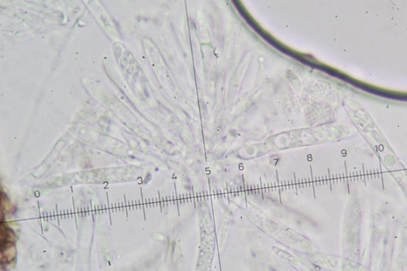
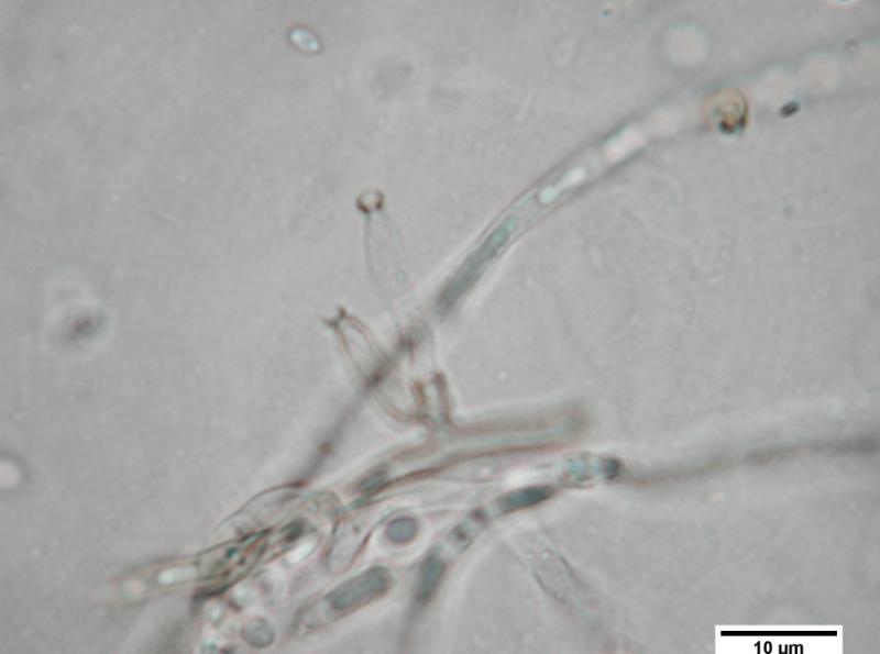
There are some indications of crosier-like structures, but I have not seen any extremely distinct examples.
Of the three GenBank sequences, only one is from an apothecium (the type) – the rest are just isolates with similar ITS sequences.
I have seen the large drop in the ascus in a number of Pyrenopeziza species – I don't think it is always present, but maybe it is an important structure produced at a particular phase in the development of their asci?
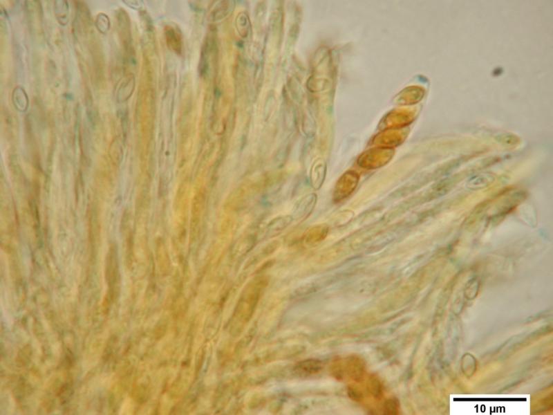
Attached are a few images of my collection – I'll be publishing this as part of a bigger phylogeny of Pyrenopeziza when I get some time to finish off the manuscript, but you're welcome to add them to your DVD/image library if you want!
Cheers,
Brian
I hope you have more Pyrenopeziza sequences for comparison than those of P. brassicae in GenBank. Good to hear that it falls in vicinity of those.
For the protologue of M. dextrinospora I can only find the remark J-, and that the pore does not blue even when KOH-pretreated. So I see a distinct difference in this point. How high is the similarity in the ITS region between your isolate and the type?
On your photo a red reaction of the spore wall is visible though I assume it would be more distinct in Melzer's. I never saw such a reaction in this group, but I did not compare M. dextrinospora so far, so did not pay special attention to it. In Luis' photo in IKI a clear reaction of the spores I cannot see. Also in "M. aquosa" no such reaction is visible on a photo by Piotr Perz (Pyrenopeziza aquosa, PP0090427-001 008.jpg). He cultured the fungus and obtained medium brown phialides with conidia similar as yours (Pyrenopeziza aquosa, PP0090427-001, 08-08-09 MEA-2.jpg). But the oil drops in ascospores and conidia is enough different in my opinion between this aquosa and ulicis not to merge them in one. But I have some problems in this group with variation, especially in what I called subviridula.
Your sample actually fits better this ulicis, and I would not mind about these warts on the spores which I saw not always. I do not believe that M. dextrinospora has partly 1-septate spores on the drawing. Such "pseudosepta" easily occur in dead material and have nothing to do with true septa. Rather they might be due to the nucleus that separates the two plasma portions around it. When spores get bicellular, then the oil drops move to the centre, this is a very common phenomenon.
Your fungus clearly possesses croziers, this is well-figured on your photo. So the type of M. dextrinospora remains to be reexamined for this feature. Whether the anamorph associated with the M. aquosa type belongs to it or not remains uncertain to me. Couldn't it be one of those dematiaceous hyphomycetes related to pyrenomycetes?
Are you sure that the isolate from New Zealand by Peter Johnston was not from apothecia?
Thanks for sharing your experience on this topic!
Zotto
thx a lot for your very interresting and detailed answers.
regards,
Ralph
Yes, I have Pyrenopeziza sequences from collections distributed throughout a moderately large clade of species (as indicated by unique DNA sequences in GenBank), although not as many collections as would be desired. Much more work needs to be done on this group, especially since many are economically important plant pathogens.
As you say, the reported ascus pore reaction of the Mollisia dextrinospore protologue was negative! I misremembered it as red for some reason, and didn't double check before posting... apologies!
The ITS sequence I have for my collection was essentially identical to the type strain (GenBank AY259134?; CBS 401.78), differing in only three unconnected single nucleotide polymorphisms. At that level of similarity my collection is either the same species, or belongs to a species complex that can't be distinguished from the type using the ITS region. Here's a link to the type strain details:
Yes - the anamorph in the Mollisia aquosa protologue could well be some other hyphomycete! I'm ordering the new Genera of Hyphomycetes soon, so I'll see if I can track it down when it arrives.
Peter Johnston's isolate from New Zealand was cultured from decayed vines of Actinidia deliciosa (kiwi-fruit), and identified by DNA sequencing only: http://nzfungi.landcareresearch.co.nz/icmp/icmp_data.asp?ID=&icmpVAR=18083
My own collection was from a dead decorticated piece of wood – I think it was Hedera helix, but I'm not sure. It was collected near Bognor Regis, England.
Cheers,
Brian
If you need some material, I can always send some to you.
regards,
Ralph
Unfortunately I haven't got the time to sequence/examine your specimen (or even some of mine) at the moment :( .
Hopefully in the months to come I'll be able to carry on with Pyrenopeziza and other Helotialean genera. The best way to do this would be to obtain a large number of well characterised collections from people like yourself throughout the world, and get a much bigger and more comprehensive phylogeny. So it would be good if you can keep your collection safe for me, or for anyone else with sequencing facilities who wants to sequence it!
Cheers,
Brian
Regarding Peter's isolate I am used that he collects discomycetes rather than making isolations, but surely I am not aware of all his work. Did you ask him about it or ist it clear for you from the database that no apothecia were involved?
Though the species is probably not host specific: I think that Hedera could be identified from the wood anatomy, having rather broad radial rays and small single pores arranged in tangential rows.
http://www.wsl.ch/dendro/xylemdb/index.php?TEXTID=2588&MOD=1
Zotto
Neobulgaria alba sp. nov. and its Phialophora-like anamorph in native forests and kiwifruit orchards in New Zealand (2010). : Mycotaxon, Volume 113, July-September 2010 , pp. 385-396(12)?.
http://dx.doi.org/10.5248/113.385?
The ICMP database makes no mention of apothecia, and the isolates are derived from what appears to be a culture-base survey performed by M. Manning (most were originally identified as Cadophora spp.). My default assumption is that no apothecia were collected since I would have thought it would be annotated if there were. But you never know...
And the Xylem Database looks like a very useful resource.
Cheers,
Brian
The paper on Neobulgaria alba I have as printed book, but not as pdf. I asked him also.
Zotto

amitiés
Chris
 2010-113-385-0001.pdf
"We had this fungus only in cultures grown from discoloured vascular tissue from living plants. There were no teleomorph fruiting bodies, the anamorph was Phialophora-like in morphology. The identification of ICMP 18083 as Mollisia dextrinospora was made purely on the basis of an ITS sequence match to AY259134 (voucher CBS 401.78, identified by Dick Korf)."