
30-05-2026 21:12
Philippe PELLICIERSur branche de mélèze (Larix) près de la neige,
31-05-2026 10:35
Hulda Caroline HolteHello,I collected this species growing on a rather
25-05-2026 16:35
 Bernard CLESSE
Bonjour à toutes et tous,J'ai trouvé récemment,
29-05-2026 15:35
daniel FERREBonjour à tous,Je voudrais votre aide pour cette
28-05-2026 16:15
James MitchellHello,Does anyone have the original publication of
28-05-2026 11:06
Thomas Læssøehttps://svampe.databasen.org/observations/10596750
23-05-2026 11:44
 Charles Grapinet
Hello, I am having trouble identifying this copro
25-05-2026 16:44
François BartholomeeusenHi forum members,During an excursion organised by
26-05-2026 21:25
Dirk GerstnerHello everyone, I'm completely stumped by this li
26-05-2026 22:44
Ethan CrensonHi all, I think I have Incrucipulum capitatum her
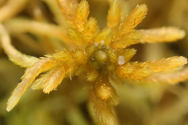
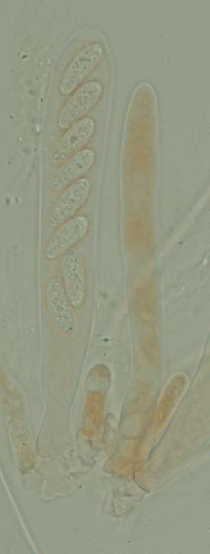
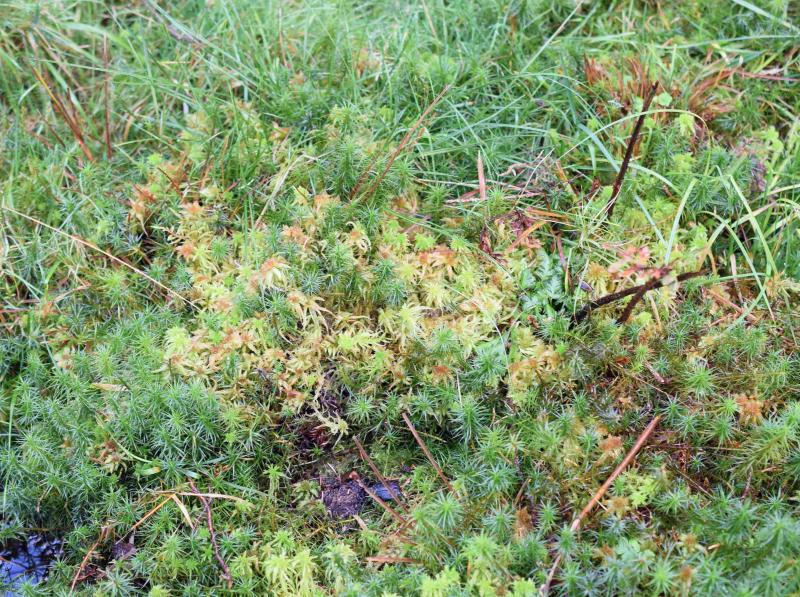
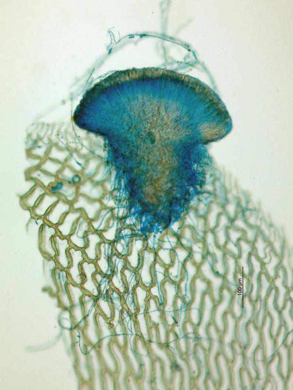
I've compared this specimen with the description in the paper Redhead, Spicer (1981, Discinella schimperi). It seems fits well. Only spores in mine some smaller (measured in alive apos), and the host is another (Sphagnum magellanicum). Yet i was not able to observe medullary excipulum (probably since apothecia small and it is not very developed).
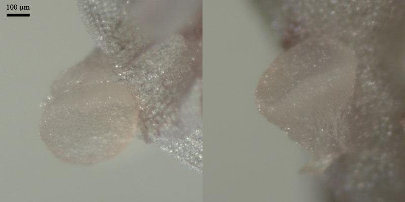
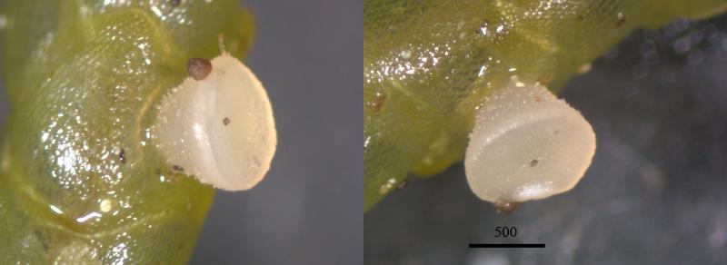
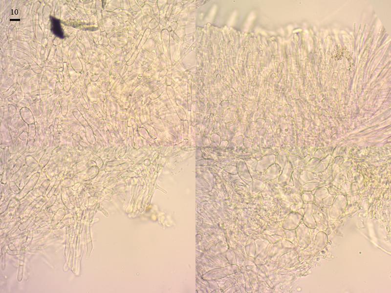
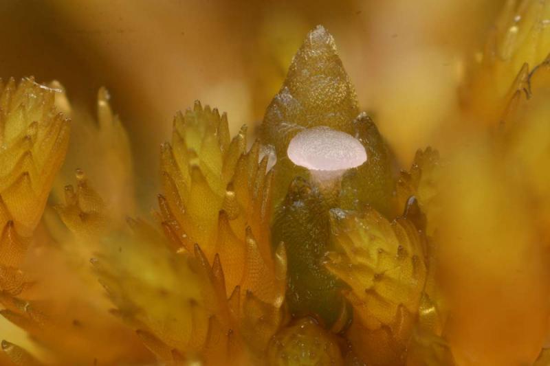
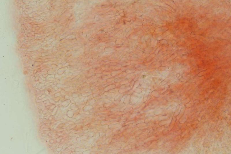
Apothecia turbinate, translucent, white to pink, to 0,5 mm wide, smooth, sited at leaves (phyllids) in capitula of S. magellanicum.
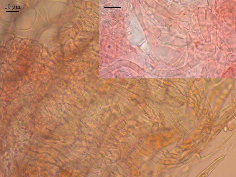
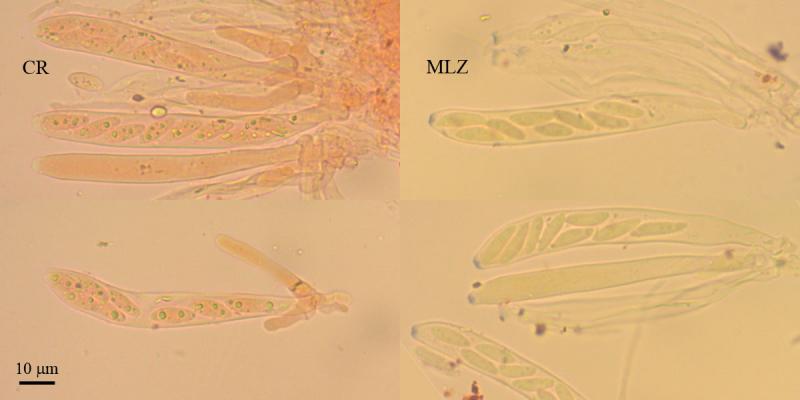
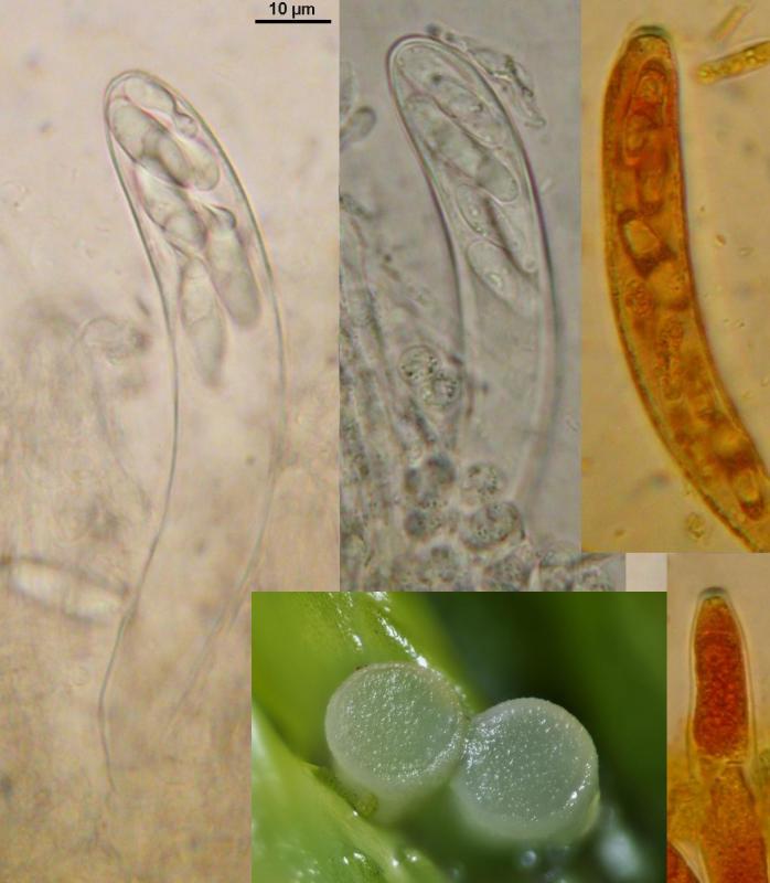
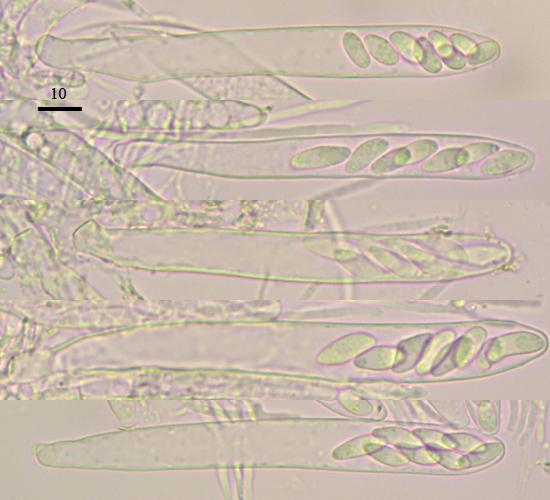
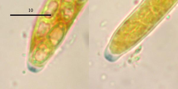
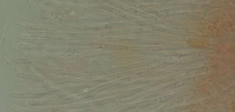
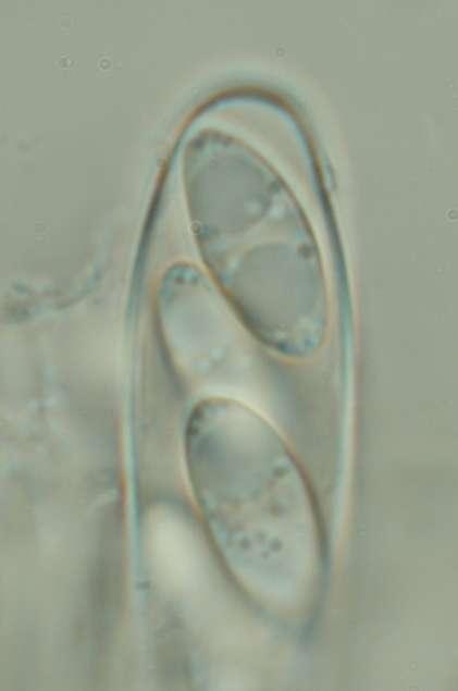
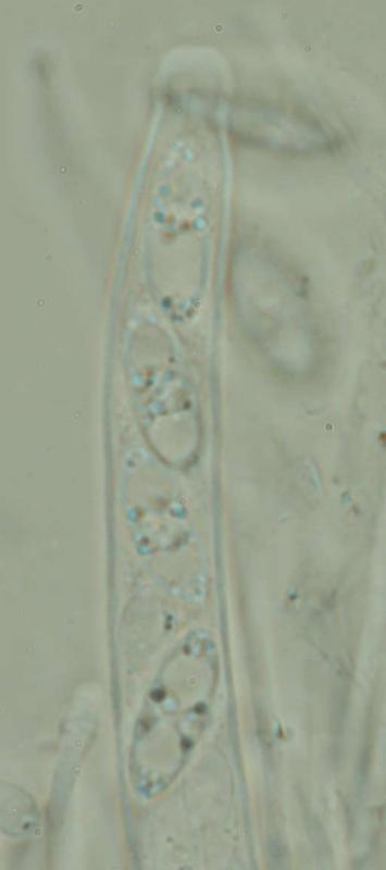
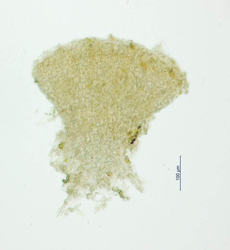
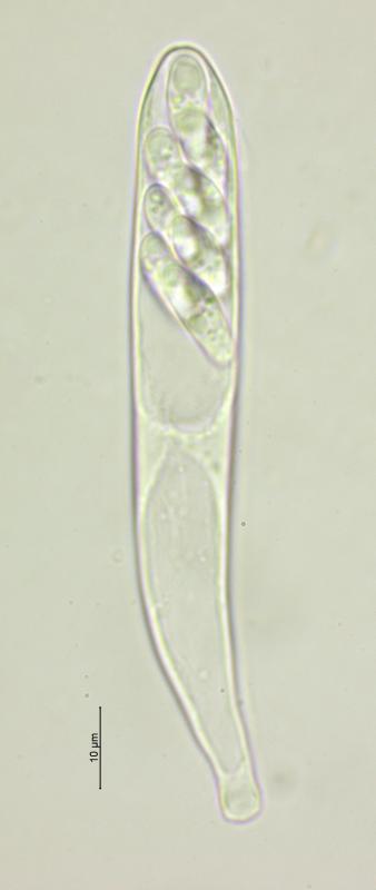
Excipulum from textura prismatica (oblita?), with dextrinoid reaction in MLZ (KOH), cells at the base larger , more ellipsoid (20 x 10), narrower to the edge, end cells obtuse, cohered (15 x 5); receptacle attaches to the substrate by the bunch the hyphae from cylindrical cells (15 x 5); asci clavate, with pronounced clamp, with CR+ content, with amyloid pore, 85 (78-93) x 10,5 (9,2-11); spores fusoid, with obtuse ends, with amorphous oil content in fresh apothecia, with several medium and small guttules in KOH, mature spores 1-2 septate, septum thin, scarsely seen, content CR+, 15,3 (13,7-17,2) x 5,3 (4,8-5,8) (Q=,9; N=16, spores from alive apth.); paraphyses cylindrical, slightly bulged in different parts, segmented, branched in 2-4 parts or unbranched, 1,5-3 mk broad.
Collected 06.08.2012, N61,063892° E69,455695°, ombrotrophic bog.

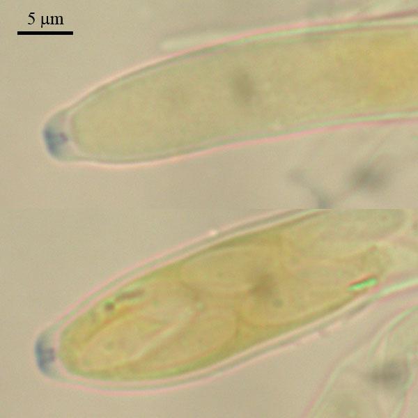
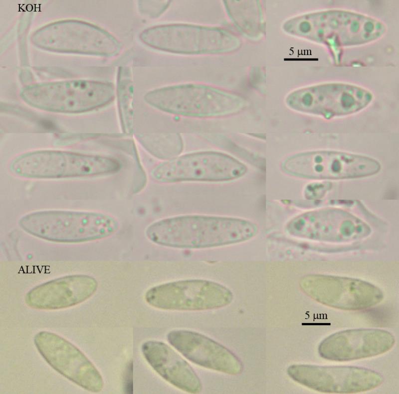

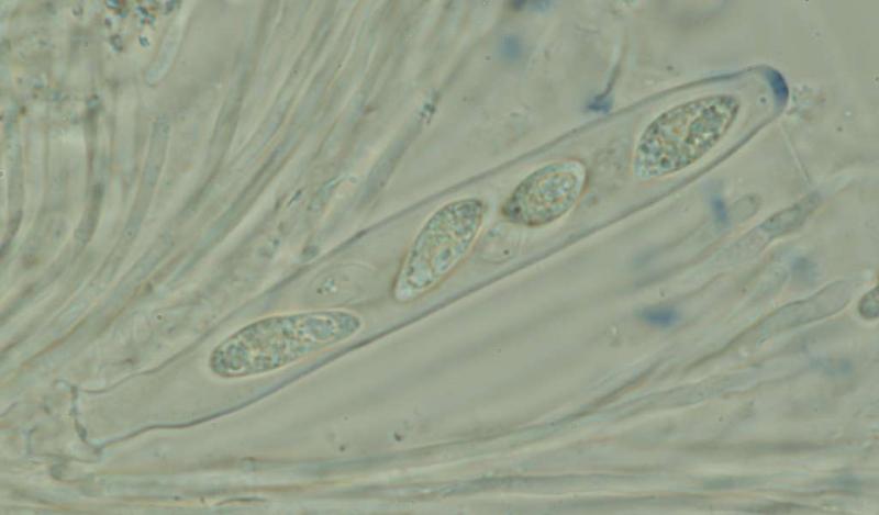
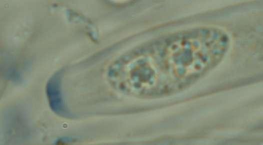
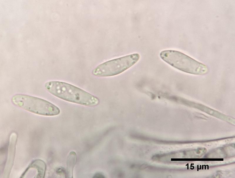
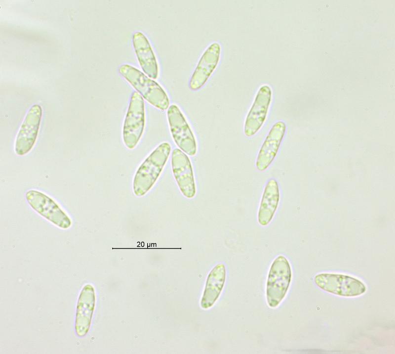
this is great!! I once saw Discinella schimperi when I was in Lapland, thanks to Marie Davey from Finland, who said that it exclusively grows on Sphagnum squarrosum. I made the atached spore photos which show these striking refractive vacuoles in the living spores which I can also see in your pics in water.
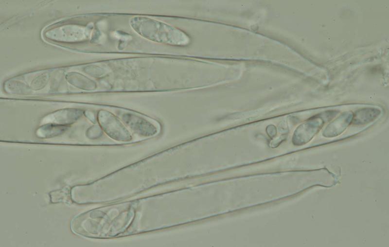
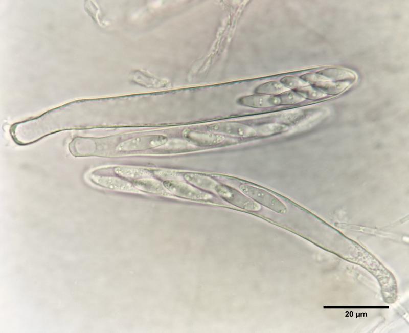
What is not the case in your specimen: the asci always contained 4 large and 4 smaller spores:
"Always 4 large spores with VBs and 4 smaller spores without VBs in each living ascus. Large sp. *16-19 x 5.3-5.7 µm, fusoid, containing very elongate 1-2refractive large VBs, const. 2 per spores."
so maybe we have different species?
Zotto
there are no close species described (?) - as i could see from Redhead's paper and IF - i checked Discinella spp., but it is difficult to check all Helotium spp. Yet i am not imformed at all in this group, since it is difficult to do conclutions. Do you think it may worth to describe sp. nov.? There are about 10 apos in the collection, i could microscopy once more to get clear picture (if you want, point to the features which should be checked more precisely). All previous micro pictures are there: http://www.flickr.com/photos/bog-fun/sets/72157631521757540/?
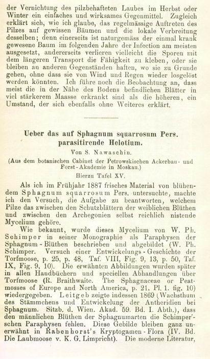
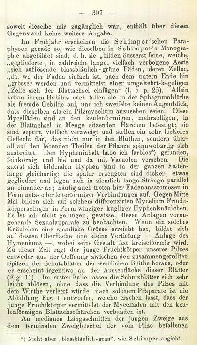
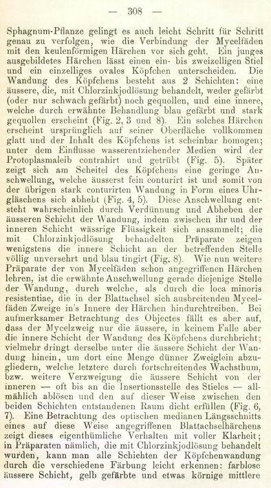
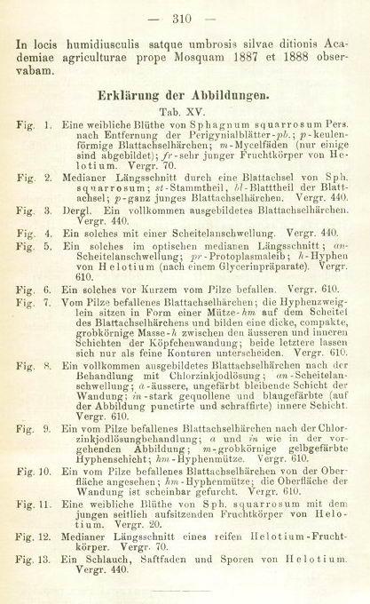
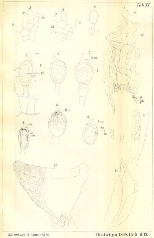
H. schimperi was described by Nawashin near Moscow, on Sphagnum squarrosum.
No mention of heterospore asci, the drawing shows 5 or 6 spores in the living ascus, all containing these large vacuoles. Spore size 18-21 x 5-6, so your spores look shorter. Asci in Nawashin are 90-100 x 10-13 µm (alive), not clearly larger than yours (in dead state).
I do not see strong evidence for a different species.
The generic placement. I see some similarity with Pezoloma, and a sequence analysis (Stenroos et al. 2010) show it to be near that genus, but also near Mitrula and Epiglia.
We could genetically show (unpublished) that Discinella and Pezoloma are very close, so that genus is not bad for schimperi.
Zotto

It will be interesting to compare D. schimperi from the both sphagna. The ecology of these two species quite different, S. squarrosum is minerotrophic and common in fens and bogged forests, and S. magellanicum is ombrotrophic and inhabits bogs. But i did not look at S. squarrosum, though it is common there as well.
This type of ring is similar as in Ascoocoryne or Peziloma but very different from Hymenoscyphus (Helotium).
Zotto
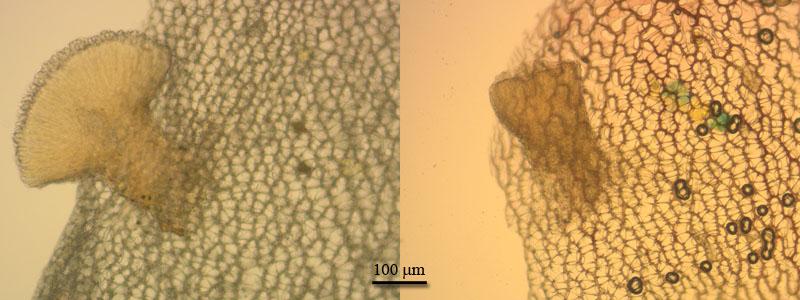
I am very surprised about D. schimperi. It is very specific, hyphae always in one special cell at shoot apex, always on S. squarrosum.
Would be nice to know whether such a cell is present in magellanicum and whether the species is behaving as in S. squarrosum.
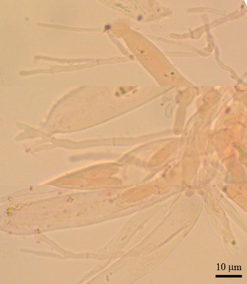
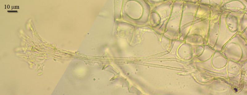
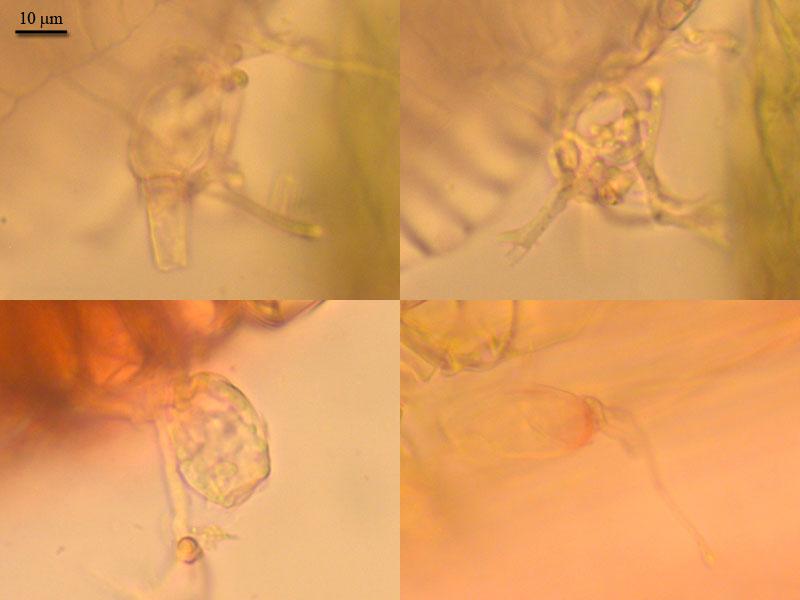
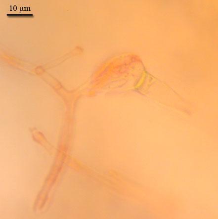
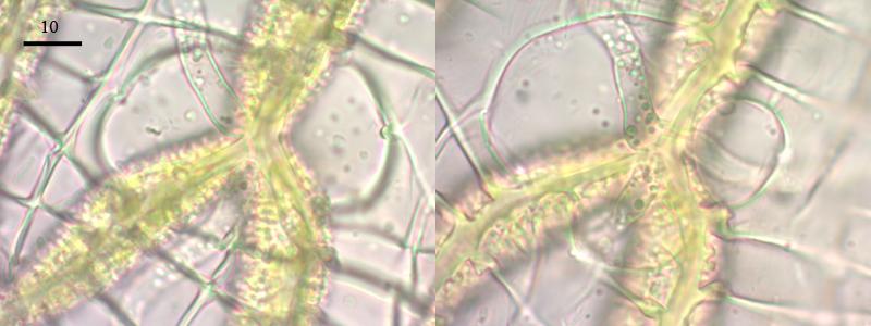
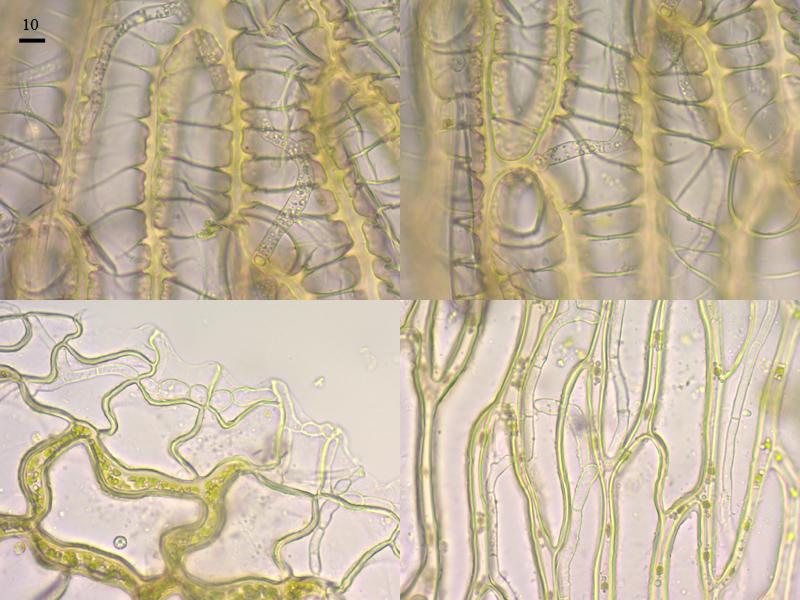
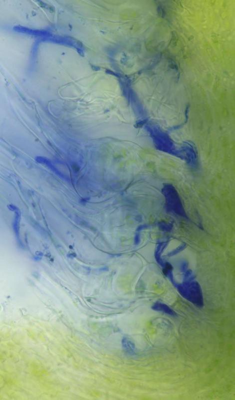
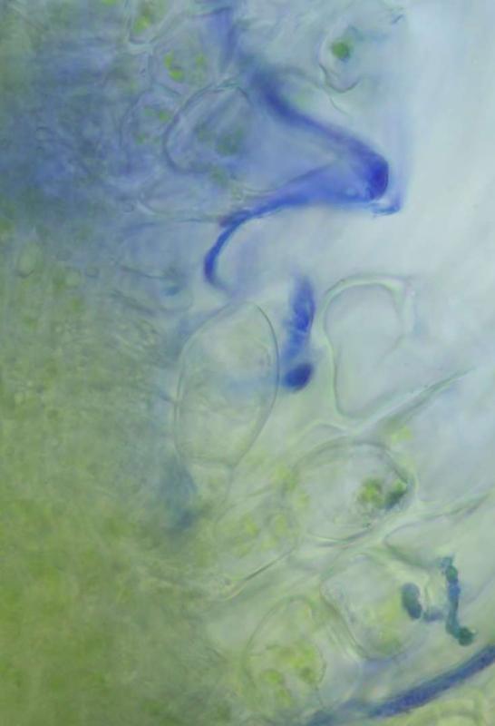
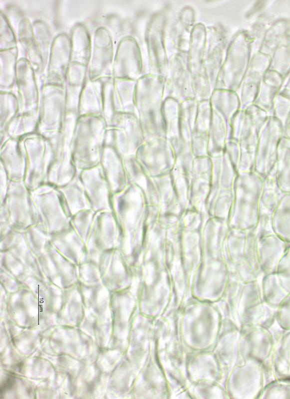
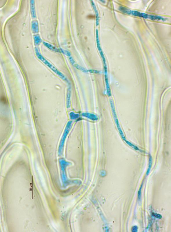
Apothecia attaches to upper part of leaves (phyllida) by the weft of cylindrical hyphae. Hyphae radiate from stipe base, several go down to reach the base of the leaf. Other hyphae spread in leaf tissue, they seen inside cells, along and penetrating green (chlorophyllous) cells, often in places of cell contacts.
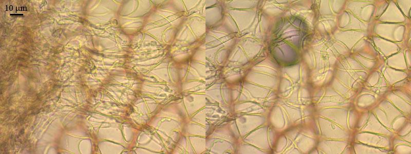
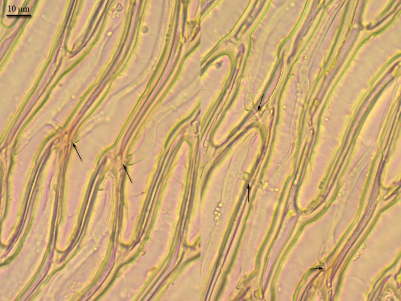
I was not able to trace all way hyphae does from leaf tip to it's base and further down to base of branch (where mentioned mucilaginous cells are placed). Separately, i looked these cells in capitula. Hyphae were spread there abundantly as well. Part of them were inside cells. Others were free, in bundles (2-3), ending with higly branched entangled agglomerages. Some of these were seen branching close to muciaginous cells. Mucilagenous cells with such agglomerages outside also were seen, as well as single hyphae attached as cap to it's tip and penetrationg inside.
There was only one capitula which i was studying (unfortunatelly i separated branches with apothecia right after collection, being not informed that other parts may be important). Mounts were not very clear.
But it seems close to what was described on S. squarrosum after such short examination. How to interpret all this... i don't know.


Zotto
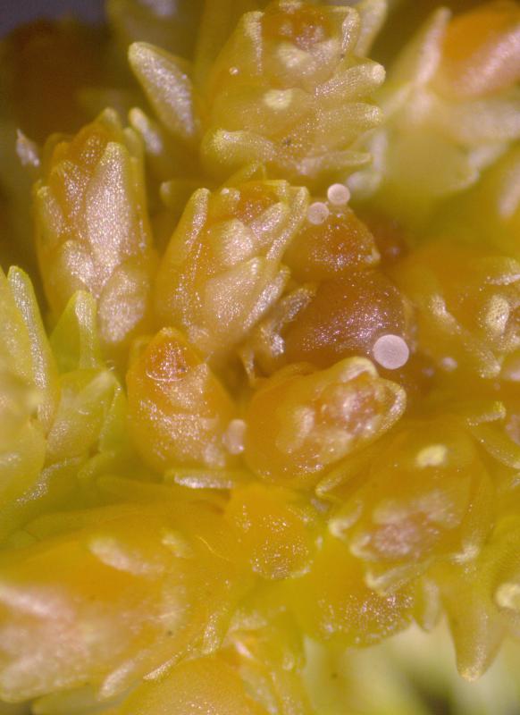
But now i opened a book on Sphagnum biology and see, these balls must be antheridia actually. Antheridia "nestled among the leaves near the tips of the capitulum branches". So, it is antheridia infected, probably.
What is mucilaginous cells, then? - haven't found anywhere in books about sphagnum. There is a citation in Bryophyte biology (2009): "Juvenile leaves hide delicate filaments that line the insertion of the leaf. These hairs originate from the leaf initial and secrete a mucilage of polysaccharides that may be essential in preventing the delicate growing apices from dehydrating".
I have not understand the description by Nawaschin yet, since it is in German and Google translation worked quite littered. May be, there will be more clarity.
Zotto

Chris
PS - when downloaded it looks fine - honestly!
 1888-v37-p306-0001.pdf
Another collection of D. schimperi has showed up here. But now it parasiting on another sphagnum - S. papillosum (also ombrotrophic bog, 40 km from previous collection).
The measurements of asci, paraphyses and spores coinside with previous collection.
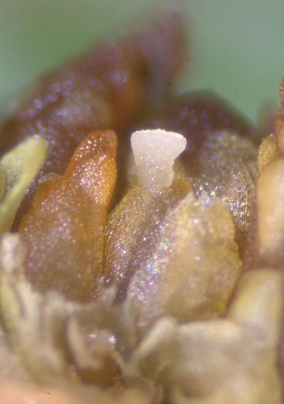
Apothecium (single apos collected) turbinate, 650 mk high, 900 mk broad, whitish, watery.
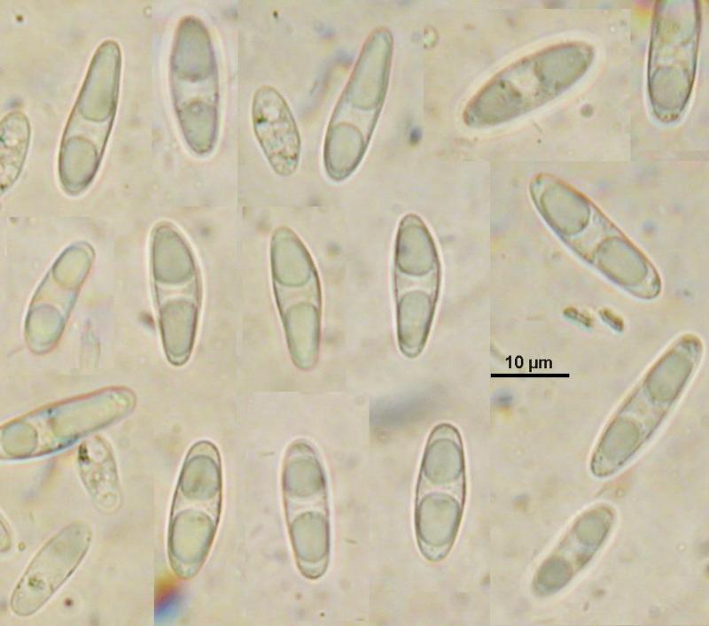
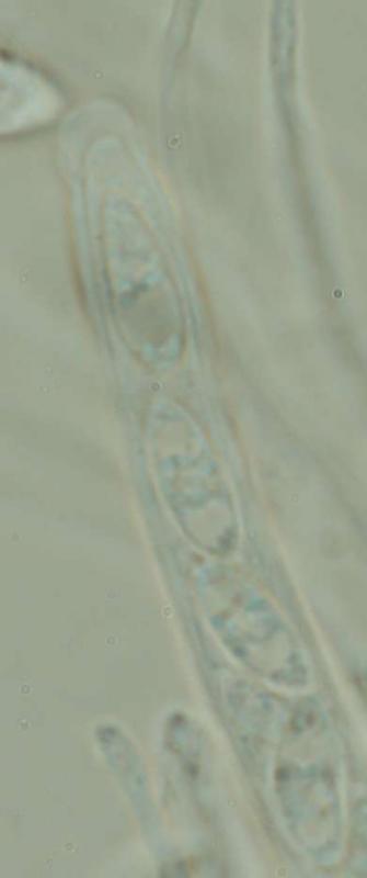
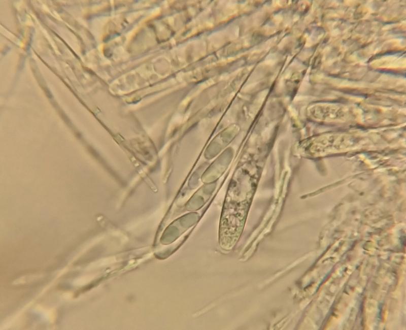
Asci clavate, with crozier, with euamyloid cylindrical pore, 83.2–113.5 x 9.2–13.4; paraphyses cylindrical, rarely branched, 2 broad, with pale round to elongated vacuoles; spores ellipsoid, with pale irregularly oils at the ends, 14.7 (13–17.7) x 4.8 (4.4–5) (n=12).?

Zotto
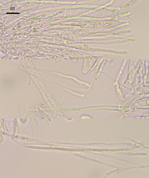
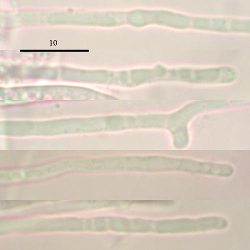
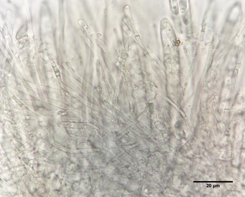
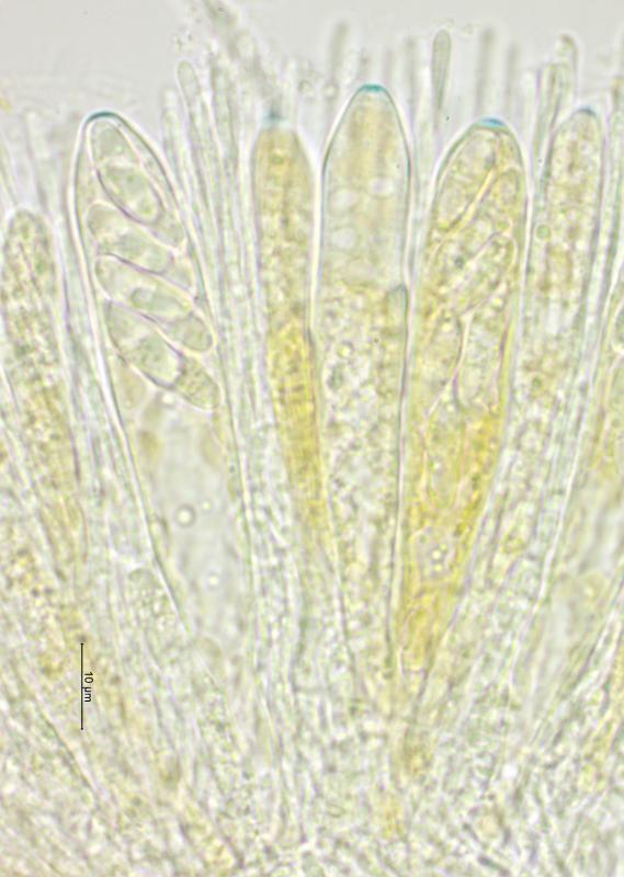
paraphyses filiform, straight, rarely branched, 1.6-2.5 µm wide below, 2.0-3.5 µm wide above, walls slightly wavy
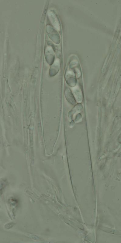
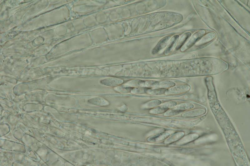
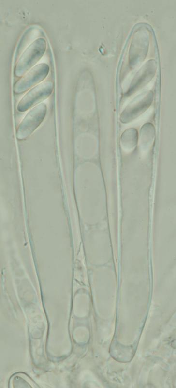
asci cylindrical, K/I+, small tholus, 8-spored but often only 4 (or 2) spores reach maturaty, 4(-6) spores aborted
ascospores ellipsoid, one-celled, 13-17 x 5-6 µm, to 20 x 6 in two-spored asci, several large dropdlets 1-4 µm in diameter (weakly refractive) and several small droplets c. 0.5 µm in diameter (strongly refractive); ascospores germinating shortly after discharging, germinates spores sometimes with one or two septa
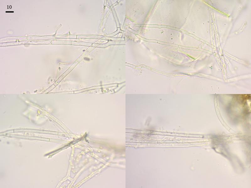
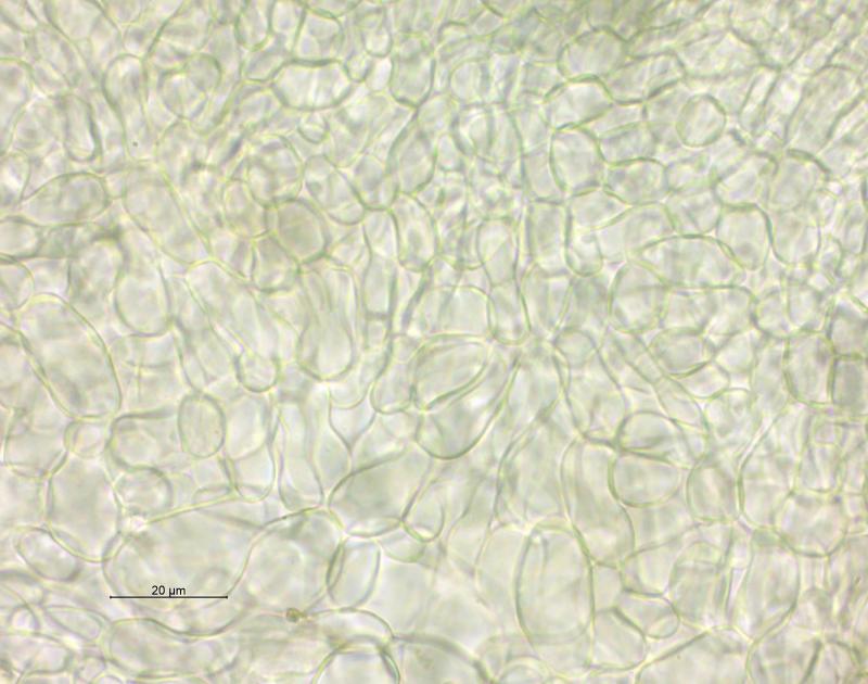
hyphae on surface of Sphagnum leaves going down to the tiny spaces between young leaves, squeezing into the leaf axils of very young leaves near the shoot apicies, often growning around the axillary hairs but without entering these cells;
no infection seen, hyphea remain outside the Sphagnum cells only rarely growing through hyalocysts (dead cells)

I collected this species in the Czech Republic in N Bohemia - Jizerske hory Mts (Isergebirge) on Sphagnum centrale (I am not completely sure with ID, but I hope it is). It was sampled in autumn 2018 on an open raised bog at altitude of almost 1000m, few months ago I sent the specimen to Zuzana Sochorova for revision for sure
best Zdenek
Luis Quijada would be interested in these finds.
All the best,
George

That is quite striking and certainly looks different to mine.
I have material on S. squarrosum but not the capacity to sequence it myself at this point. I know Luis Quijada is interested in this group, maybe he will be able to sequence some. I think he is on holiday now but I will email him.
In your fresh material were you able to find where the hyphae localise? I was thinking a character of this Sphagnicolous group was the distinctive infection on the mucilage cells but perhaps not.
All the best,
George

Despite searching, I only found one apothecium last week at the site. That said, the apo is quite large. I can try to get sequence data from half of it and still have a bit left.
I have the real H. schimperi as well so I will try with both. Morphologically, they are quite similar. I have not yet tried to look for the infection in this one though.
Dennis (1962) said that H. vasaensis was closer to H. schimperi than to anything else he had seen. I agree. In Stenroos et al. (2010), they show a few Hymenoscyphus spp. from Sphagnum as sister to H. schimperi. It is likely they also collected this vasaensis-like one as it does not seem to be uncommon, generally. I recently saw images of an identical collection from Bosnia & Herzegovina.
Bests,
George
Voici également une récolte sur Spaghnum (pâturage boisé à 1'000m), apothécies 0.3 - 0.7 mm de diamètre, blanchâtres/roses, stipitées, Ascospores 4 - 5.25 µm, hyalines, non septées, lisses. Asques 93 - 105 x 10 - 13 µm, avec crochet, J+, contenant 8 spores (moins avec l'âge). Paraphyses droites, hyalines.
Elisabeth
Best wishes,
George
Merci pour vos commentaires.
Elisabeth
cordialement
So far, it is only published on Sphagnum squarrosum. I have only found it once myself on this host, but have collected the related Helotium vasaense on other Sphagnum species. The most recent publication is Döbbeler et al. 2023. Bryophilous ascomycetes of North America – an overview of the recorded species. Herzogia 36 (2), 2023: 305–370.
Kind regards,
George
