
22-04-2026 01:06
 Richard VALERI
Bonjour à tous.Je vous présente cette Nectria s.
21-04-2026 13:36
Gernot FriebesHi,I am out of ideas for this one. I collected Sal
21-04-2026 13:19
Gernot FriebesHi,this Lophodermium on Typha has ascospores measu
21-04-2026 13:05
Gernot FriebesHi,this hyphomycete feels familiar but I was not a
20-04-2026 22:00
 Malcolm Greaves
These pale yellow, hairy ascos were growing on cul
19-04-2026 21:23
Steve ClementsBonjour, I found this anamorphic fungus on old pl
19-04-2026 20:46
Steve Clements1 mm diameter approx spherical conidiophores on pl
12-04-2026 17:56
 Hardware Tony
Found on dead stems in February earlier this year
Discomycete on leaves
Josep Torres,
02-10-2025 09:16
 Hello.
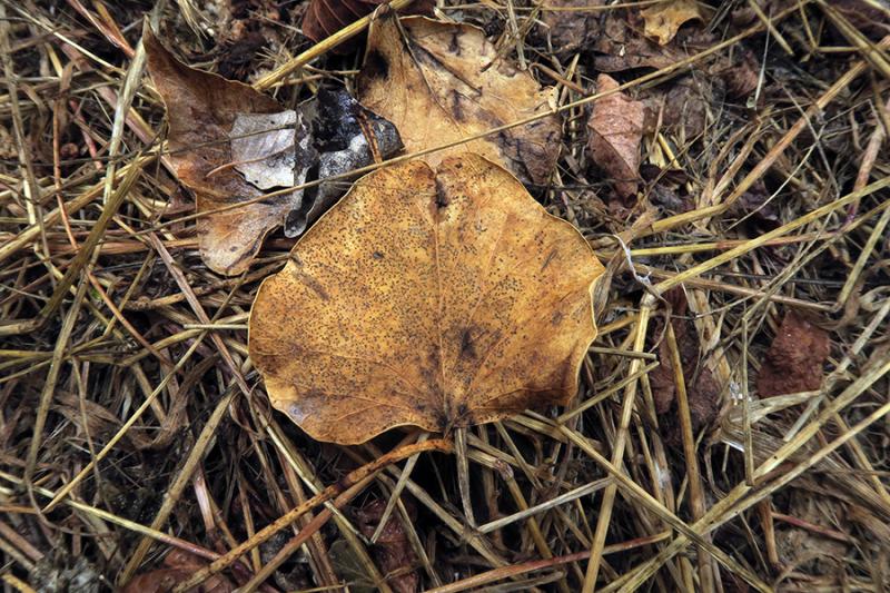
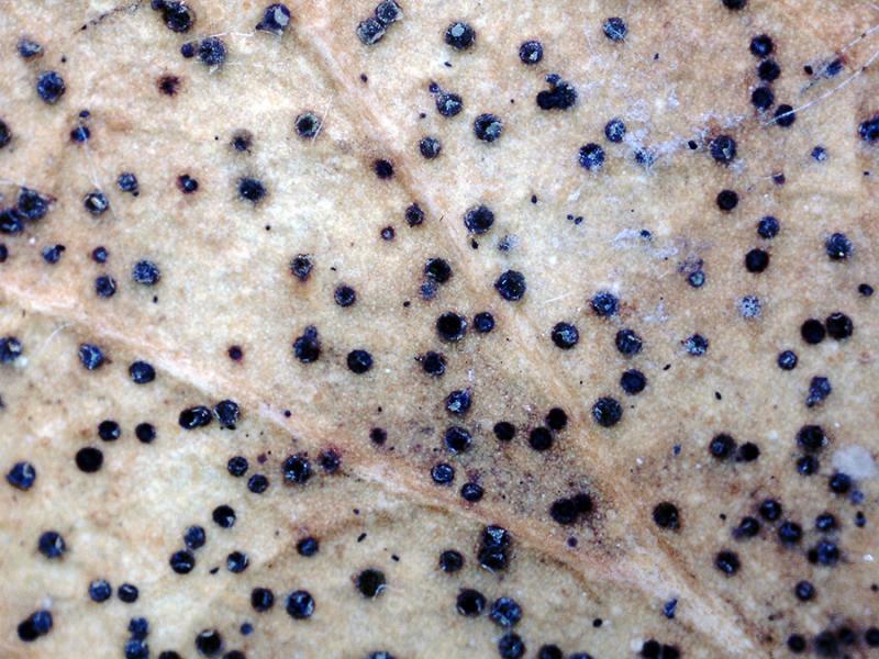
Some tiny apothecia sprouting in a scattered but abundant manner on decaying leaves in the riverine forest under poplars (Populus).
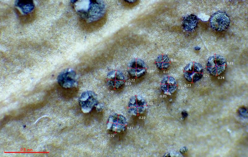
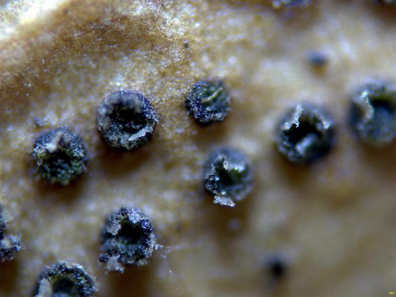
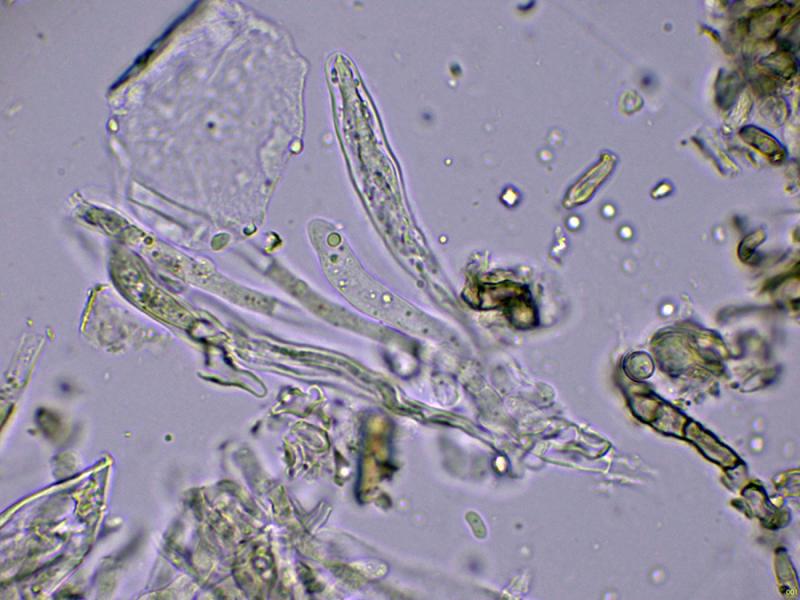
The apothecia are blackish, only 0.2 to 0.3 mm in diameter, without a clearly distinct margin.
Under microscopy, I couldn't see any structures that could correspond to marginal hyphae; if they were present, they would be very few.
The excipule is very sparse, globose in texture, angular.
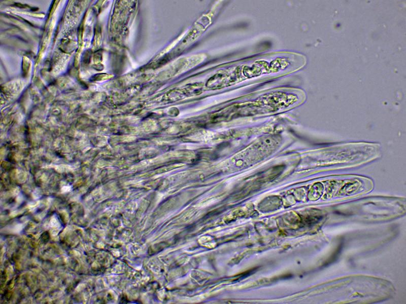
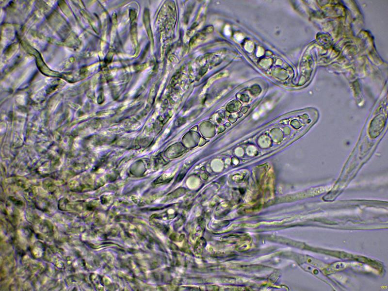
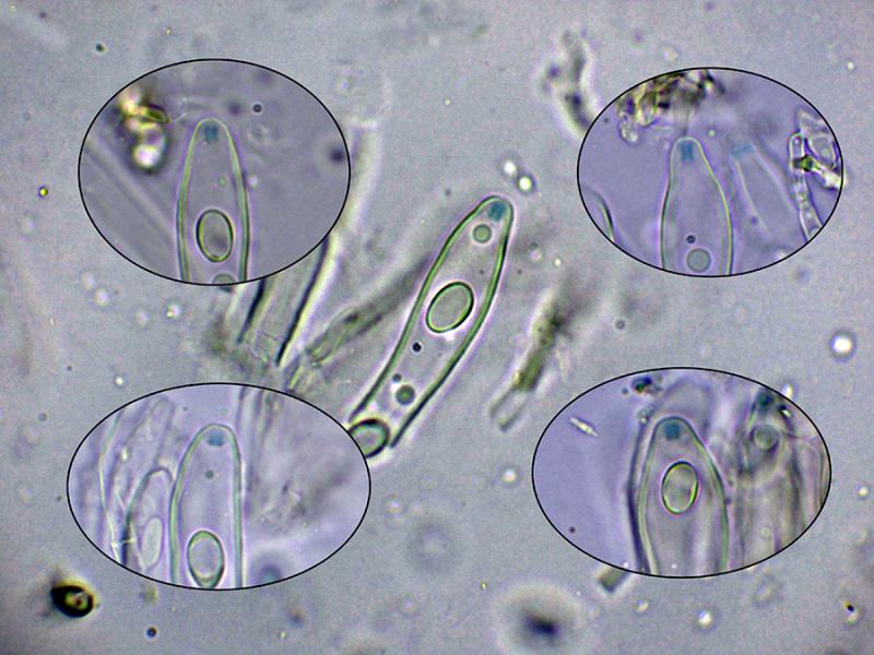
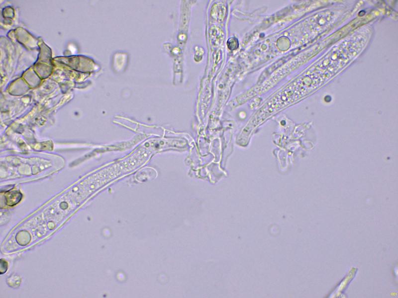
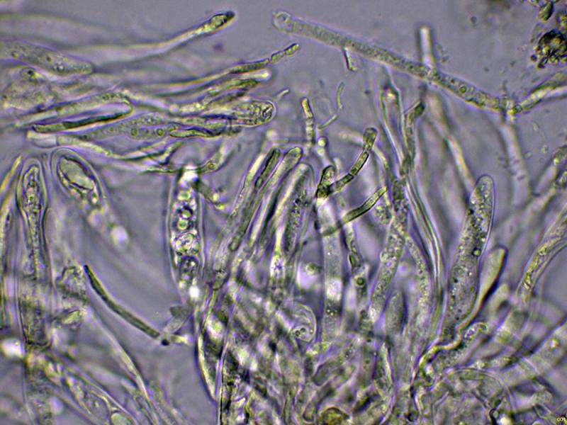
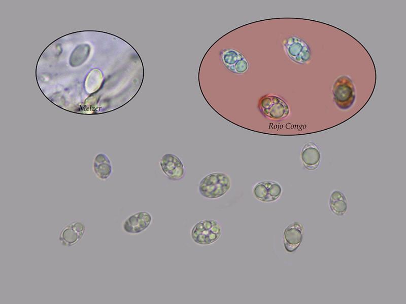
Octosporic asci, uniseriate, thick-walled, apparently without clear uncinules at their base, with measurements in water of (50.1) 56.1 - 70.8 (79.2) × (8.6) 9.3 - 11.4 (11.6) µm., Me = 64.9 × 10.4 µm., and with an amyloid apical apparatus to Melzer, cylindrical to slightly widened at its upper part, with measurements of 2.6 - 3.1 (3.4) × (2.1) 2.12 - 2.29 (2.3) µm. As a curiosity, I could not observe this apical apparatus in the preparations with Lugol. The paraphyses are filiform, septate, with a slight widening at the apex, protruding above the level of the asci.
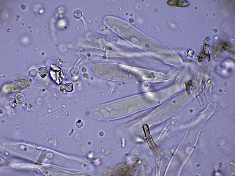
Ellipsoidal ascospores, with one or more lipid droplets inside, inamyloid in the Melzer test, and with measurements in water of (8.2) 8.8 - 11.2 (11.4) × (5.4) 5.9 - 8 (8.4) µm, Me = 10 × 6.8 µm; Qe = 1.5.
Based on microscopy and the amyloid reaction of its apical apparatus, the closest I could find would be Drepanopeziza, but its appearance doesn't match.
Any feedback from you would be welcome.
Thank you in advance.
Best regards.
Josep Torres,
02-10-2025 09:18
Re : Discomycete on leaves
The rest of the images.
Hans-Otto Baral,
02-10-2025 09:23

Re : Discomycete on leaves
You have Trochila craterium on Hedera leaves I think, unless these are not Ilex leaves.
Josep Torres,
02-10-2025 09:26
Re : Discomycete on leaves
Thank you, Zotto, for your prompt response. Ivy (Hedera helix) is abundant in the area, and it's most likely the Trochila craterium you're suggesting.
Best regards.
Best regards.