
22-04-2026 01:06
 Richard VALERI
Bonjour à tous.Je vous présente cette Nectria s.
21-04-2026 13:36
Gernot FriebesHi,I am out of ideas for this one. I collected Sal
21-04-2026 13:19
Gernot FriebesHi,this Lophodermium on Typha has ascospores measu
21-04-2026 13:05
Gernot FriebesHi,this hyphomycete feels familiar but I was not a
20-04-2026 22:00
 Malcolm Greaves
These pale yellow, hairy ascos were growing on cul
19-04-2026 21:23
Steve ClementsBonjour, I found this anamorphic fungus on old pl
19-04-2026 20:46
Steve Clements1 mm diameter approx spherical conidiophores on pl
12-04-2026 17:56
 Hardware Tony
Found on dead stems in February earlier this year
Microscopic ascomycete.
Josep Torres,
24-09-2025 09:22
 Hello.
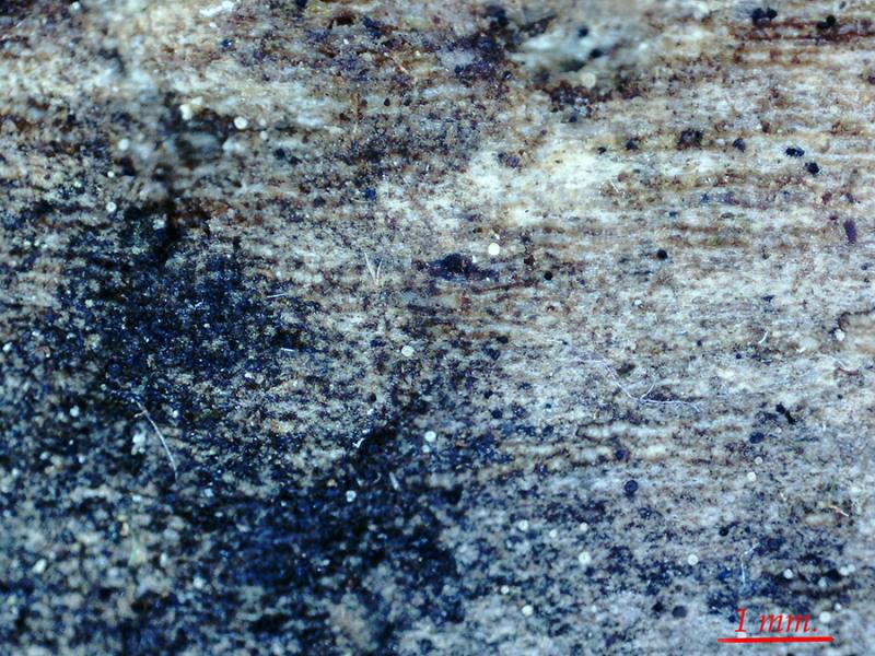
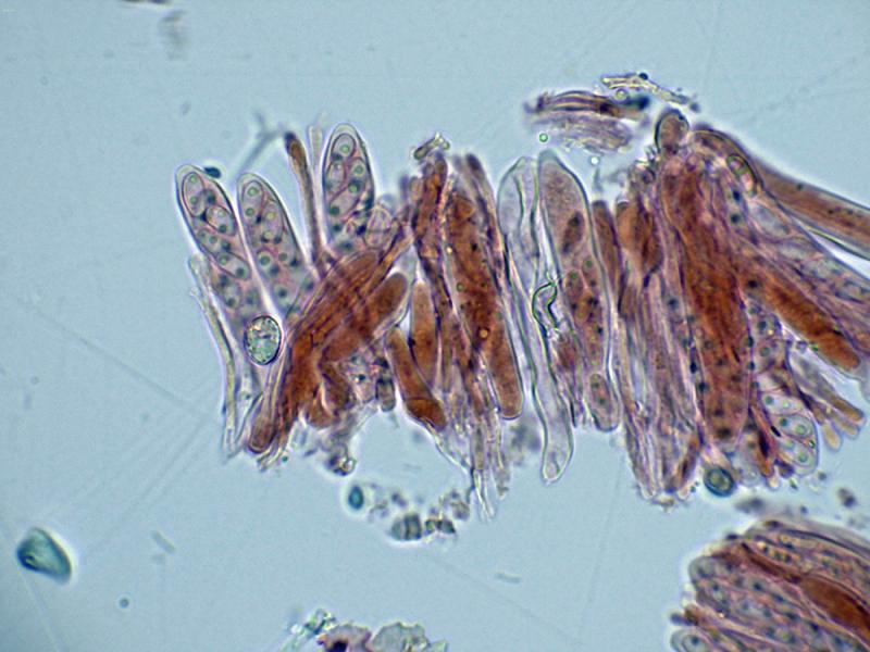
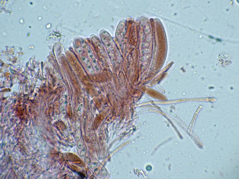
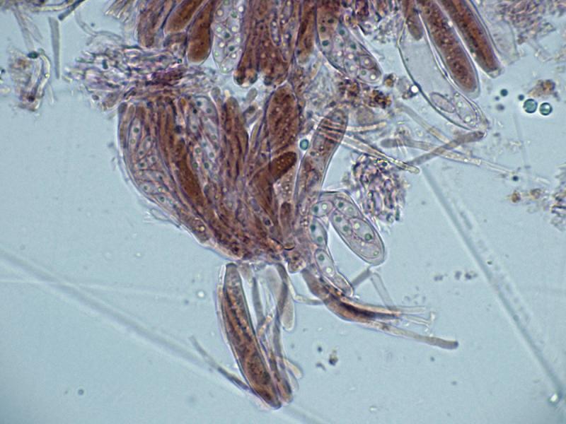
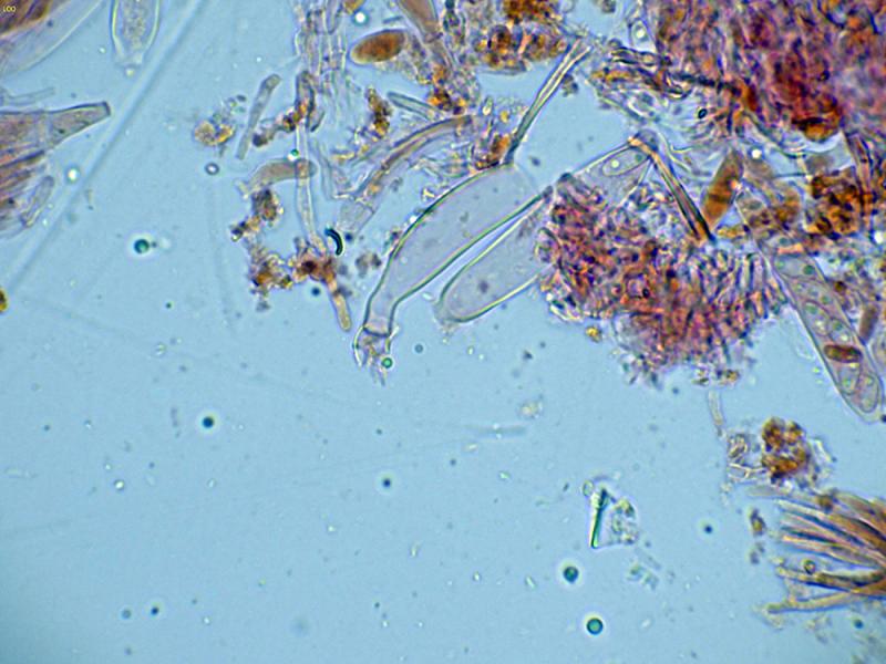
Very tiny apothecia, sprouting in a scattered manner, located on the surface of a semi-submerged trunk of Fraxinus sp.
Measuring only 0.07 to 0.11 mm in diameter, they are whitish in color and have a slightly hairy outer edge.
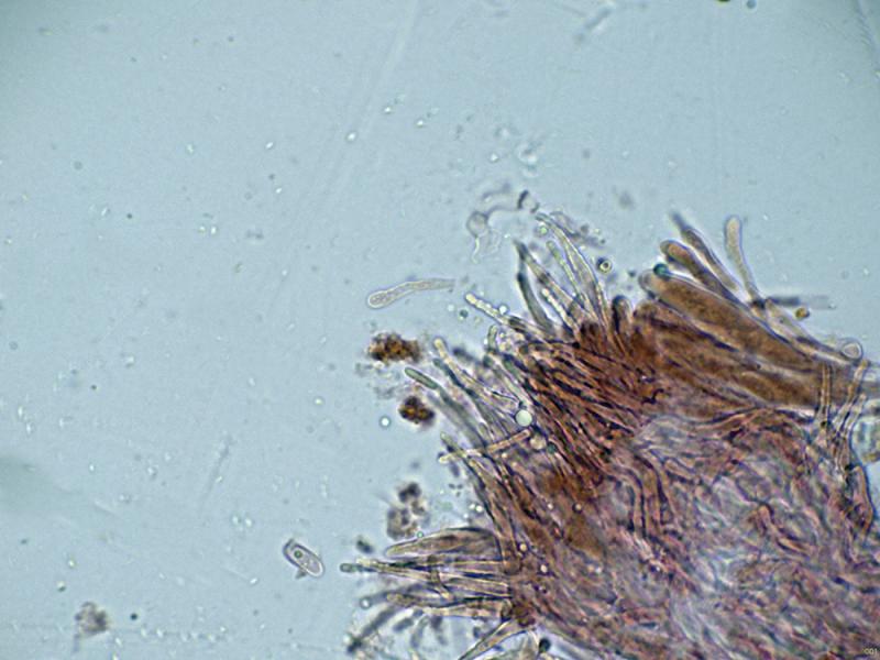
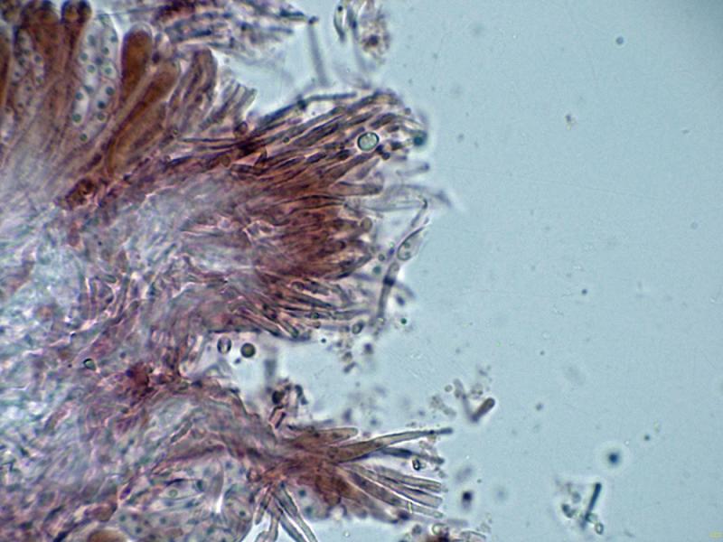
Marginal hairs with moderately pointed ends and inlaid surfaces.
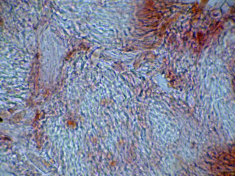
The medullary exciple hyphae are intricate, and below these, hyphae are already tending toward globose angularis.
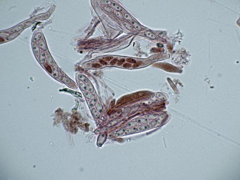
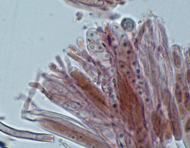
Octosporic asci, apparently without uncinules at their bases, mostly biseriate, and measuring (35.7) 40.9 - 52 (52.4) × (7) 7.7 - 9.1 (9.6) µm.
I can't provide data on the behavior of the asci to the Melzer test because I only managed to prepare a single sample with a single apothecium. The next day, the remaining apothecia had literally disappeared after dehydration.
Ellipsoidal ascospores with two large lipid droplets at the poles, with measurements of mature ascospores within the ascus of:
(5.9) 6.6 - 8.9 (9.3) × (2.9) 3.2 - 4.3 (4.7) µm
Q = (1.5) 1.8 - 2.4 (2.8) ; N = 30
Me = 7.6 × 3.6 µm; Qe = 2.1
The paraphyses are filiform, slightly or not at all widened at the apex, not protruding above the level of the asci, with one or two septa and a width of 2 to 2.55 microns.
Considering their characteristics, I think they are likely a Hyaloscypha, and among them is a Hyaloscypha microscopica, which, although the name would be appropriate, I have been unable to find any information about the species.
Any feedback from you would be most welcome.
Thank you in advance.
Best regards.
Hans-Otto Baral,
24-09-2025 10:17

Re : Microscopic ascomycete.
Will be difficult without the iodine reaction. Perhaps Hyaloscypha intacta which has inamyloid asci (in Lugol).
H. microscopica was not ever redescribed. SVrcek 1985 only combined it in Discocistella and Huhtinen 1990 did not study the sparse material.
I did not look up Velenovsky but assume that the hairs are not tapering.
Josep Torres,
24-09-2025 14:20
Re : Microscopic ascomycete.
Thanks, Zotto.
Considering its tiny size, Hyaloscypha intacta seems like a good option. This has been mentioned here before. Here's a link to a study very similar to my proposal, which you yourself considered Hyaloscypha intacta.
http://ascofrance.com/forum/16979/hyaloscypha-hyalina#
Best regards.
Considering its tiny size, Hyaloscypha intacta seems like a good option. This has been mentioned here before. Here's a link to a study very similar to my proposal, which you yourself considered Hyaloscypha intacta.
http://ascofrance.com/forum/16979/hyaloscypha-hyalina#
Best regards.