07-09-2025 11:34
 Zuzana Sochorová (Egertová)
Hello,I have identified this fungus as Hymenoscyph

05-09-2025 18:53
 Robin Isaksson
Hi! Spores 1 septate; 12-13 x 3um Hairs 35-75
05-09-2025 09:32
 Nicolas VAN VOOREN
Bonjour, hi everyone,Do you know where the fungari
03-09-2025 21:59
Philippe PELLICIERLa Léchère, Col de la Madeleine, alt 1970m, au s
04-09-2025 20:11
Åge OterhalsSaccobolus on dear droppings. Can any of you confi
03-09-2025 12:44
 Enrique Rubio
Hi to somebody.I would like to know your opinion o

31-08-2025 19:41
Enrique Rubio
Hi to someone.I need to download this issue of Sve
 Hello.
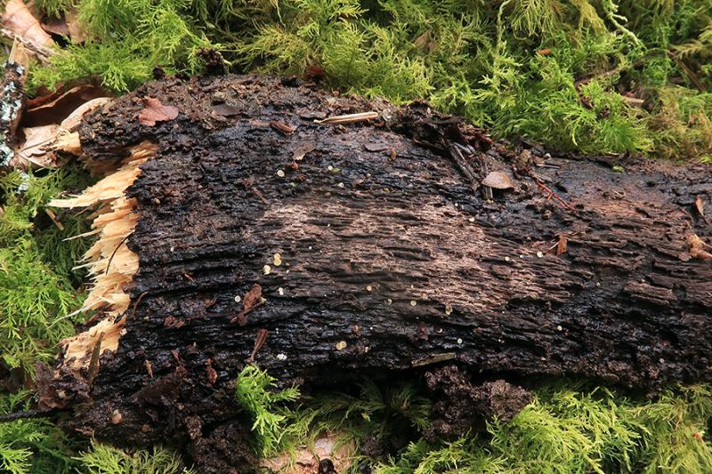
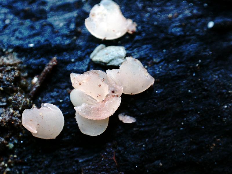
Tiny pinkish discomycetes, photographed and collected on August 17, sprouting scattered on the surface of a fallen spruce (Abies alba) trunk.
Apothecia attached to the substrate only by the central part, without a distinct stem, with a diameter between 1.17 and 1.75 mm, whitish with pinkish tinges.
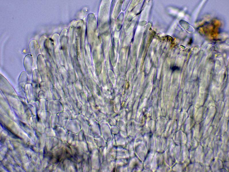
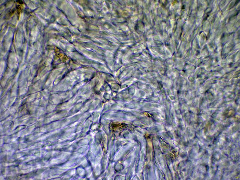
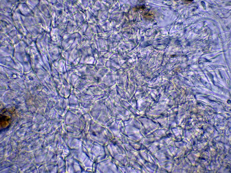
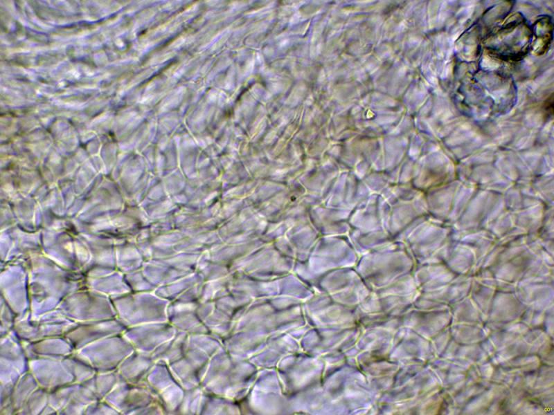
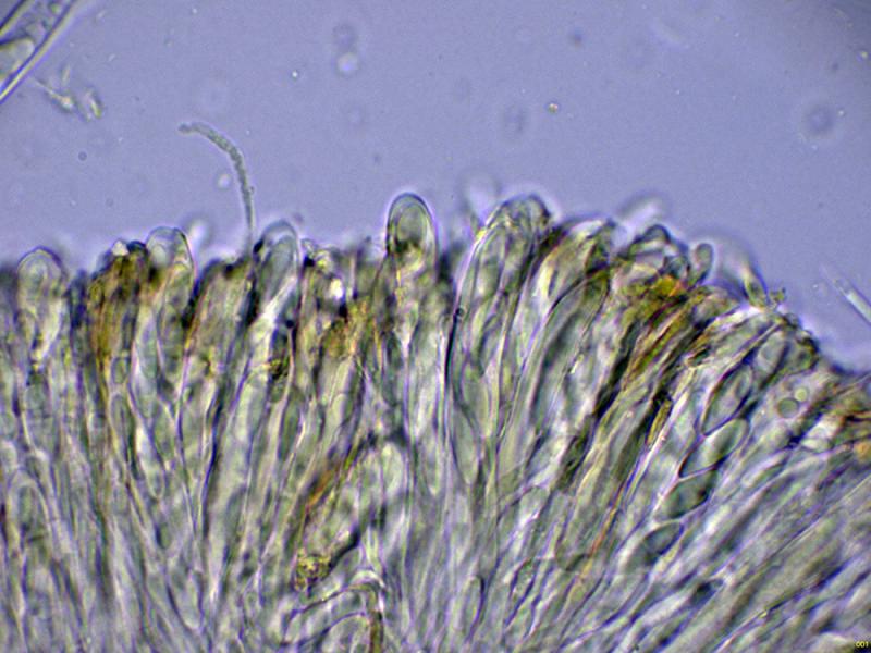
The medullary exciple is filamentous, with an intricate texture, and the ectal exciple has a globose-angular texture.
Short, septate marginal hairs with rounded ends.
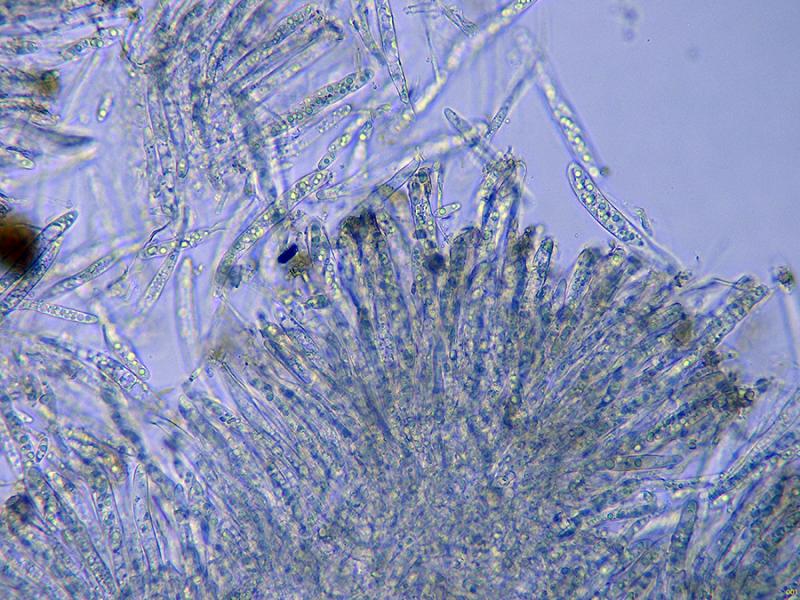
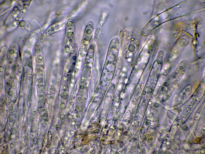
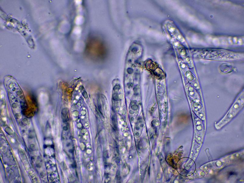
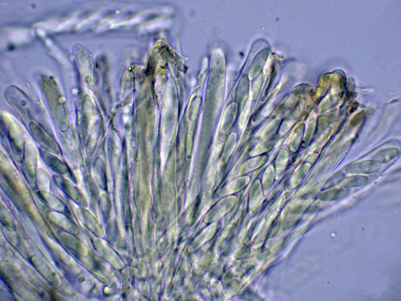
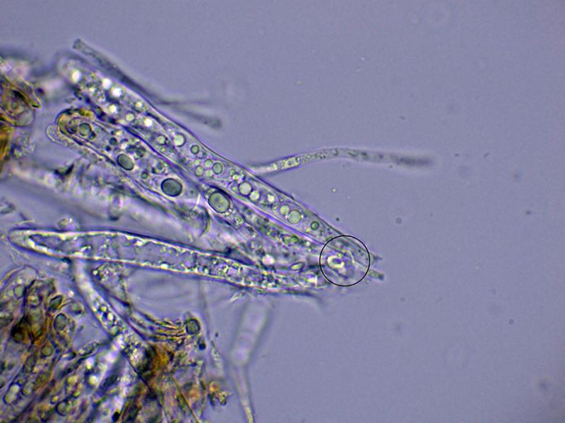
Octosporic asci, in some cases with uncinules.
The paraphyses are septate, filiform, unbranched, and protrude very slightly above the level of the asci.
No significant reaction of the asci with Melzer's reagent has been observed.
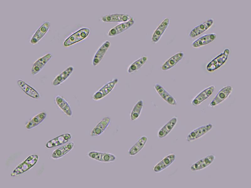
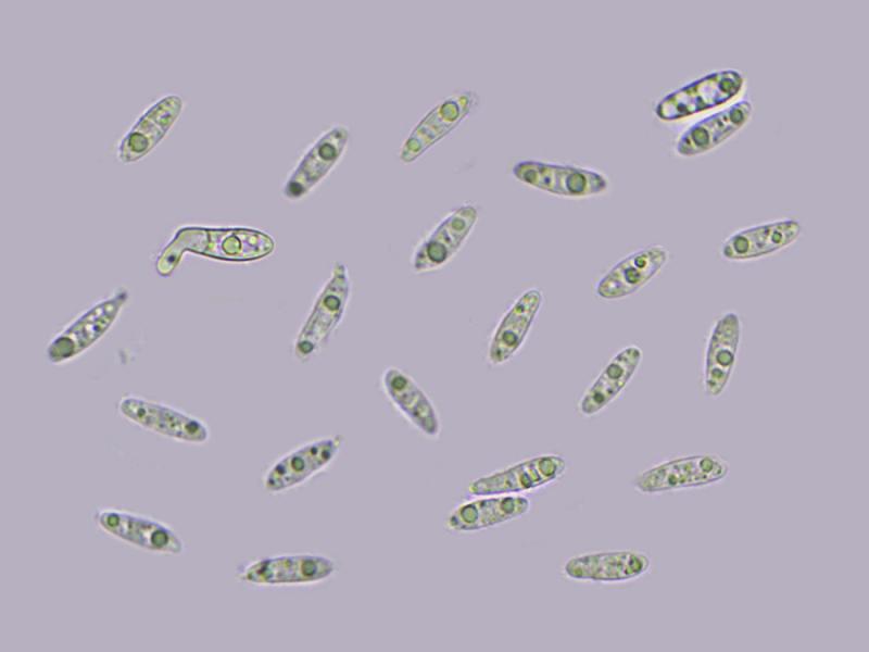
Cylindrical ascospores, with one or two large lipid droplets inside and other smaller ones scattered throughout. Between one and three septa can be discerned in some of these ascospores, with measurements in water of:
(11.5) 12.5 - 15.2 (15.6) × (3.8) 4 - 4.8 (5) µm
Q = (2.4) 2.9 - 3.6 (4) ; N = 30
Me = 13.9 × 4.3 µm ; Qe = 3.2
These specimens were found very close to what appears to be Hymenoscyphus imbrebis (there were three specimens on a nearby trunk), so at first I thought they were probably the same, although the microscopy doesn't match at all. Based on its microscopic characteristics, I'm thinking of Calycellina, and within Calycellina, the best fit is Calycellina subtrabinella, but there are too many conflicting characteristics, such as the host, which I haven't been able to find information on whether it can grow on coniferous wood, and the lack of reaction to Melzer's, as well as the spore width, don't agree.
Any feedback from you would be welcome.
Thank you in advance.
Best regards.