
05-03-2026 10:07
Hulda Caroline HolteHello, I found and collected this species growing
07-03-2026 13:06
 éric ROMERO
Bonjour tous, Sur cône d'épicea fortement imbu,

08-03-2026 14:05
 Thierry Blondelle
Bonjour à tous,Sur 3 récoltes supposées de H. l

05-03-2026 16:30
François BartholomeeusenDear forum members, On the 2nd of February 2026,
06-03-2026 09:41
 Alain GARDIENNET
Hi forum, I'm now looking for another reference c
Ascobolus michaudii with possible bycatch Pyxidiophora arvernensis?
François Bartholomeeusen,
18-05-2025 16:49
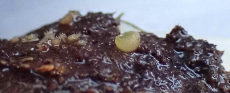
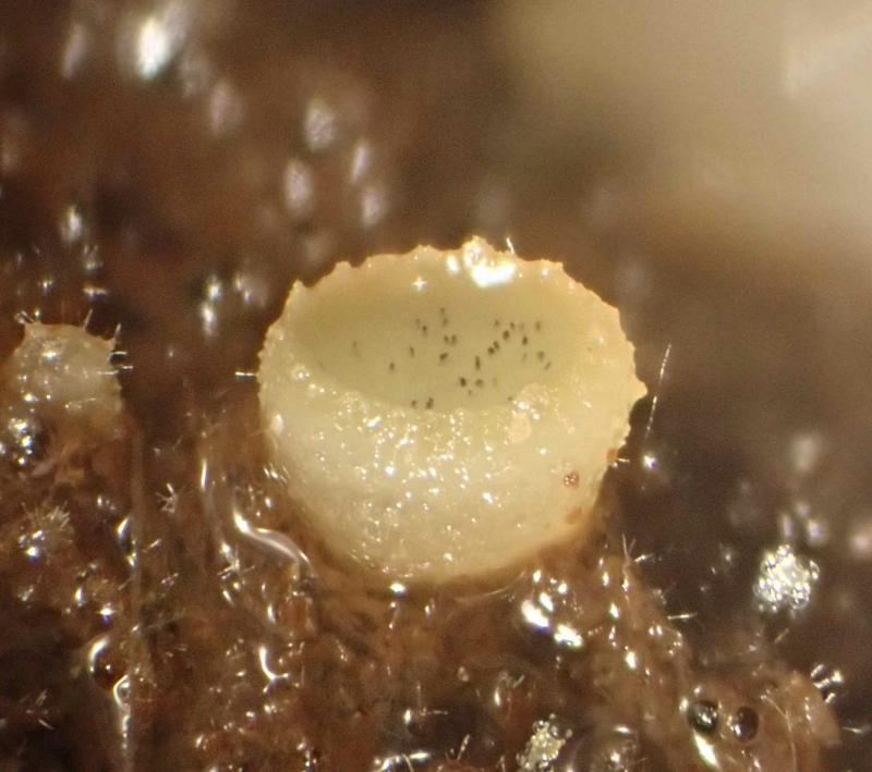
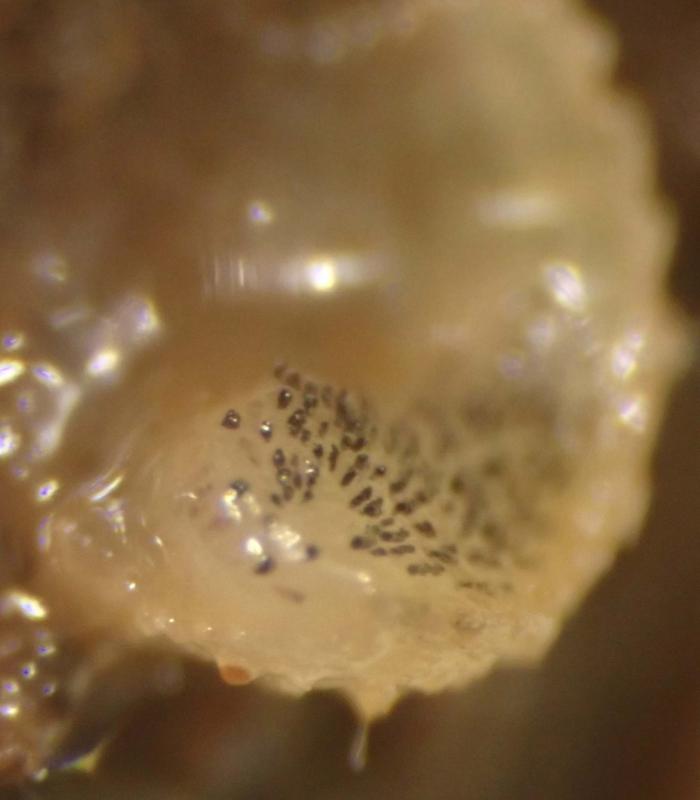
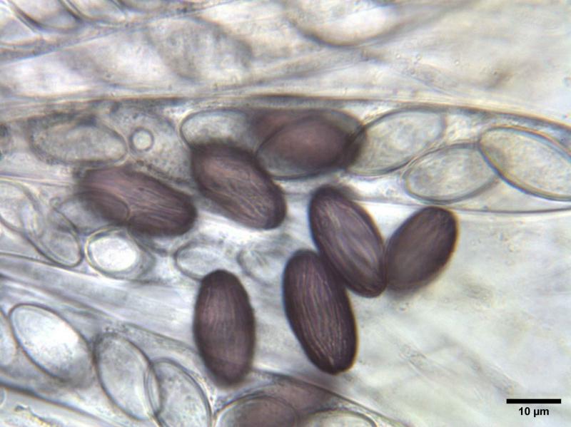
On cow dung, between fruiting bodies of Lasiobolus papillatus, I found almost cylindrical ascomata with a diameter < 2 mm. The disc is yellowish with dentate margin.
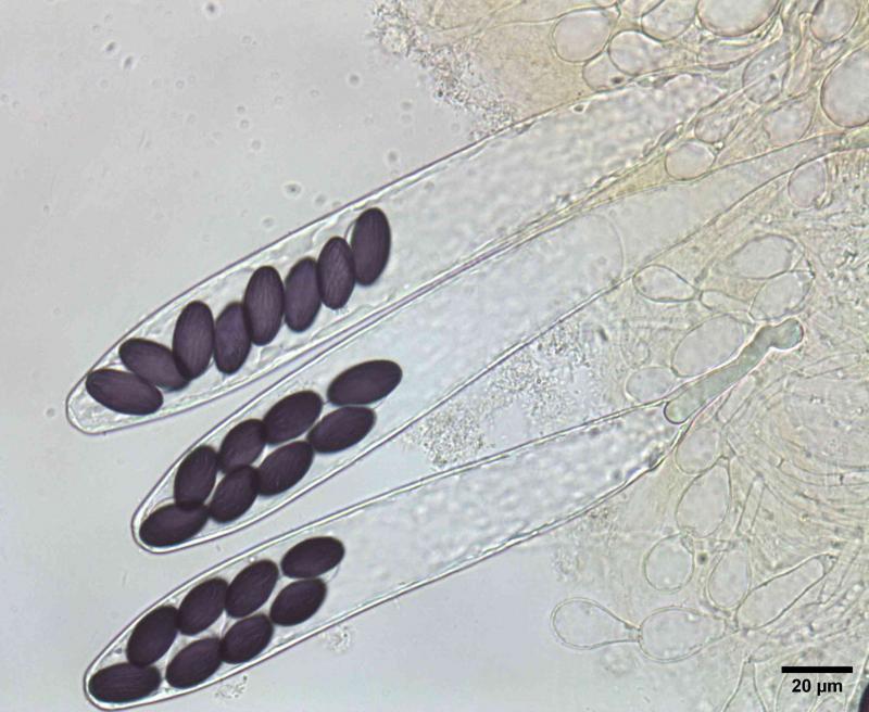
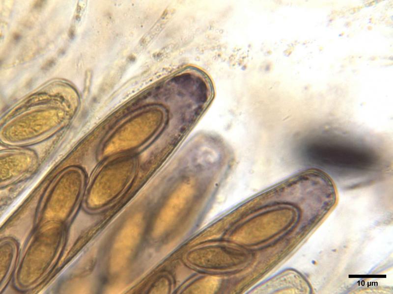
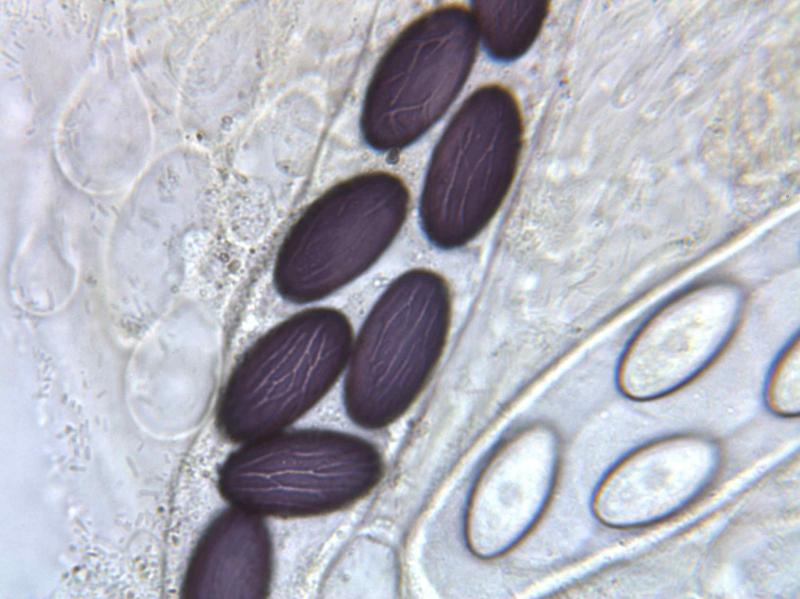
Spores: elliptical with gelatinous coating, with longitudinal anastomosing lines, from hyaline to purple; (18.6) 20 - 24.3 (24.8) × (10.4) 11 - 11.9 (12.3) µm; Q = (1.6) 1.8 - 2.1 (2.2) ; Me = 22.3 × 11.4 µm ; Qe = 2
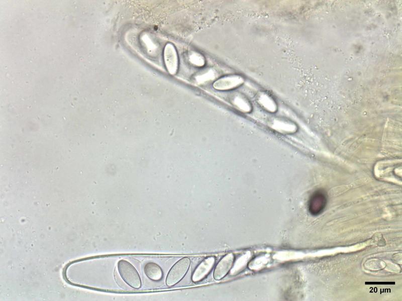
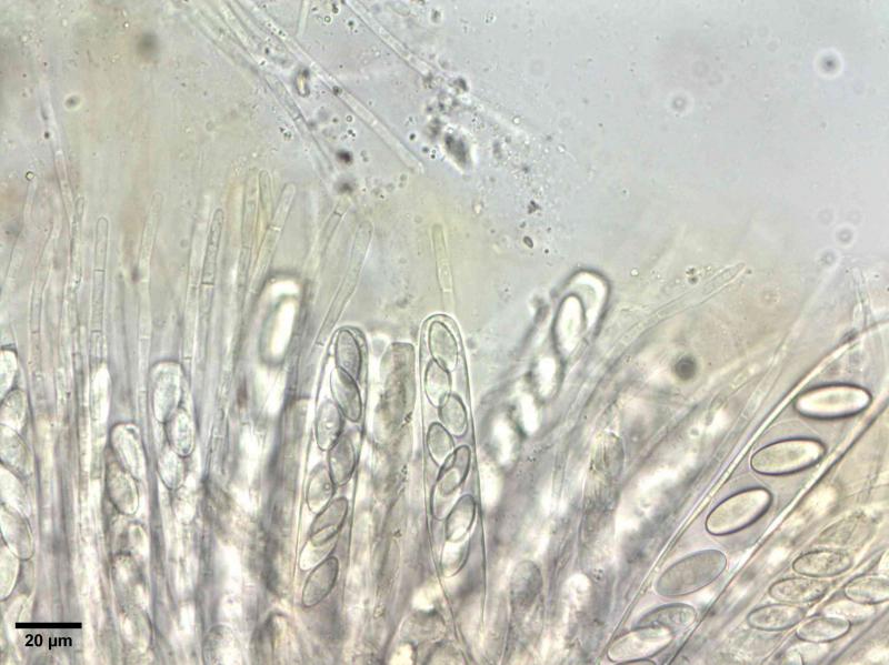
Asci: elongated club-shaped, Me = 210.5 × 27.4 µm ; Qe = 7.7; slight purple discolouration in Melzer:
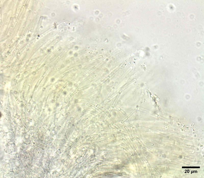
Paraphysis: filiform, with multiple septa, width about 3.5 µm in a yellowish substance. After some days in moist environment I observe fortoulisme.
Ectal excipulum: textura globulosa.
Could this be Ascobolus michaudii?
Thanks in advance,
François Bartholomeeusen
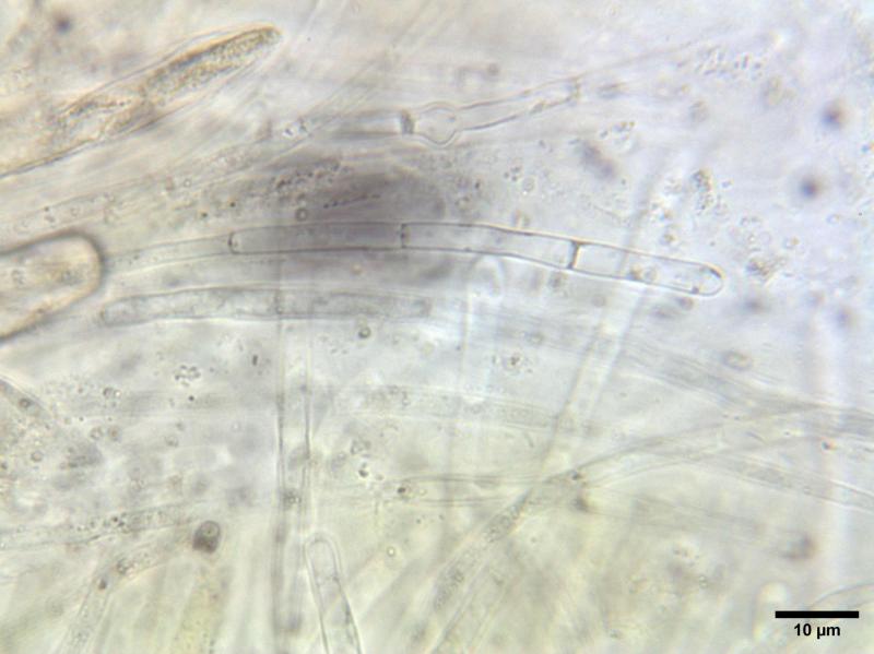
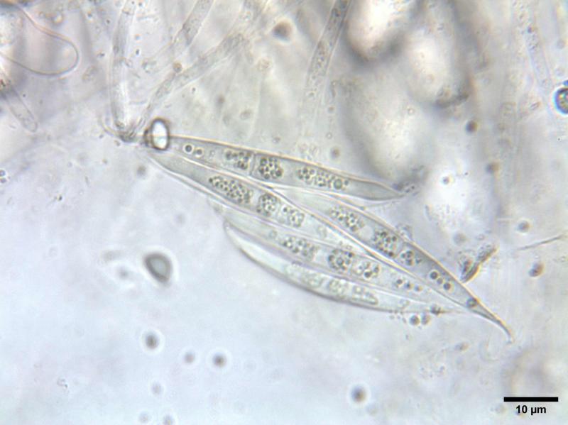
N.B.: on two photos during microscopic examination I found clustered spores with the following dimensions: (52.3) 52.33 - 58 × (4.4) 4.42 - 5.15 (5.2) µm: Me = 55 × 4.9 µm ; Qe = 11.6.
I found only these spores and no perithecial beak. Could these spores belong to Pyxidiophora arvernensis (Laboulbeniomycete)?


Peter Welt,
19-05-2025 10:55

Re : Ascobolus michaudii with possible bycatch Pyxidiophora arvernensis?
Certainly a species of the genus Pyxidiophora. However, not P. arvernensis, since the asci have eight spores. Probably P. spinuliformis (Speg., 1909) N. Lundq., since all other species have fewer than eight spores.
See: Doveri, F. & B. Coue (2006) - First record of Pyxidiophora badiorostris from France. Doc. Myco
l. 34/ 133- 134: 33-41.
See: Doveri, F. & B. Coue (2006) - First record of Pyxidiophora badiorostris from France. Doc. Myco
l. 34/ 133- 134: 33-41.
The article is available online.
Best regards, Peter
François Bartholomeeusen,
19-05-2025 15:06
Re : Ascobolus michaudii with possible bycatch Pyxidiophora arvernensis?
Dear Peter,
Thank you very much for your response. I think you are right. I also used the key in that publication, but I was unsure about the number of spores and their length. I suspect the spores are not fully mature.
Thanks again, now I only need to obtain confirmation about Ascobolus michaudii!
Many greetings,
François