10-06-2026 13:41
 François Freléchoux
Bonjour à nouveau, Voici une trouvaille d'hier.
10-06-2026 12:54
Steve ClementsBonjour encore, Pouvez-vous m'aider, s'il vous pl
10-06-2026 11:53
Steve ClementsBonjour, This disco is abundant on dead stems of
10-06-2026 10:45
François Freléchoux
Bonjour à nouveau, Encore une détermination qui
08-06-2026 10:16
 Spooren Marco
I don`t have a clou about this fungus,it is not in
10-06-2026 09:24
François Freléchoux
Bonjour, J'imagine que cette détermination ne do
09-06-2026 18:32
Camille MertensSur morceau de roseau immergé 0,5 - 0,7 mm de dia
08-06-2026 17:00
François BartholomeeusenGood day everyone, On June 5 2026, I collected de
07-06-2026 15:10
 William Slosse
Hello everyone,On 05-06-26, I found following asco
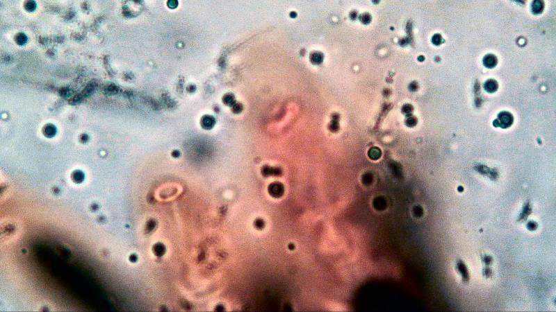
 When we are investigating characters of species under a light through microscope we do observe that in a 2D picture.
So we have to think in 3D but that is not always possible because our mindset cannot cope with the optical illusion we are looking at.
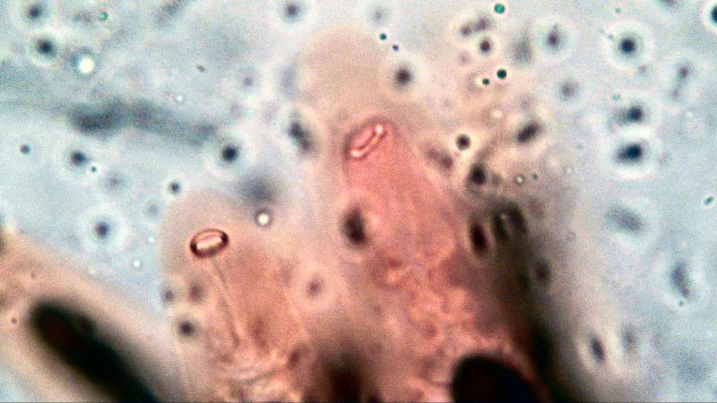
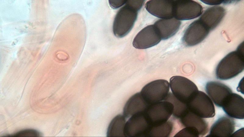
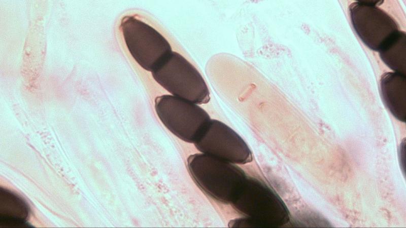
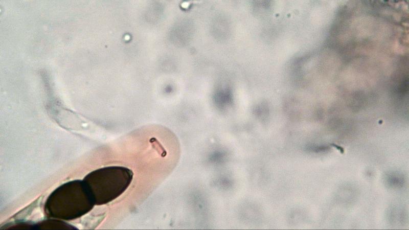
Accidentally I did find out that we can create a 3D picture by changing the focal distance from the lens to the object using a Plane Objective 100x/1.25 (photo 1 & 2). Probably by stacking photo's you will create the same effect.
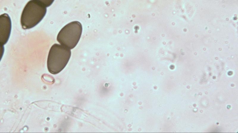
The ring is elastic and the distance when the apical system is not fully developed is as follows: Diameter of the outer circular ring is 0,9 um; total diameter is 4,6 um and inner diameter is 2.8 um. Photo-3 is a ring clearly visible with a spore ready to enter.
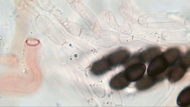
Photo 4 the ring is connected to an ampty inner wall, photo 5 is the same situation but inside a still present outer wall.
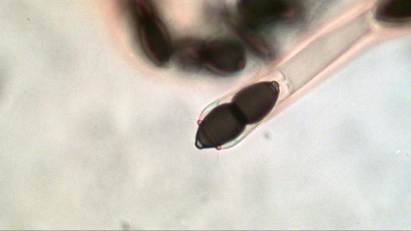
Photo 6 shows the apical ring in the end phase with spore clicked inside and the outer wall still present.
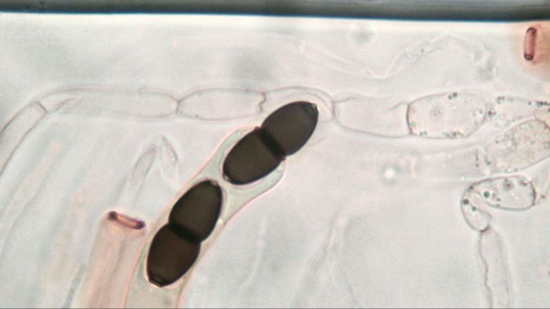
Photo 7 & 8 show spores inside the ring and outer wall gone.
The ring itself is more oval than it is circular. (photo 9)
Kind regards,
Joop