03-05-2026 10:57
 Castillo Joseba
Me mandan el material seco de Galicia (España) re
02-05-2026 12:42
Alain BRISSARDBonjour à tousJeuidi 30 avril dernier on m'a remi
02-05-2026 13:06
Pauline. PennaBonjour Please can someone help me with this id
01-05-2026 22:45
 Thierry Blondelle
Bonjour à tous, Une récolte sur bouse séchée d
28-04-2026 20:07
 Lothar Krieglsteiner
... on twig in the air at standing Ceratonia siliq
14-04-2026 05:32
Ethan CrensonHi all, A few weeks back a friend pointed out som
28-04-2026 20:33
Vitus SchäfftleinHello, I found Trochila ilicina on Ilex aquifoliu
30-04-2026 10:28
Rot BojanHello, by appearance I would say that I am dealing
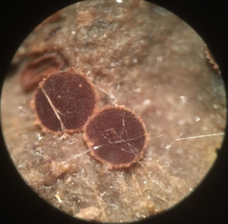
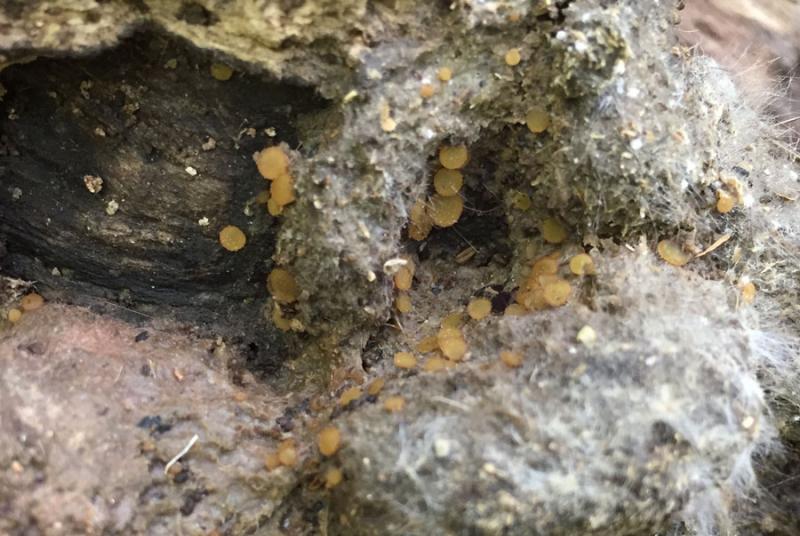
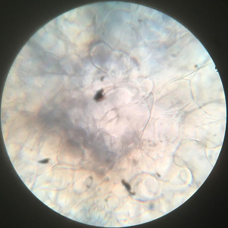
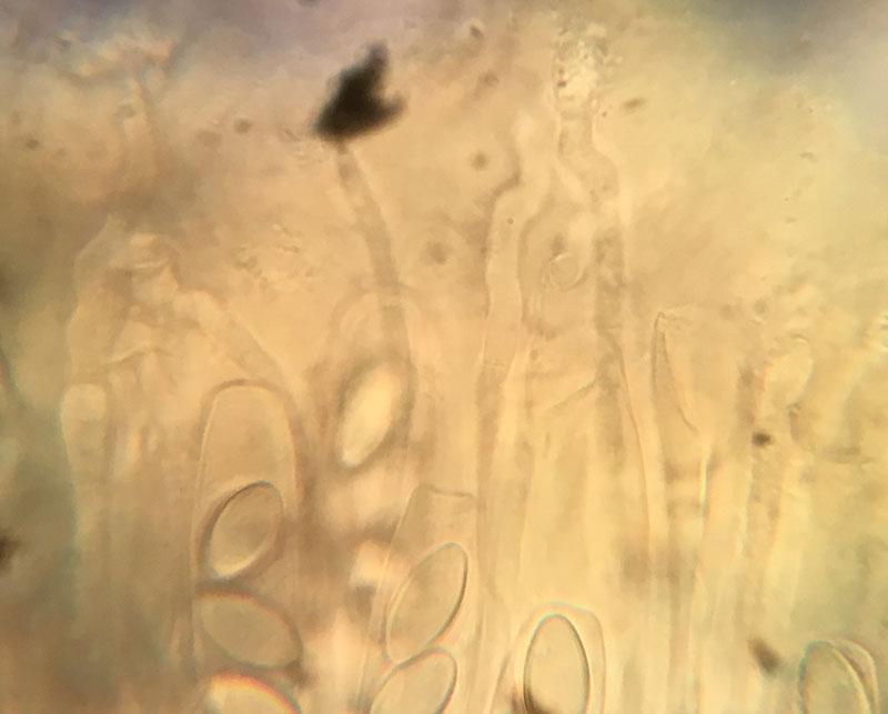
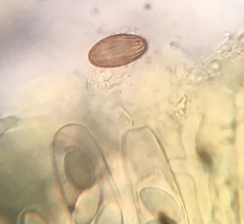
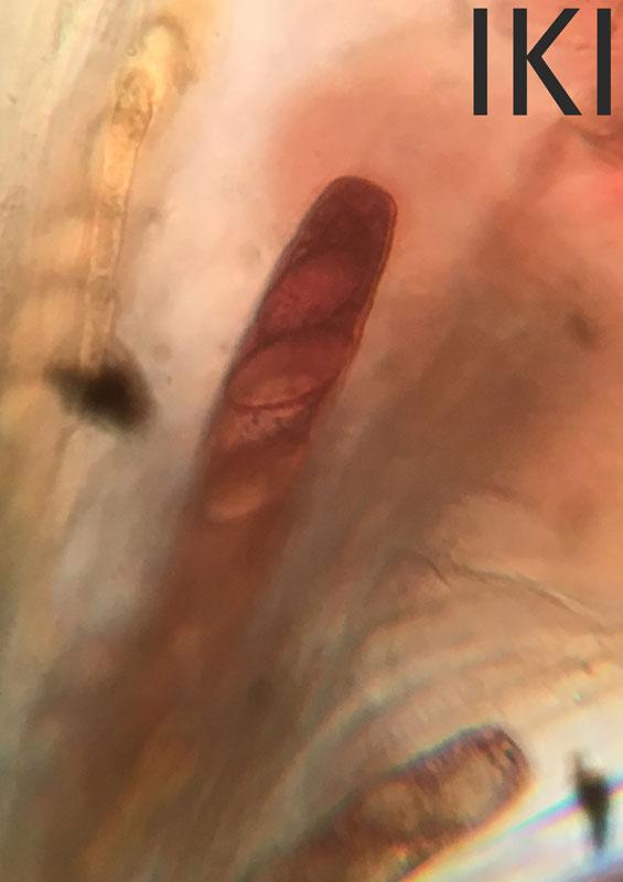
Ascobolus on raccoon droppings
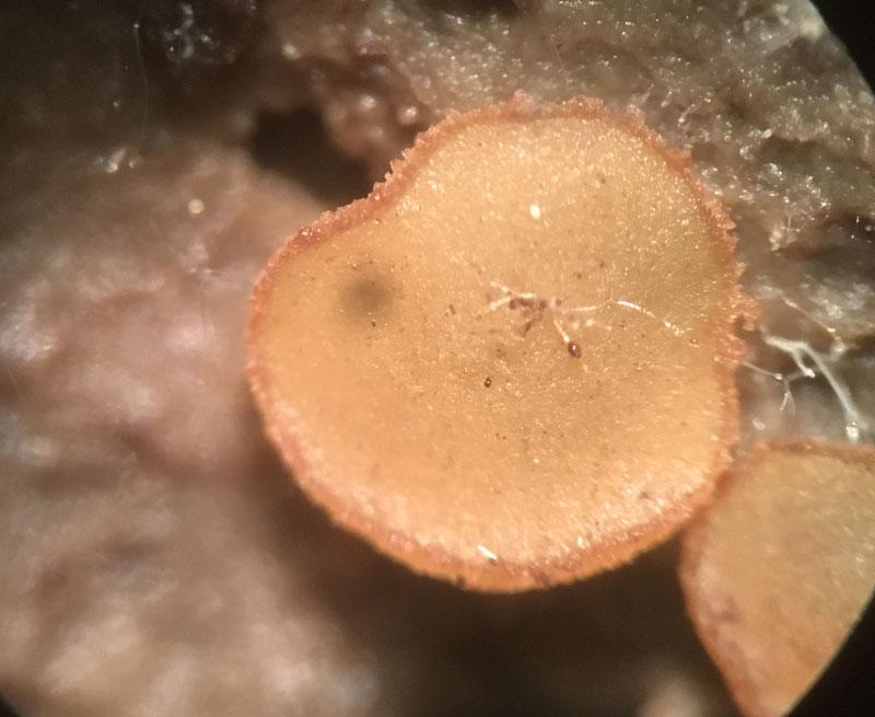
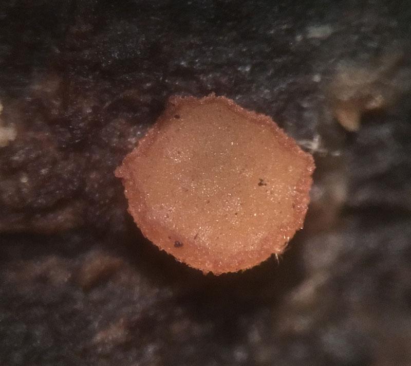
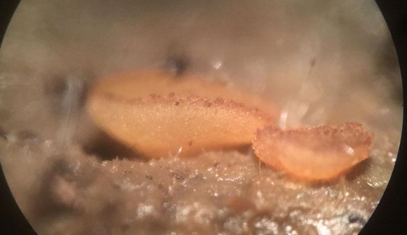
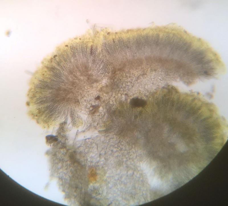
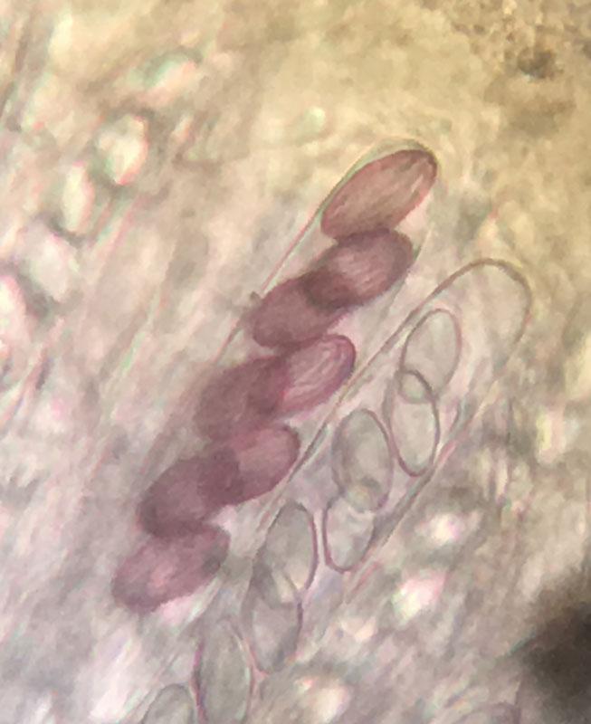
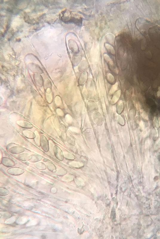
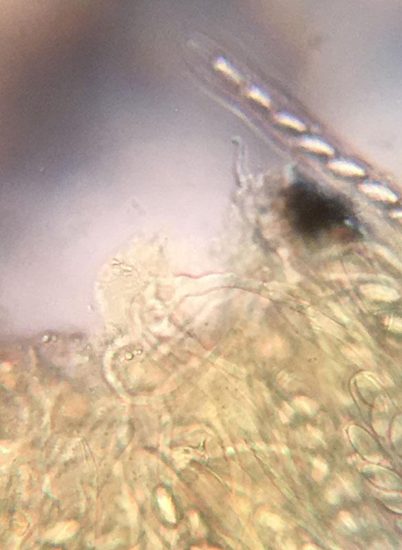
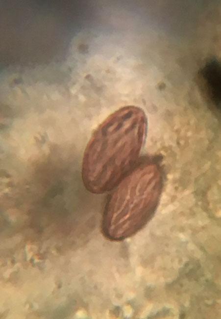
Ethan Crenson,
20-05-2019 22:07
Jacky Launoy,
21-05-2019 09:35
Re : Ascobolus on raccoon droppings
It looks like Ascobolus michaudii
Ethan Crenson,
22-05-2019 21:32
Re : Ascobolus on raccoon droppings
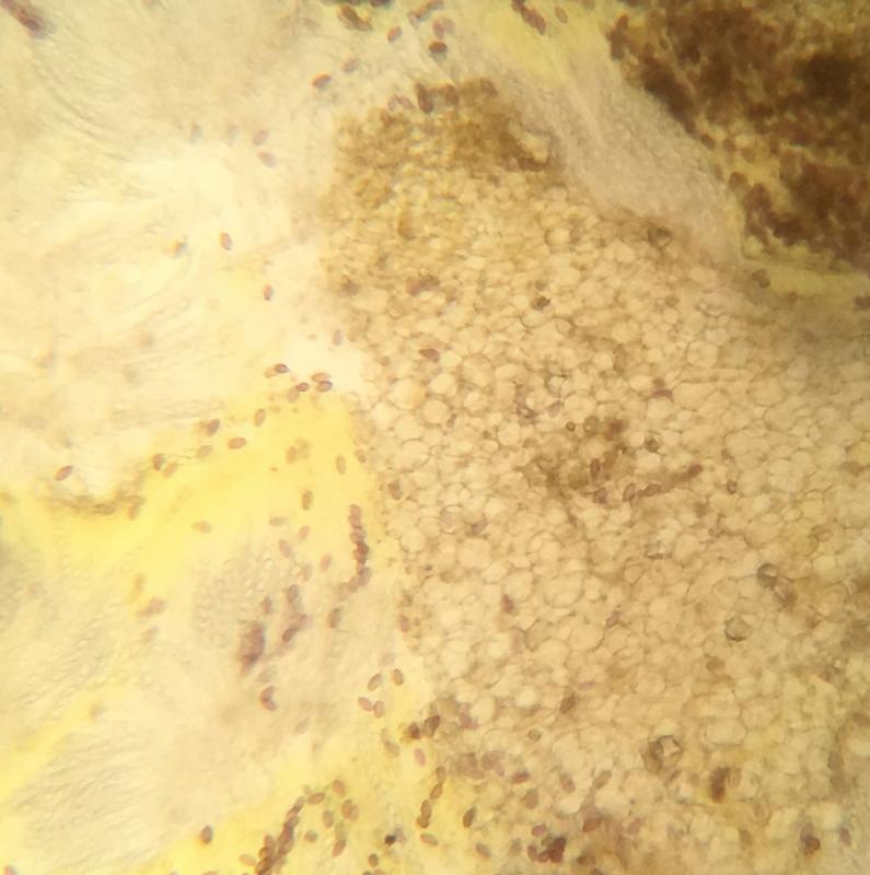
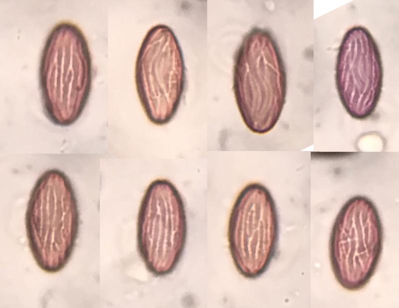
Thank you, Jacky. I am having some difficulty understanding the finer points differentiating A. lignatilis, foliicola, furfuraceus, michaudii. In another post on the topic on this forum François Valade comments that the brown walled excipulum cells (in michaudii & lignatilis) vs. hyaline or yellow cells (in foliicola)--but I'm not sure I'm translating these concepts properly. At any rate I have made more images, including more mature apothecia, mature spores and excipulum. Spores at maturity seem to be quite reliably 16-18 x 9µm, brown, with some branching grooves--even some that connect laterally. Q=1.93 The apothecia only reach 3-4 mm in diameter. Perhaps there is more detail here that could confirm A. michaud?