
06-05-2026 11:25
 Castillo Joseba
Me mandan el material seco de Galicia (España) re
28-04-2026 20:07
 Lothar Krieglsteiner
... on twig in the air at standing Ceratonia siliq
05-05-2026 22:40
Gernot FriebesHi,I believe this is a Plagiostoma growing on a Sa
04-05-2026 18:13
 Stephen Martin Mifsud
ID request for what seems to be a true aquatic fun
04-05-2026 16:39
Stephen Martin Mifsud
ID request: This specimen was collected in Malta o
28-07-2011 18:31
 Alex Akulov
Dear FriendsToday I made the pdf file of Velenovsk
04-05-2026 09:50
Castillo Joseba
Me mandan el material seco de Galicia,(España) re
02-05-2026 12:42
Alain BRISSARDBonjour à tousJeuidi 30 avril dernier on m'a remi
02-05-2026 13:06
Pauline. PennaBonjour Please can someone help me with this id
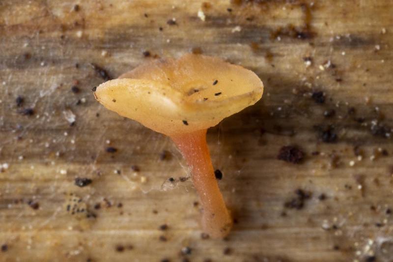
Cyathicula on Hemerocallis fulva
Johan Myhrer,
04-11-2018 18:24
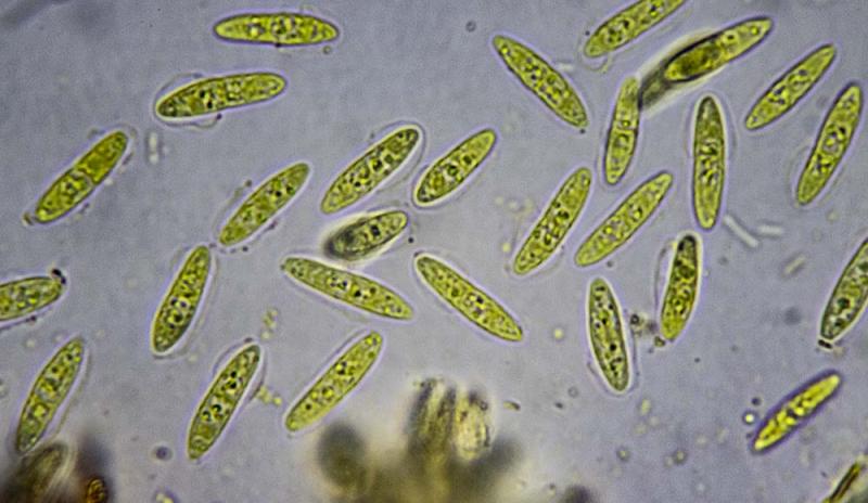
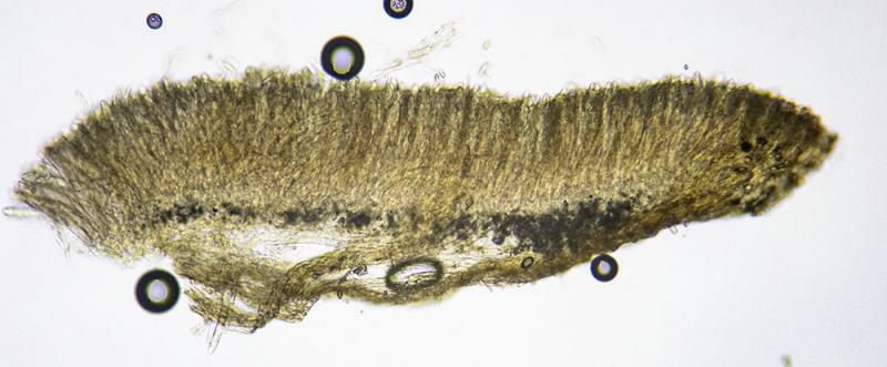
On dead flower stems of Hemerocallis fulva laying on soil in my garden (central Sweden 2018-11-04), I found these (along with typical Cyathicula coronata). Spores ~11x4µm, ascus apex IKI+ weakly blue. The latter difficult to observe because of "explosive" spore release when solution added. Could be overmatured C. coronata with septated spores? Measured spore size on coronata in this collection is ~21x4µm though. Thankful for guidance!
/Johan Myhrer

Hans-Otto Baral,
04-11-2018 18:34

Re : Cyathicula on Hemerocallis fulva
Dear Johan
I canot believe 11 x 4 µm. When I measure the spores are either 11 x 3 µm or 15 x 4.
The excipulum is strongly gelatinsed? Or could it be a Hymenoscyphus?
Explodung occurs in mature asci. usually there are immature ones and they are especially suitable for apical ring structure, of course in dead state when cell walls are thick.
As you observed abundant exploding, I assume that the figured spores are freshly ejected. This means that they were 1-septate within the living asci, a rare feature in Hymenoscyphus and also Cyathicula. But the guttulate paraphyses exclude a Calycina where 1-septate spores often occur.
Zotto
Johan Myhrer,
04-11-2018 19:14
Re : Cyathicula on Hemerocallis fulva
Might very well be Hymenoscyphus, any species that would fit? Few species recorded on Hemerocallis as it seems?
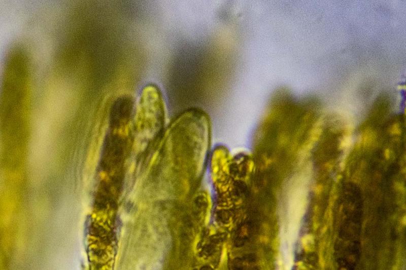
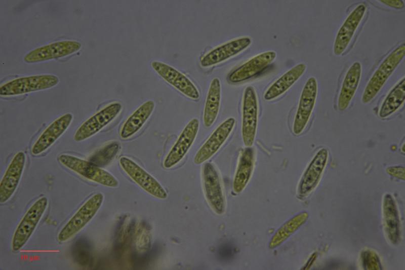
My notes from measurement of spores and picture of section:
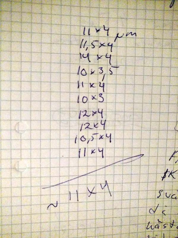
Johan Myhrer,
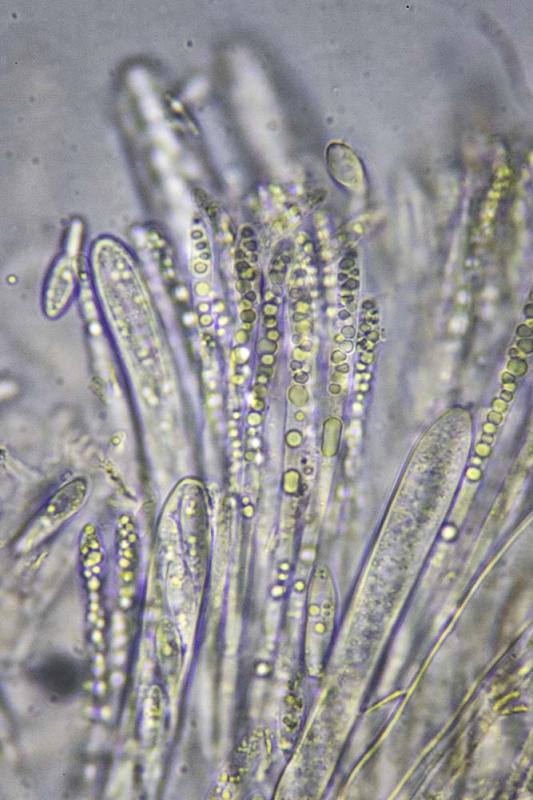
04-11-2018 19:19
Re : Cyathicula on Hemerocallis fulva
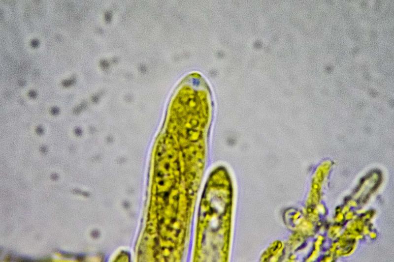
And ascus apex with IKI:
Johan Myhrer,
04-11-2018 19:33
Re : Cyathicula on Hemerocallis fulva
Second try with ascus:
Hans-Otto Baral,
04-11-2018 20:28
Re : Cyathicula on Hemerocallis fulva
A closeup of the ectal excipulum would be necessary.
I do not suspect that the fungus is specific to a genus. Maybe to monocots. But I do not have an idea. The apical ring photos are splendid, they seem to exclude Hymenoscyphus because of the conical apex.
You don't have a scale to your spore photos?
I do not suspect that the fungus is specific to a genus. Maybe to monocots. But I do not have an idea. The apical ring photos are splendid, they seem to exclude Hymenoscyphus because of the conical apex.
You don't have a scale to your spore photos?
Johan Myhrer,
04-11-2018 20:50
Re : Cyathicula on Hemerocallis fulva
No scale to measurement on photos cause I photo through trinocular microscope and measure with scaled (calibrated) eyepeace :-/
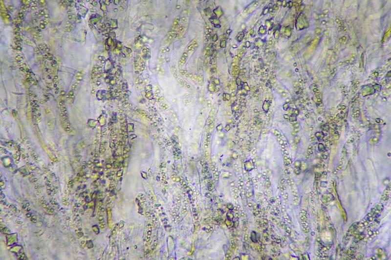
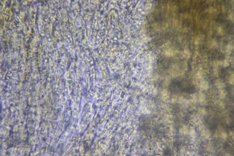
Ah, ectal excipulum:
Hans-Otto Baral,
04-11-2018 21:15
Re : Cyathicula on Hemerocallis fulva
You could easily photograph a calibration slide and prepare a scale to be included in a photo. That would enable control over measurements.
Your excipulum photo is great, it shows undulating guttulate living cortical hyphae and crystals - all typical of Cyathicula. The proper excipulum is below, however, and that could be gelatinized or not.
C. dolosella is a possibility, that has 1-septate spores in the living asci, *11-17 x 2.4-2.9. Ap margin with fine teeth
Zotto
Johan Myhrer,
04-11-2018 21:32
Re : Cyathicula on Hemerocallis fulva
Thank you for guidance Zotto! I will prepare a "calibration-picture" 'til next time. Have to give this Cyathicula a rest now though...
Johan Myhrer,
06-11-2018 15:11
Re : Cyathicula on Hemerocallis fulva
Tried a new method for spore measurement and it seems as I have to use this one in the future. Photoshop have a great; but a little complicated; measurement tool that I were not aware of!
Zotto; you where right about my measurements beeing to short, they are infact 12-16µm. So C. dolosella seems to be the one (Crocicreas dolosellum according to Swedish checklist).
/Johan Myhrer
 Cyathicula-spores-0001.txt)
Hans-Otto Baral,
06-11-2018 16:00
Re : Cyathicula on Hemerocallis fulva
O.k., and did you measure in Lugol or water? The nice photo in water must have another scale, can you find out that? because I fear that in Lugol the spores are already smaller (narrower).
I finally think that it is not dolosella. But the ectal excipulum is very difficult to squash?
I finally think that it is not dolosella. But the ectal excipulum is very difficult to squash?
Johan Myhrer,
06-11-2018 17:02
Re : Cyathicula on Hemerocallis fulva
Yes the excipulum is quiet tough. Measurement of spores in water as in attached files.
Hans-Otto Baral,
06-11-2018 17:42
Re : Cyathicula on Hemerocallis fulva
So it is mainly 14-15 x 3.8-4 µm.
Spore size of C. dolosella in Dennis 1978 is 12-15 x 2-2.5 (probably in dead state).
I think it is another species. The excipulum is not very clear, to me it looks thin-walled though difficult to separate. Cyathicula has 1-2 µm thick intercellular refractive gel.
Spore size of C. dolosella in Dennis 1978 is 12-15 x 2-2.5 (probably in dead state).
I think it is another species. The excipulum is not very clear, to me it looks thin-walled though difficult to separate. Cyathicula has 1-2 µm thick intercellular refractive gel.
Johan Myhrer,
06-11-2018 17:47
Re : Cyathicula on Hemerocallis fulva
I could not get the excipulum any clearer, this picture represents very well my observation through the eyepiece...