
06-05-2026 11:25
 Castillo Joseba
Me mandan el material seco de Galicia (España) re
06-05-2026 17:23
Thomas Læssøehttps://svampe.databasen.org/observations/10594257
28-04-2026 20:07
 Lothar Krieglsteiner
... on twig in the air at standing Ceratonia siliq
05-05-2026 22:40
Gernot FriebesHi,I believe this is a Plagiostoma growing on a Sa
04-05-2026 18:13
 Stephen Martin Mifsud
ID request for what seems to be a true aquatic fun
04-05-2026 16:39
Stephen Martin Mifsud
ID request: This specimen was collected in Malta o
28-07-2011 18:31
 Alex Akulov
Dear FriendsToday I made the pdf file of Velenovsk
04-05-2026 09:50
Castillo Joseba
Me mandan el material seco de Galicia,(España) re
02-05-2026 12:42
Alain BRISSARDBonjour à tousJeuidi 30 avril dernier on m'a remi
 Hello,
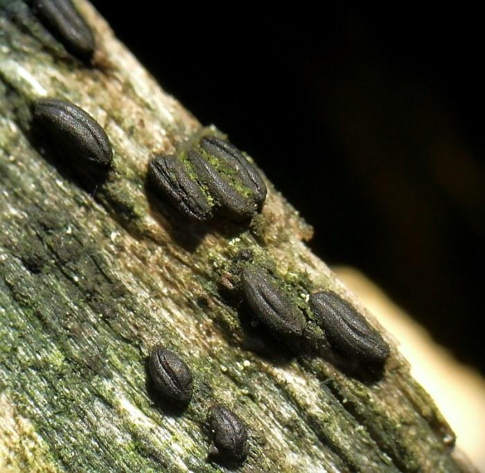
I found that in an herbarium sheet, probably on Fagus sylvatica, in France, Puy-de-Dôme (63). Do any one can confirm / infirm the ID ? I'm not conviced by the general shape...
Many thanks for the help!
Rémy
Hi Remy,
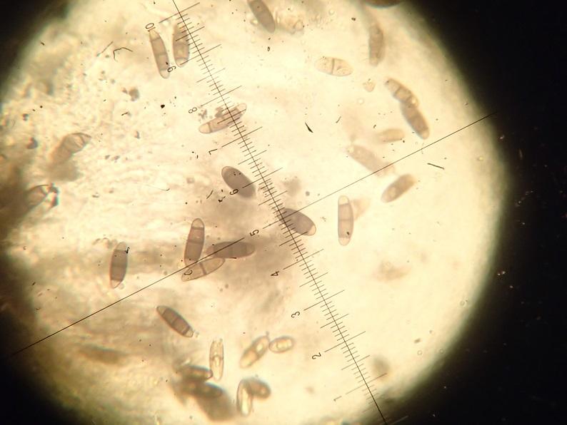
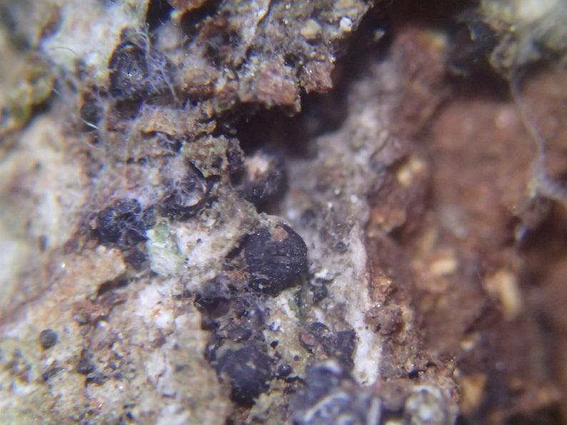
although the macrofoto is not very distinctive and does not show the form of the coffee beans I think there can be no doubt because of the very unique spores.
Regards from Lothar
Many thanks for your very quick answer !
I agree that I make very poor quality picture - not sure yet if it is because of the photographer, or the material ;)
In fact, I was not sure for this ID because many herbarium sheets I am currently reviewing (for epiphytic lichens) contains this species, and most of them are more "sphaerical" that coffee bean shaped... All are from the same locality.
Cheers,
Rémy
Hi Remy,
the growth on bark of living deciduous trees is very typical for H. pulicare. But - if the ascomata are not hysterothecia, it must be something different, anyway ...
Best regards from Lothar
Thank you for the precision ; I will try to ID other specimen in order to be sure !
Bests,
Rémy

so Hysterium pulicare would have to look.
Greetings Peter.
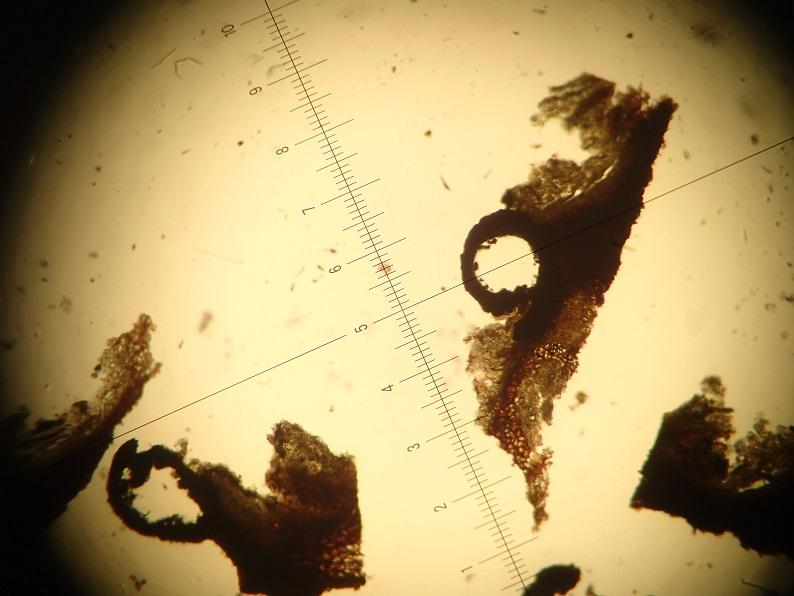
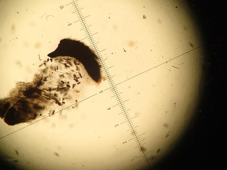
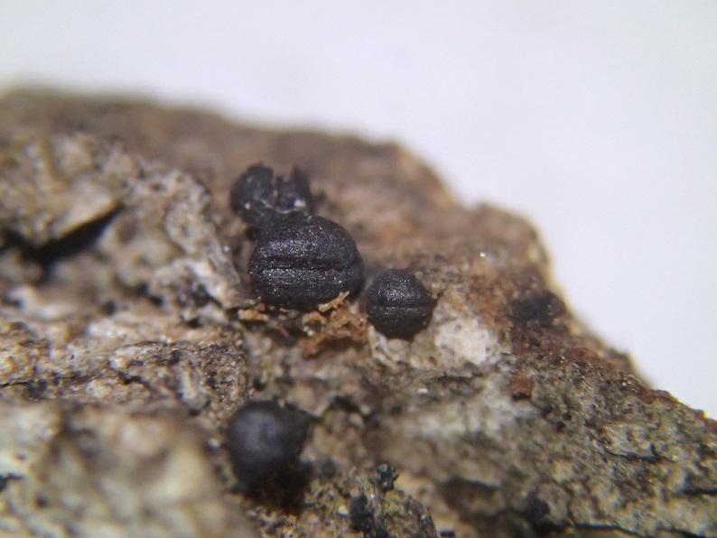
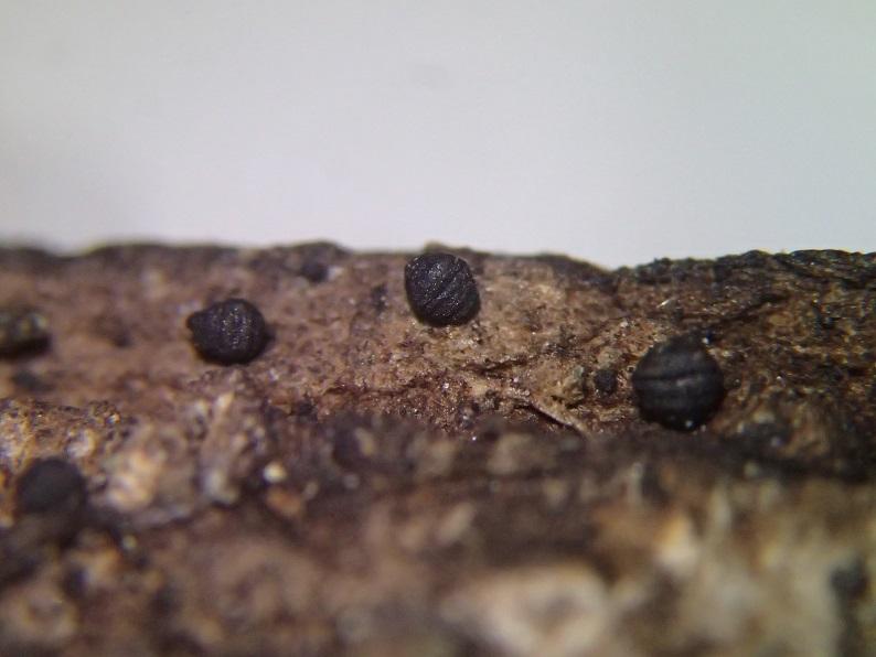
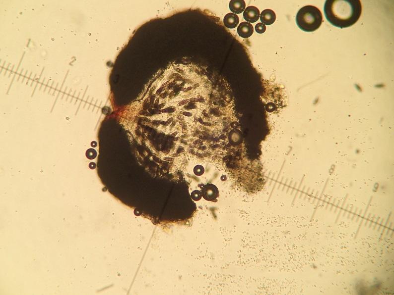
I found a new one in another herbarium sheet. The two pictures are taken on the same piece of bark... On has a coffe bean shape, and the second one not much. Is it beacause of dessication ? Or another species ?
Many thanks,
Rémy
Hi Remy,
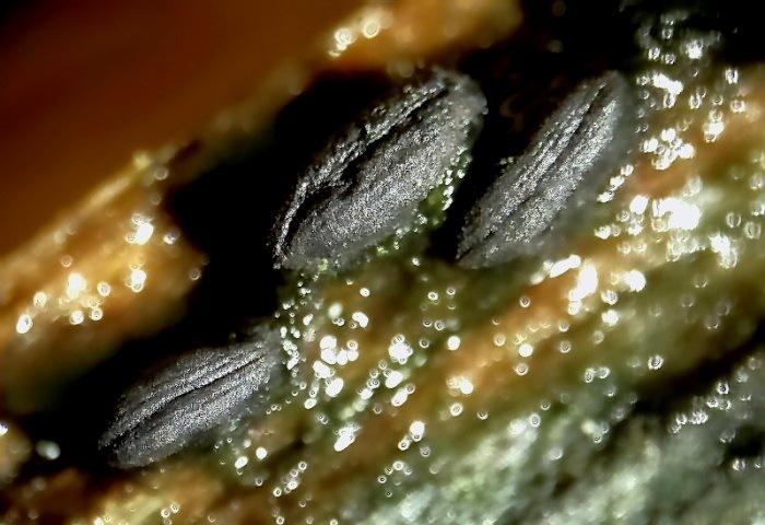
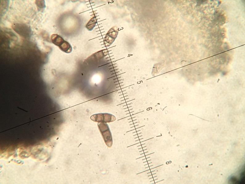
the pictures show clear hysterothecia in my opinion. Do they have the same spores (4-celled with terminal hyaline cells)? Then it ist H. pulicare.
Maybe unripe or badly developed specimens do not form the typical slit very distinctly. Peters fotos are very typical.
Regards from Lothar
this goes not without microscopic examination. If the scales of the photos 1:1 with the micro, it cannot be H. pulicare, the spores would be too small there.
Greetings Peter.
some new picture from the inside. Spores are about ~20-28µm.
Bests,
Rémy
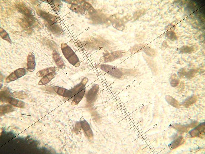
now everything is clear.
Greetings Peter.