
06-05-2026 17:23
Thomas Læssøehttps://svampe.databasen.org/observations/10594257
06-05-2026 11:25
 Castillo Joseba
Me mandan el material seco de Galicia (España) re
28-04-2026 20:07
 Lothar Krieglsteiner
... on twig in the air at standing Ceratonia siliq
05-05-2026 22:40
Gernot FriebesHi,I believe this is a Plagiostoma growing on a Sa
04-05-2026 18:13
 Stephen Martin Mifsud
ID request for what seems to be a true aquatic fun
04-05-2026 16:39
Stephen Martin Mifsud
ID request: This specimen was collected in Malta o
28-07-2011 18:31
 Alex Akulov
Dear FriendsToday I made the pdf file of Velenovsk
04-05-2026 09:50
Castillo Joseba
Me mandan el material seco de Galicia,(España) re
02-05-2026 12:42
Alain BRISSARDBonjour à tousJeuidi 30 avril dernier on m'a remi
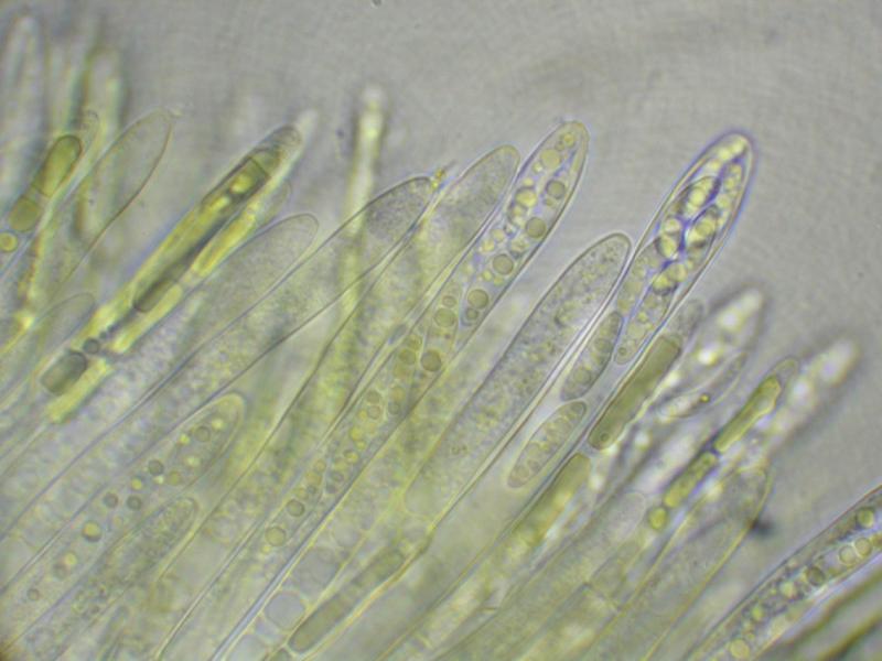
 Greetings AscoFrance!
Here is a curious discomycete, field IDed to Ionomidotis by Paula DeSanto on the recent Peck Foray in Watkins Glen, New York. This particular find is from a mixed, predominantly hardwood forest within the Meads Creek State Forest. When I got a look at the dried material and field photos, I saw enough resemblance to my own Ionomidotis collection from North Carolina (http://mushroomobserver.org/174774) to consider the possibility, but upon preparing the material for microscopy we noticed that KOH extractable pigments (3% solution) were conspicuously absent. Can it still be Ionomidotis without this reaction? Perhaps this is a member of some other genus in the Encoelioideae?
Ascus tips inamyloid, despite appearing somewhat bluish in the micrographs. No paraphyses observed.
Spores:
9.5-14×=2.5-4.5?m (x=12.25×3.325?m, Q= 2.44-5.2?m, Qm=3.781?m, m=20, s=1)
13.5 x 3 ; 4.5
13 x 4 ; 3.25
13 x 2.5 ; 5.2
12.5 x 3.5 ; 3.57
14 x 4 ; 3.5
13 x 3 ; 4.33
9.5 x 3 ; 3.17
12.5 x 4 ; 3.13
13 x 3.5 ; 3.71
11 x 4.5 ; 2.44
13.5 x 3 ; 4.5
13.5 x 3 ; 4.5
10.5 x 4 ; 2.65
12 x 3 ; 4
12.5 x 2.5 ; 5
12.5 x 3 ; 4.17
9.5 x 3 ; 3.17
14 x 4 ; 3.5
11.5 x 3 ; 3.83
10.5 x 3 ; 3.5
Many thanks!
-Danny N.
PS: The images are all apparently too large for the site :( Please find them on Mushroom Observer here: http://mushroomobserver.org/218595

I am reminded of a Chlorencoelia, but the two species for which I have images, C. versiformis and C. torta) have distinctly amyloid asci. The spores would fit.
I am sure that the paraphyses would be seen when squashing the hymenium. If you had pictures from fresh material the genus Chlorencoelia would show a striking feature in the paraphyses (vacuolar bodies, see attach).
Zotto
Also, I believe the fact that the bottle of Melzer's used was labelled "Melzer's Replacement" may have something to do with the lack of observed blueing. Will use a more reliable reagent for the second set of micrographs.
These vacuolar bodies are a useful character at the family level. They are rather typical for the family Cenangiaceae as we now circumscribe it, but absent from the Cordieritidaceae which inbclude many ionomidotic species.
many thanks!