
27-04-2026 18:48
Tony MoverleyCollected 23rd April 2026, Norfolk, EnglandSwarms
27-04-2026 17:41
 Lothar Krieglsteiner
.. Algarve, same leaf than the last post.ô The con
27-04-2026 18:05
Lothar Krieglsteiner
... still attached at standing tree. The green con
27-04-2026 17:16
Lothar Krieglsteiner
.. Algarve, moist lying.The conidiomata look like
27-04-2026 12:54
Steve ClementsBonjour. Ce petit champignon blanc rûˋsupinûˋ et
27-04-2026 09:59
Pauline. PennaBonjour Can anyone advise me on these pycnidia fo

22-04-2026 20:54
 Enrique Rubio
Hi to everybody.This Pyrenopeziza grew in moist le
24-04-2026 03:16
 David Chapados
Found while looking at something else from wood in
Hymenobolus agaves anamorph
Miguel ûngel Ribes,
12-03-2013 00:39
 Good night
Perhaps someone remember this Hymenobolus agaves: http://www.ascofrance.fr/search_forum/10909
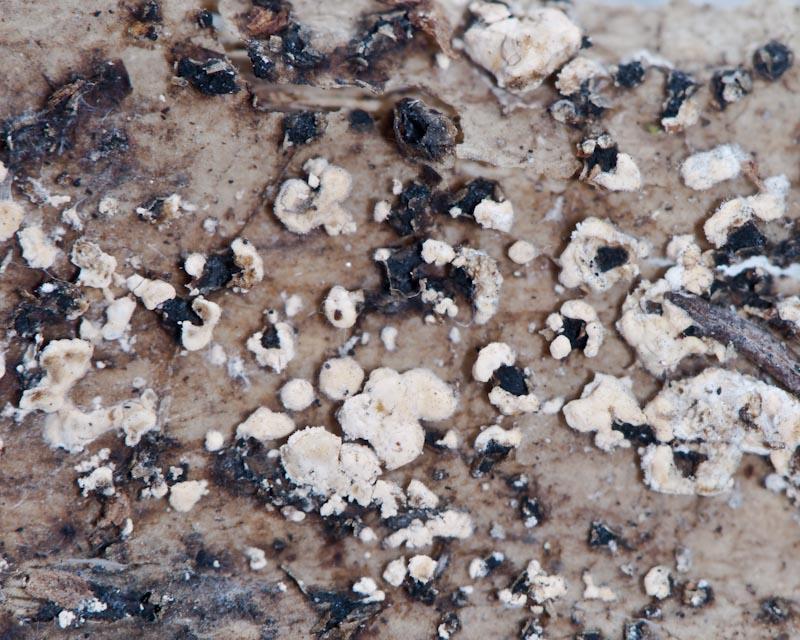
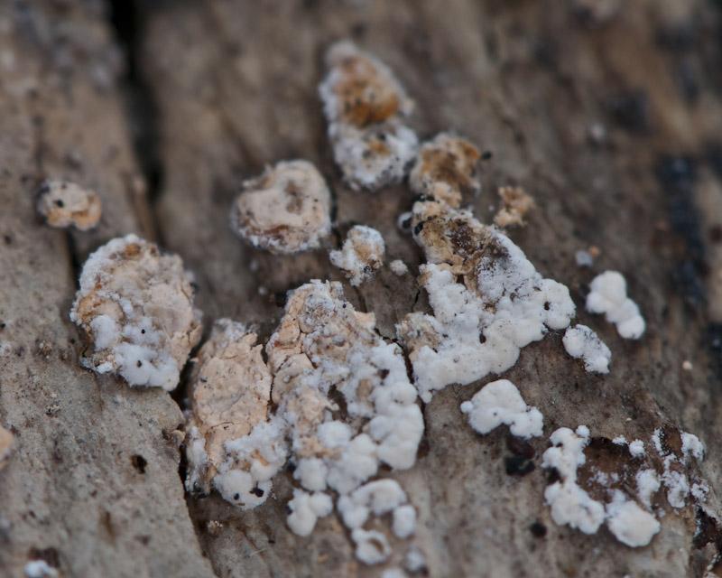
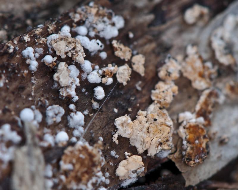
Rubûˋn has foung more collections in another Canary Island, La Gomera. In some collections, between H. agaves apothecium, are growing too a white-orange anamorph, 2-5 mm broad, relatively hard (it is posible to cut it).
Is it posible the anamorph of H. agaves? How to study this anamorph?
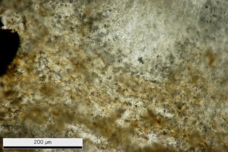
This are general views.
Thank you.
Miguel ûngel Ribes,
12-03-2013 00:44
Re : Hymenobolus agaves anamorph
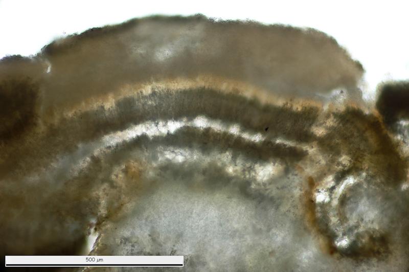
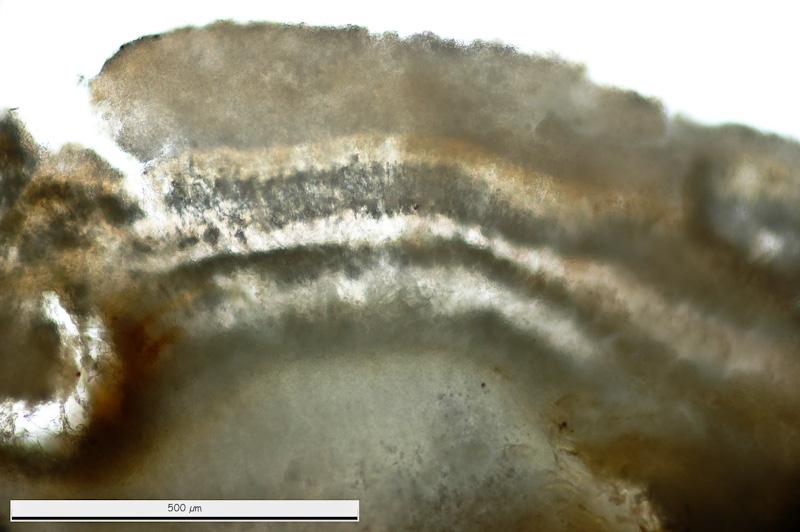
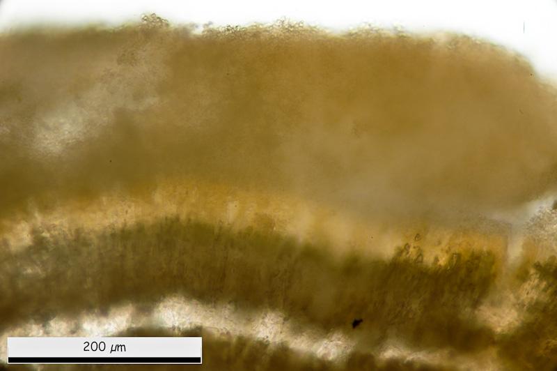
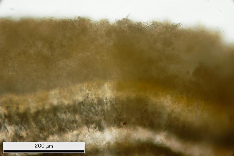
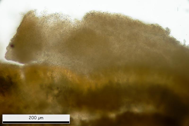
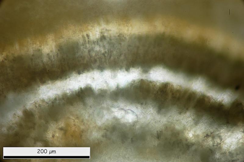
General micro views. It is posible to see some layers. External one with more-less rounded cells.
Miguel ûngel Ribes,
12-03-2013 00:50
Re : Hymenobolus agaves anamorph
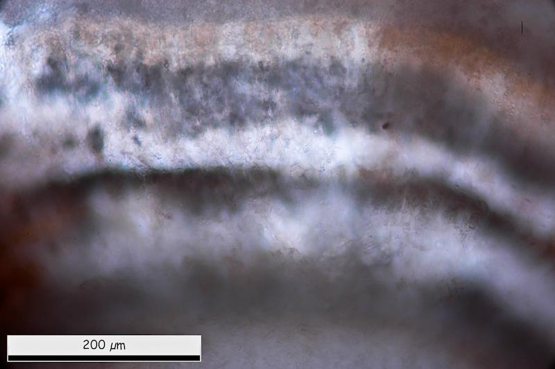
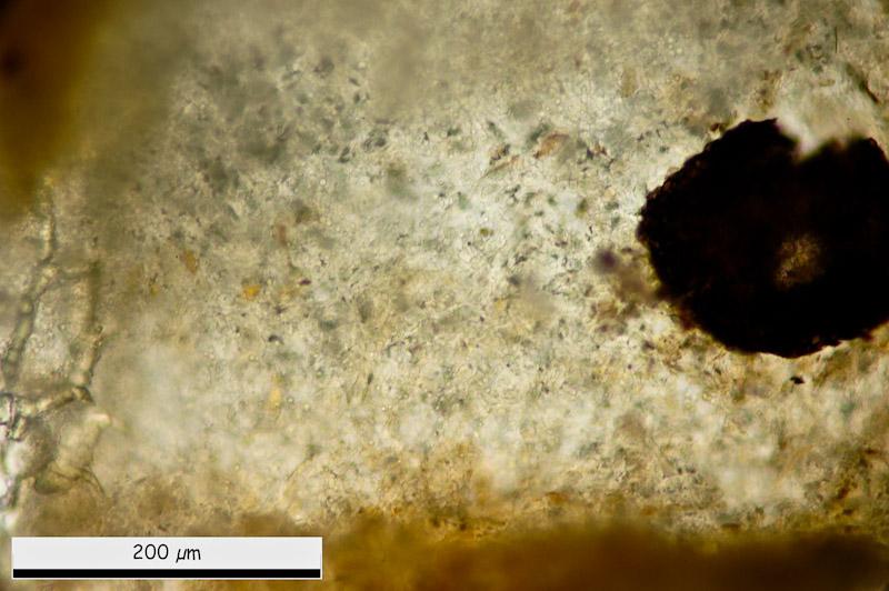
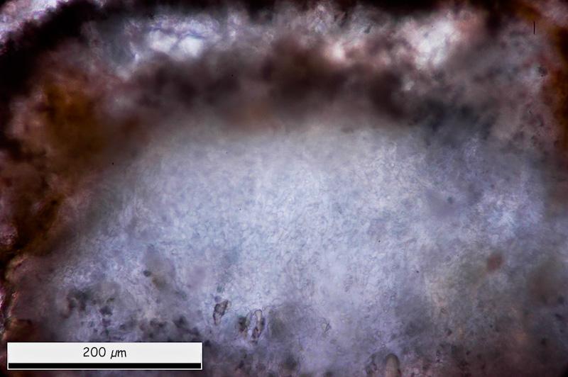
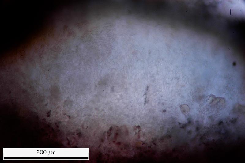
Inside, tow black lines. And in medular area a white area with globose-angular structure mixed with cilyndrical cells.
Miguel ûngel Ribes,
12-03-2013 00:53
Re : Hymenobolus agaves anamorph
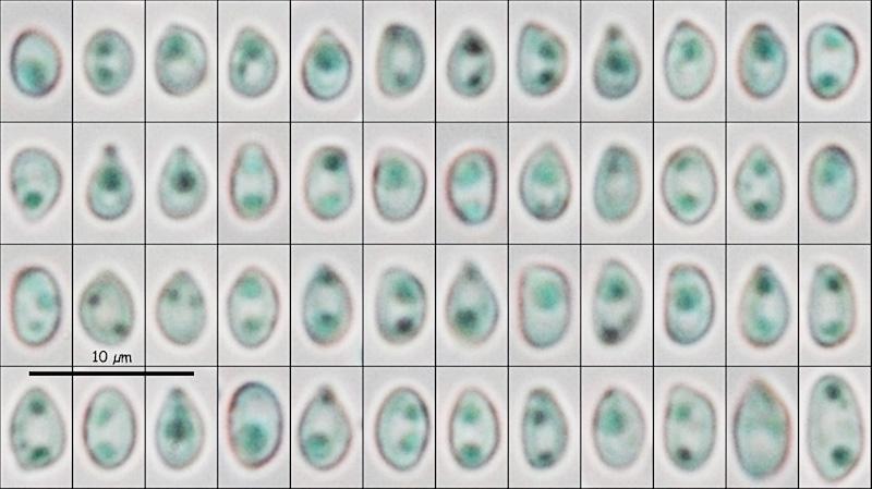
Eliptical conidiospores in water:
(4.06) 4.49 - 5.21 (6.63) x (2.87) 2.94 - 3.35 (3.57) ôçm
Q = (1.28) 1.40 - 1.70 (1.86) ; N = 52
Me = 4.88 x 3.14 ôçm ; Qe = 1.56
(4.06) 4.49 - 5.21 (6.63) x (2.87) 2.94 - 3.35 (3.57) ôçm
Q = (1.28) 1.40 - 1.70 (1.86) ; N = 52
Me = 4.88 x 3.14 ôçm ; Qe = 1.56
Miguel ûngel Ribes,
12-03-2013 00:55
Re : Hymenobolus agaves anamorph
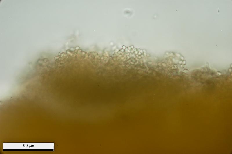
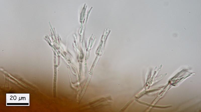
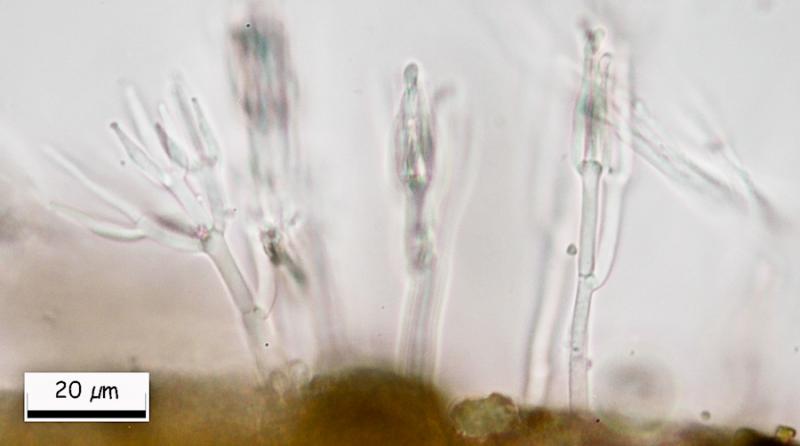
And finally, at the margin, this conidial structure.
Than you in advance.
Miguel û. Ribes
Than you in advance.
Miguel û. Ribes
Hans-Otto Baral,
12-03-2013 08:06

Re : Hymenobolus agaves anamorph
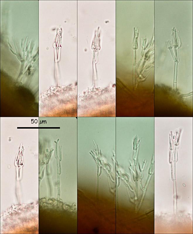
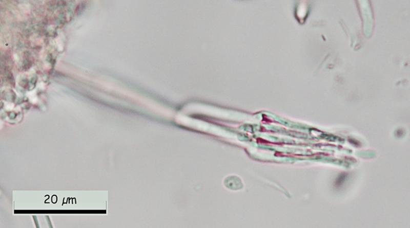
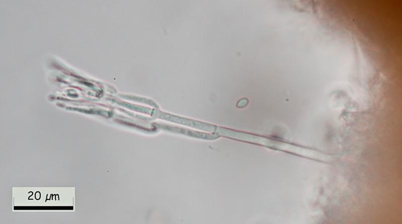
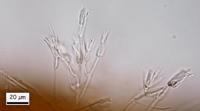
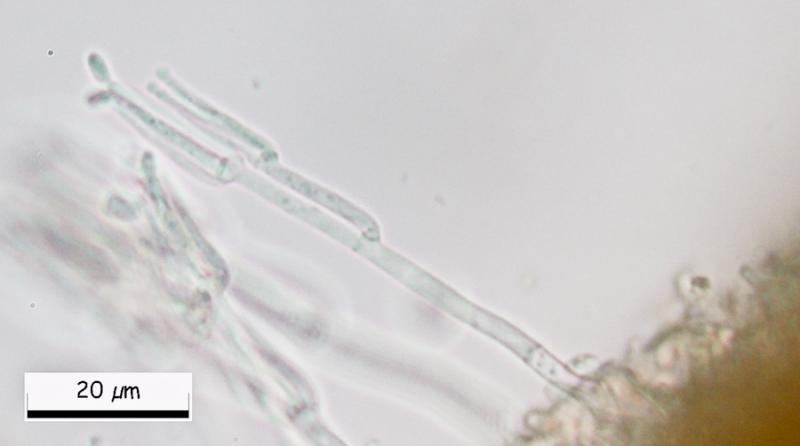
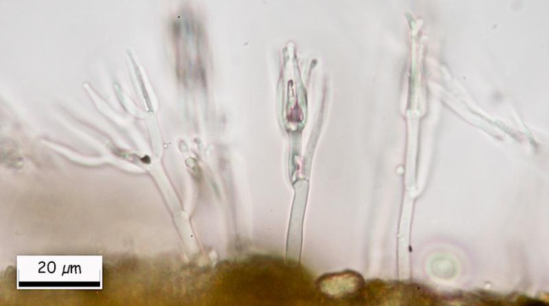
Great, Miguel! Could you please show us a closeup of the conidiogenous cells, were the conidia emerge? I assume they are phialidic. Then we can search in Genera of Hyphomycetes, or someone has an idea.
I have given the previous Hymenobolus specimen for sequencing, I am curious where it could belong.
Zotto
I have given the previous Hymenobolus specimen for sequencing, I am curious where it could belong.
Zotto
Miguel ûngel Ribes,
12-03-2013 11:27
Re : Hymenobolus agaves anamorph
Here there are.
Hans-Otto Baral,
12-03-2013 22:56
Re : Hymenobolus agaves anamorph
Hi Miguel
Walter Gams answered me that this is ô clearly aô Clonostachys, probably Clonostachysô solani (Harting) Schroers & W. Gams, which is quite common, often fungicolous, and the anamorph of a Bionectria. So certainly not belonging to Hymenobolus.
Zotto
Walter Gams answered me that this is ô clearly aô Clonostachys, probably Clonostachysô solani (Harting) Schroers & W. Gams, which is quite common, often fungicolous, and the anamorph of a Bionectria. So certainly not belonging to Hymenobolus.
Zotto
Miguel ûngel Ribes,
13-03-2013 00:15
Re : Hymenobolus agaves anamorph
Hi Zotto, Superb.
Thank you again to resolve this puzzle.
See you.
Thank you again to resolve this puzzle.
See you.