01-05-2026 22:45
 Thierry Blondelle
Bonjour à tous, Une récolte sur bouse séchée d
28-04-2026 20:07
 Lothar Krieglsteiner
... on twig in the air at standing Ceratonia siliq
14-04-2026 05:32
Ethan CrensonHi all, A few weeks back a friend pointed out som
28-04-2026 20:33
Vitus SchäfftleinHello, I found Trochila ilicina on Ilex aquifoliu

30-04-2026 10:28
Rot BojanHello, by appearance I would say that I am dealing
27-04-2026 18:48
Tony MoverleyCollected 23rd April 2026, Norfolk, EnglandSwarms
27-04-2026 20:52
Lothar Krieglsteiner
Found on hanging tiwg of Olea europaea in dried-ou
28-04-2026 22:51
 Bernard CLESSE
Bonsoir à toutes et tous,Pourriez-vous m'aider à
29-04-2026 08:01
Lothar Krieglsteiner
... on twig attached to small tree of Citrus auran
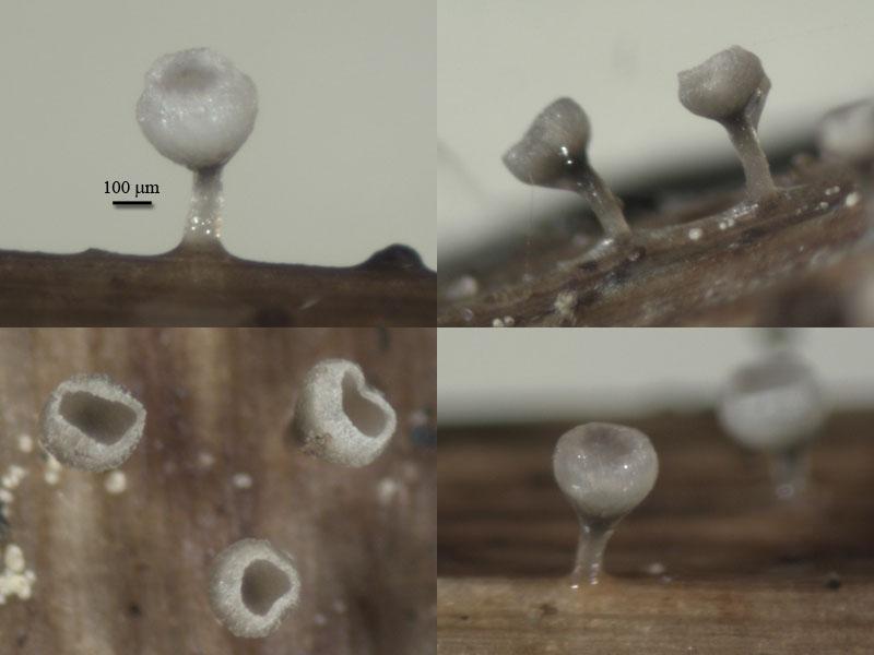
Apothecia goblet-shaped, receptacle deep-cupulate, to 0,5 mm in diam, stipe thin (100 mk), the same high as cup, all frb up to 1 mm high; stipe brownish, translucent, receptacle brownish at base, lighter to white at margin (when dry edge powdery from incrustation), edge rised under hymenium surface forming narrow collar.
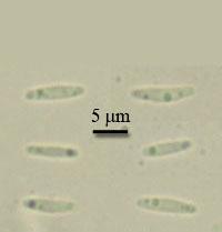
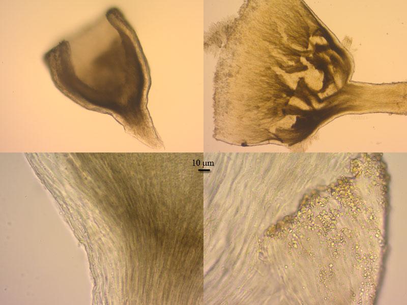
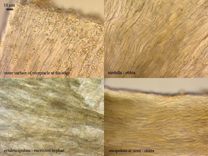
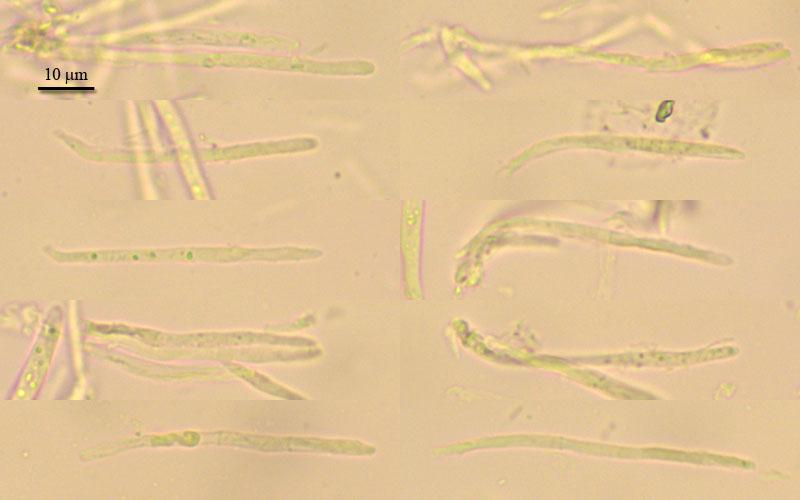
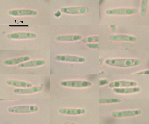
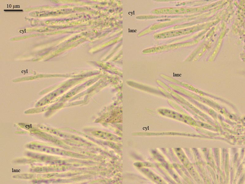
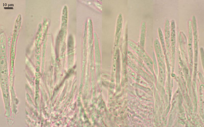
Excipulum from textura oblita, but outer layer of receptacle formed by porrecta, hyphae with rough walls (brown); margin from textura oblita, with abundant crystals; asci clavate, with crozier, with small euamyloid pore, 33,5-43 x 4,2-5,2; paraphyses lanceolate (not clear difference in two types), septate at base, slightly exceeding the asci, up to 3 mk broad in largest part; spores narrow-ellipsoid, with small guttules, 8 (7-9,4) x 1,7 (1,5-2,2) (N=18).
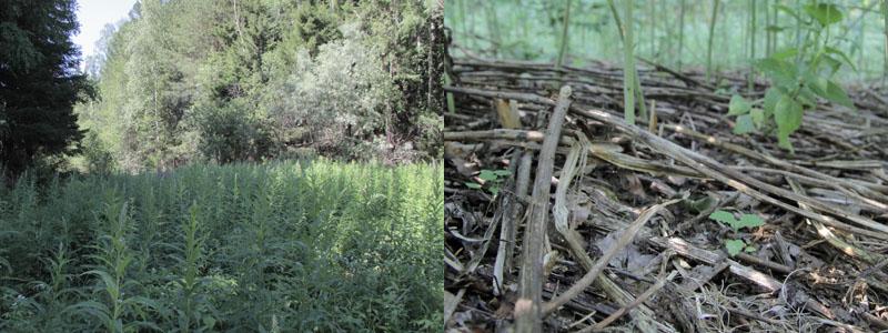
On dead stems of Glyceria triflora at forest edge, N61,090492 E69,480253, 26.06.2012.
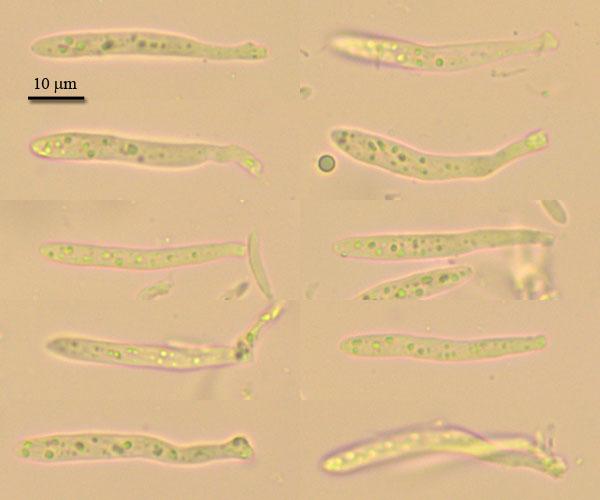

You do not have any micropics in vital state? Here I suspect multiguttulate paraphyses as typical of Cyathicula.
Useful should also bi a photo of the apical rings in IKI. If you compare their shape with those of Crocicreas gramineum, we could perhaps see a distinct difference.
You say paraphyses lanceolate, but I see also cylindrical ones.
Zotto
i will send you vital photo,
there are pictures of ring, it it differrent.
right, i was confused with paraphyses, they were badly seen in previous specimen. Since all hymenial parts smaller, differences not so clear. But now i checked again and think there are also two types, lanceolate and narrow (these rarely seen).
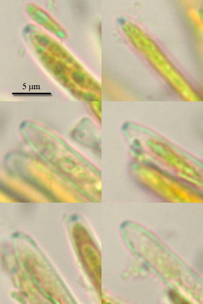
i am not sure about VBs since lack of experience seing them in vital, but what would you say?
On your spore photo I think that two spores are alive (lower left, central right). You say KOH, is this true for all spore photos?
I will compare with Cyathicula starbaeckii.