
02-06-2026 14:33
 Nicolas VAN VOOREN
Hello.I'm searching for a PDF copy of the followin
02-06-2026 17:58
Louis DENYBonjour forum, Sur feuille de Populus tremula, en

28-07-2011 18:31
 Alex Akulov
Dear FriendsToday I made the pdf file of Velenovsk
11-09-2025 16:57
 Jason Karakehian
Our revision of Marthamycetales (Leotiomycetes) is
12-11-2019 10:32
 Miguel Ángel Ribes
Hi againExactly at the same place than my previous
25-12-2019 17:54
Valencia Lopez Francisco JavierHola a todos/asEstas supuestas pezizas estaban en
12-07-2015 00:05
 Nedim Jukic
This one from the same locality as the previous on

30-05-2026 21:12
Philippe PELLICIERSur branche de mélèze (Larix) près de la neige,
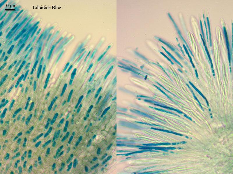
there is another specimen which goes under "subretincola", but it was collected from branches of Chamaedaphne dwarfshrub. It is regular on this substrate, was noticed several times.
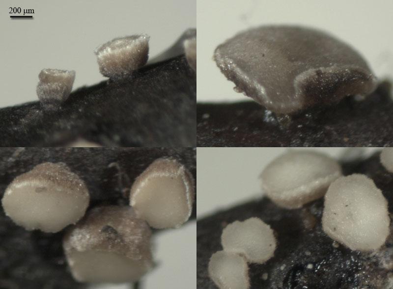
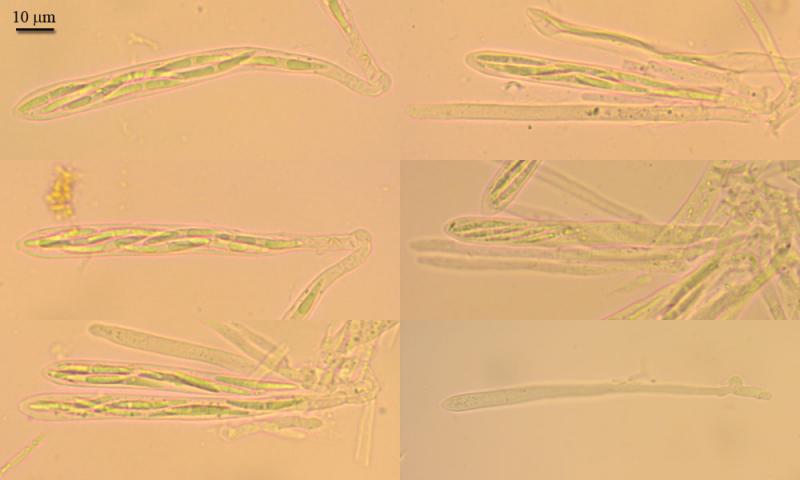
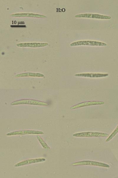
Apothecia there some smaller, up to 1,5 mm, and hymenium hasn't green or olive tinge in color. The main difference in micro is croiziers, here more developed. The structure of tissues is similar to previous specimen, crystals present, KOH reaction of paraphyses also absent, spores some longer, pictures of them were done vitally in water, since small oil droplets now seen.
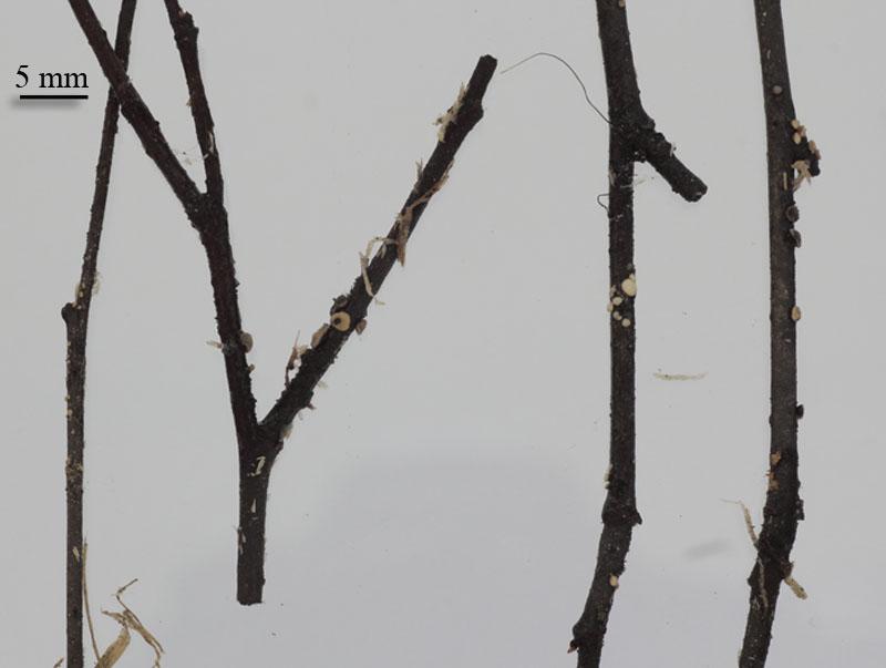
Apothecia from turbinate to cupulate, sessile, thick, up to 1,5 mm diam; outer surface brown at base, lighter at the edge, velvety, edge under high magnification ciliate; hymenium surface plane to concave, yellowish-gray; attached to bark by base without subiculum.
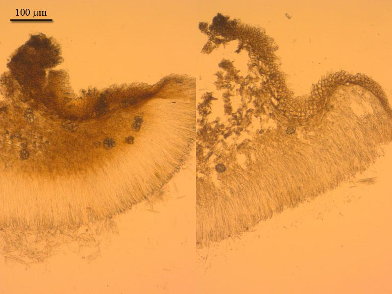
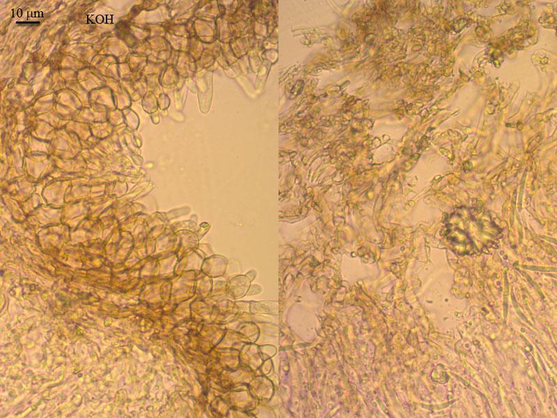
Ectal excipulum from textura globosa, cells with brown walls, 15 mk broad, in 3-5 layers; medulla from textura intricata, loosely arranged hyphae 2,5 broad, with yellowish exudates; crystalls in medulla abundant; hairs at flanks from 2-3 globose cells at base and upper clavate segment, up to 30 mk; edge hairs from 5 or more prismatic cells and upper cylindrical segment, about 50 x 5 mk; asci cylindrical, with clamp, pore euamyloid, 72,8-98 x 7-8,7; paraphyses cylindrical, exceeding the asci about 15 mk, segmented in basal part, scarsely branched, upper segment with VBs (torquose stain), KOH -, 3,5-4,6 broad; spores narrow-fusoid, 22 (20-23,7) x 2,5 (2,2-2,6) (Q=8,85; N=17).


yes , Zotto is of course right.
But it is evident that this is no Mollisia in the current sense and there is a synonymous name for it Dibeloniella citrinella. Also the molecular studies show that "Mollisia" ramealis is quite far away from other mollisioid and pyrenpezizoid taxa. May be it is not even a Dermateaceae.
best regards,
Andreas
you are right with the synonymy with Dibeloniela citrinella, see my drawing of the type HB 6471. From a morphological point of view, Nannfeldt should have placed the species in Belonopsis because of the crystals, isn't it? And isn't Mollisia ligni also quite an untypical Mollisia?
Judging from the phylotrees I have copied into a directory, I see that Dermea/Pezicula and Mollisia and allies incl. Vibrissea are quite unrelated (f.ex. Peterson & Pfister 2010, Cyttaria; Schoch et al. 2009, suppl., Fig. 6D; Wang, Binder et al. 2006). So we should better speak of Mollisiaceae.
I regret I do not have your phylotree of Mollisia, I only remember your lecture in Melle, ssuming that it is unpublished. I cannot imagine that it makes sense to exclude M. ligni from the Mollisiaceae (which presently includes also Hysteropezizella, though this is another question of a possible split at family level). Do you have any idea what could be related to it?
A question in this concern would be which genus might be the more primitive, plesiomorphic one, Mollisia or Pyrenopeziza. Do you have an idea? In the case that Mollisia is more primitive, then the VBs should have got lost in Pyrenoeziza, and M. ramealis might represent a rather ancient taxon in the family.
Zotto