28-04-2026 20:07
 Lothar Krieglsteiner
... on twig in the air at standing Ceratonia siliq
14-04-2026 05:32
Ethan CrensonHi all, A few weeks back a friend pointed out som
28-04-2026 20:33
Vitus SchäfftleinHello, I found Trochila ilicina on Ilex aquifoliu

30-04-2026 10:28
Rot BojanHello, by appearance I would say that I am dealing
27-04-2026 18:48
Tony MoverleyCollected 23rd April 2026, Norfolk, EnglandSwarms
27-04-2026 20:52
Lothar Krieglsteiner
Found on hanging tiwg of Olea europaea in dried-ou
28-04-2026 22:51
 Bernard CLESSE
Bonsoir à toutes et tous,Pourriez-vous m'aider à
29-04-2026 08:01
Lothar Krieglsteiner
... on twig attached to small tree of Citrus auran
29-04-2026 10:44
Lothar Krieglsteiner
growing at moist, drying-out soil at the side of a
 Hi to everyone, bonjour à tous
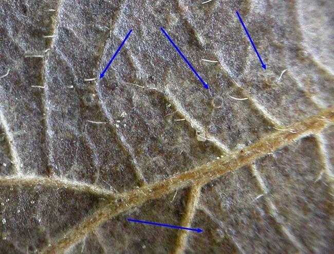
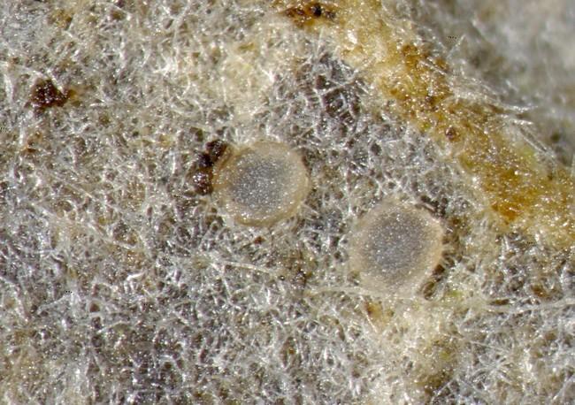
Quelqu'un pourrait il m'orienter pour cette récolte sur feuille de ronce ?
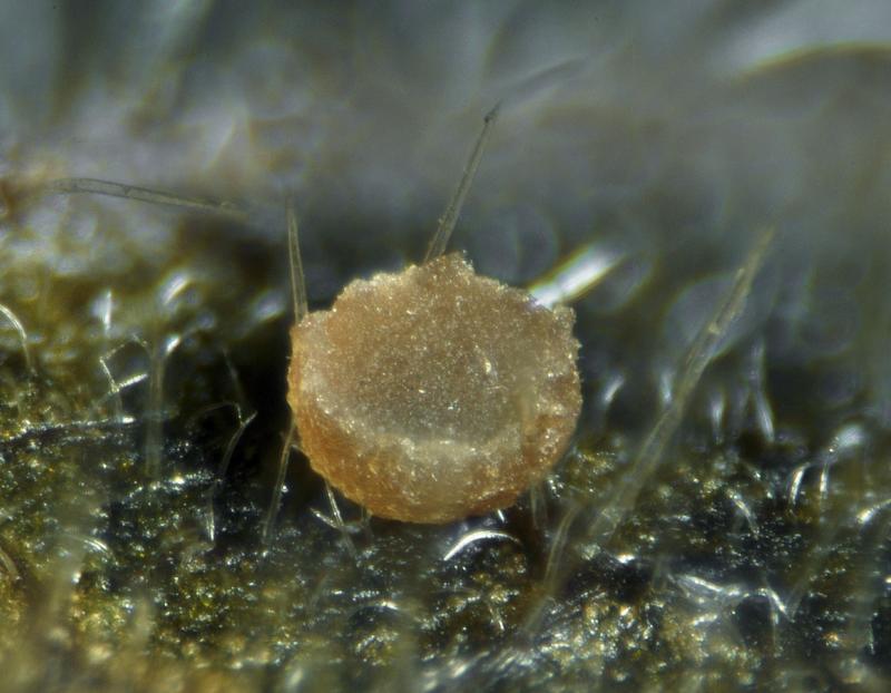
Would someone give me a clue for the following collection on dead Rubus leaves ?
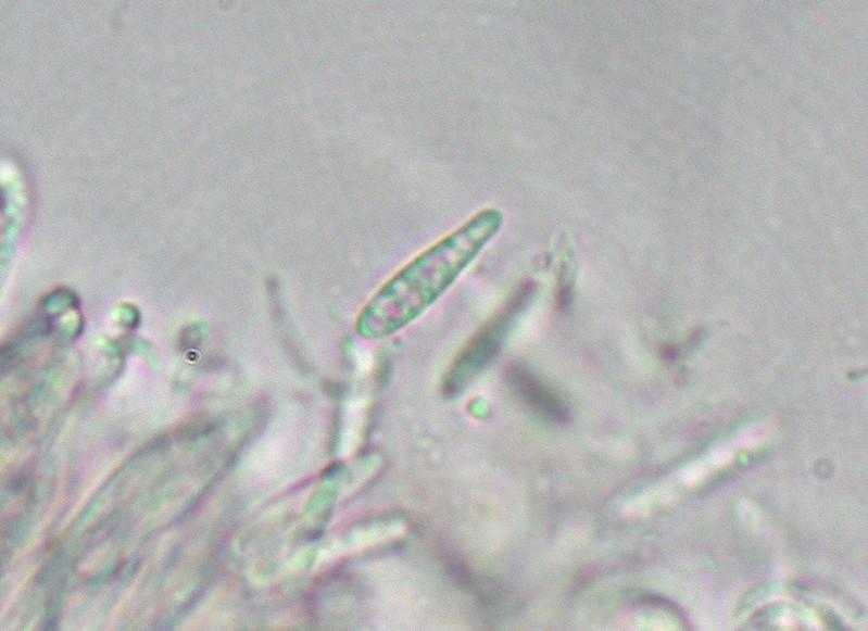
Apos urcéolées, semi immergées dans l'epiderme de la feuille , diamètre 0,1-0.2 mm, gris foncé.
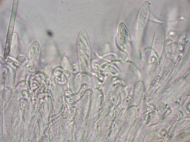
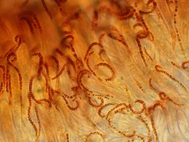
Asques H+, IKI bleu, 43_52 x 6,5-7
Spores 11-15 x 2-2,2 , clavées, G = 0,5-1 par de très fines guttules à ch. pôle
Paraphyses spiralées, dépassant largement les asques, remplies de vauoles réfringentes bleu-vert dans le CRB
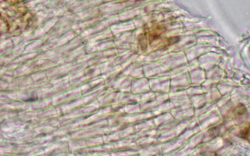
EE text prismatica , prolongé par des poils de type ''Phialina''
Merci beaucoup par avance.
Amitiés
Michel
cheers
Thomas

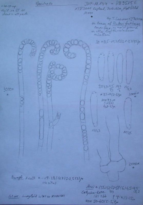
Spores clavate to conical; spores smooth; spores without septae; spores in ligth microscope hyalin; spores not reacting to reagents; spores length 11 µm, width 2 µm; ascus wall unitunicat; ascus apical apparatus J+; ascus length 65 µm; with 8 spores; paraphyses as long as the asci; paraphyses cylindrical; paraphyses with coiled apices; paraphyses width 2 µm; textura prismatica; outer side smooth; excipulum hyalin. On/with members of Rosaceae; substrate/habitat leaves. [Description auto-generated from character input in MycoKey.]

I also just looked at my drawing.... you have certainly the same, substrate is also identical.
There are a couple of described species with curled paraphyses, but they do not perfectly fit:
Pezizella orbiliodes Feltgen on Petasites, but the spores are smaller and the excipulummstains blue in iodine (not so in our fungus - you can test).
Naevia vitellina on Aegopodium is actually similar but the apothecia are elongate and break through the epidermis, though our fungus might also be erumpent.
Naevia lutescens was found in 2000 on Lamiastrum in Luxembourg, there the asci were simple-septate.
Calloria circinella on Cypripedium in China : also simple-septate and spores different.
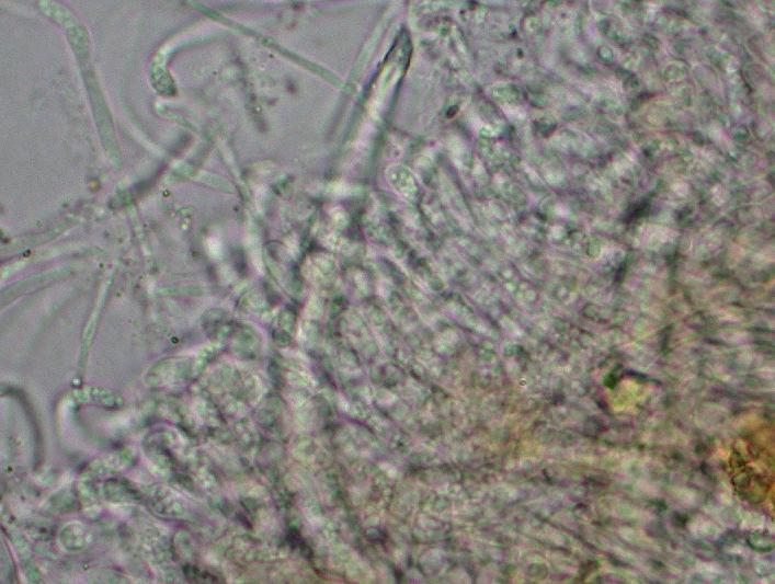
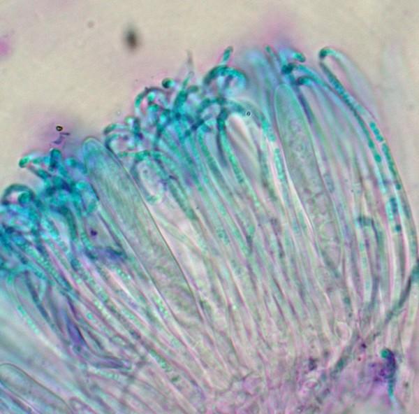
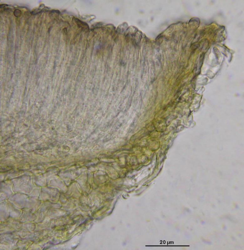
Michel, did you actually see Phialina-hairs? Any photo? My section looks like this (live and in KOH)
Zotto
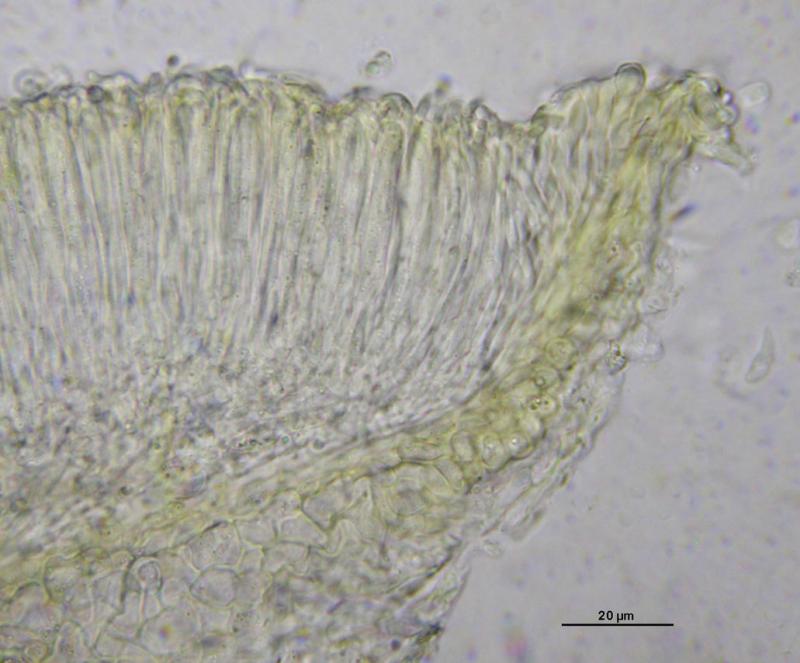
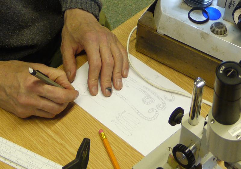
I'm glad to hear from you. Great document, this image of Zotto'hands drawing !
My fungus looks very similar to yours and I agree it doesn't quite match any of the known species cited though P. (or N.) orbilioides appears the closest among them
Sorry Zotto the phialina type terminal cells of the EE concern another fungus .
Here are images of the ectal and the margin ''cells'' , up to 3,5 µm (only 2 µm fot the paraphyses) large and full with vbs. Only one free spore , which shows the same figure as yours .
My collection data : MH 50512 La Barre de Monts , Vendée, inner side of Rubus fructicosus leave , soc F. dumorum.
Amitiés
Michel