
27-04-2026 20:52
 Lothar Krieglsteiner
Found on hanging tiwg of Olea europaea in dried-ou
27-04-2026 18:48
Tony MoverleyCollected 23rd April 2026, Norfolk, EnglandSwarms
27-04-2026 17:41
Lothar Krieglsteiner
.. Algarve, same leaf than the last post. The con
27-04-2026 18:05
Lothar Krieglsteiner
... still attached at standing tree. The green con
27-04-2026 17:16
Lothar Krieglsteiner
.. Algarve, moist lying.The conidiomata look like
27-04-2026 12:54
Steve ClementsBonjour. Ce petit champignon blanc résupiné et
27-04-2026 09:59
Pauline. PennaBonjour Can anyone advise me on these pycnidia fo

22-04-2026 20:54
 Enrique Rubio
Hi to everybody.This Pyrenopeziza grew in moist le

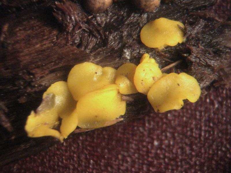
Hello, forum!
There is Bisporella collected on Populus tremula.
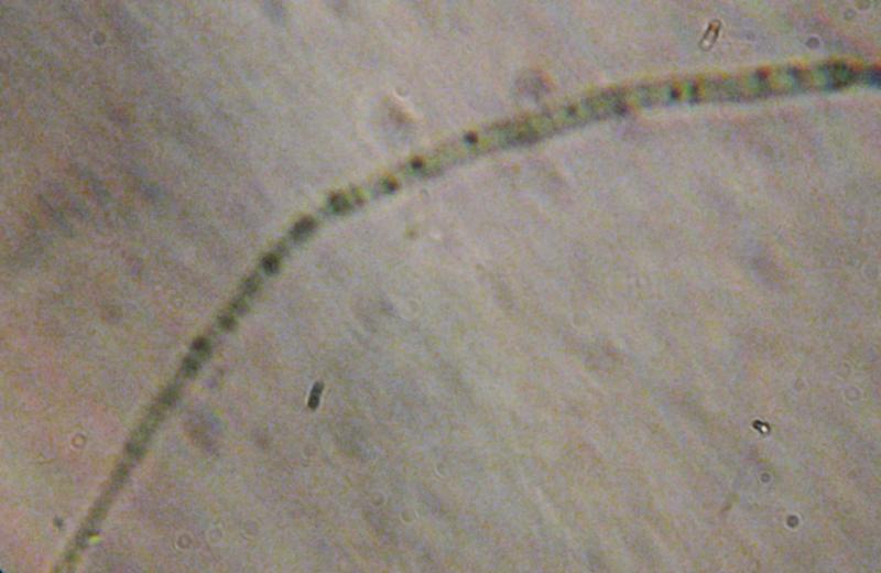
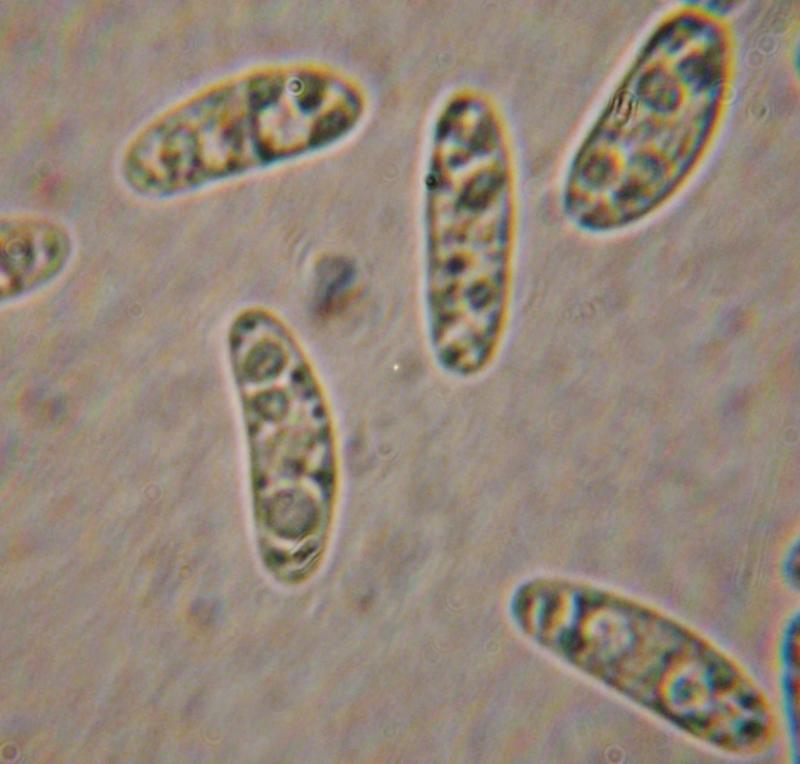
Micro:
In statu vivo:spores ellipsoid, 14,9-19,3-3,6-5,5 um, with 2-3 large and many small guttules.
Paraphyses guttulate (photo is of low quality and I can attach the drawing if needed)
Asci with croziers, 8-spored, blued in IKI (see photo)
Is it B. citrina? If it is so, why paraphyses are guttulate?
Sincerely, Irina
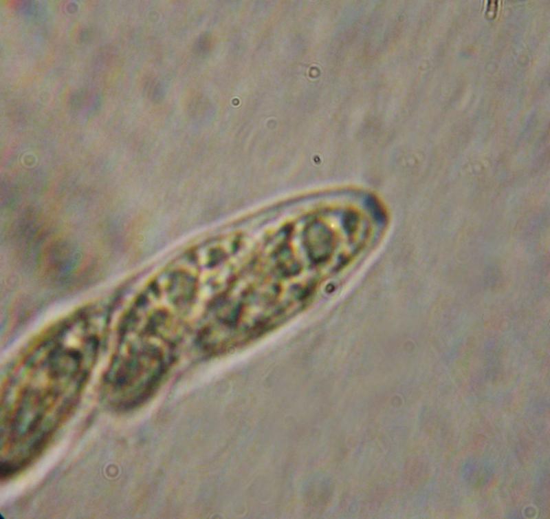

the paraphysis is strange, it should contain a homogeneous content in the upper part. Otherwise I also think B. citrina. A section is always good, excipulum should be gelatinized and the hyphae with a almost vertical orientation.
Zotto

Yes, but be careful, maybe you ovelooked very elongated (with homogenous content) vacuolar bodies in apical cells (the microphoto is very blur) because sometimes they can be of +/- whole cell length. Considering small "guttules" they could be isolated/concentrated yellow carotenoid pigment - the situation I observed in some other ascomycetes with carotenoid pigments where pigment can be separated from VB's and/or LB's. In such cases VB's and LB's are often highly refractive and hyaline or nearly so while pigment are decidedly yellow to orange to red and low refractive if at all...
At least asci are clearly of Calycina type :-) !!
Cheers,
Neven
Thank you, Zotto and Neven!
Really, in the upper part paraphyses were not-guttulate (probably, gelatinous, but I cannot say it exactly). In the lower part paraphyses contained many small rounded guttules (it's not seen from photo but I saw them very clearly).
Sincerely,
Irina