28-04-2026 20:33
Vitus SchäfftleinHello, I found Trochila ilicina on Ilex aquifoliu

30-04-2026 10:28
Rot BojanHello, by appearance I would say that I am dealing
27-04-2026 18:48
Tony MoverleyCollected 23rd April 2026, Norfolk, EnglandSwarms
14-04-2026 05:32
Ethan CrensonHi all, A few weeks back a friend pointed out som
27-04-2026 20:52
 Lothar Krieglsteiner
Found on hanging tiwg of Olea europaea in dried-ou
28-04-2026 22:51
 Bernard CLESSE
Bonsoir à toutes et tous,Pourriez-vous m'aider à
29-04-2026 08:01
Lothar Krieglsteiner
... on twig attached to small tree of Citrus auran
29-04-2026 10:44
Lothar Krieglsteiner
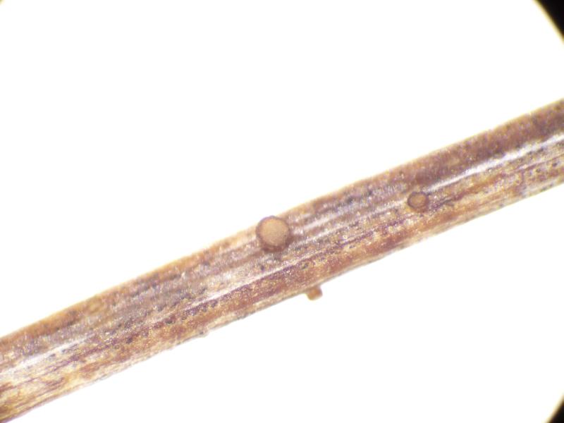
growing at moist, drying-out soil at the side of a

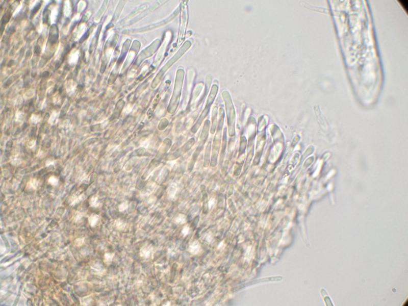
Apothecia sessile; cup-shaped; ca 300 µm diam.; hymenium pale grey; exterior brownish orange.
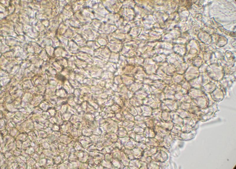
Excipulum brown textura globulosa.
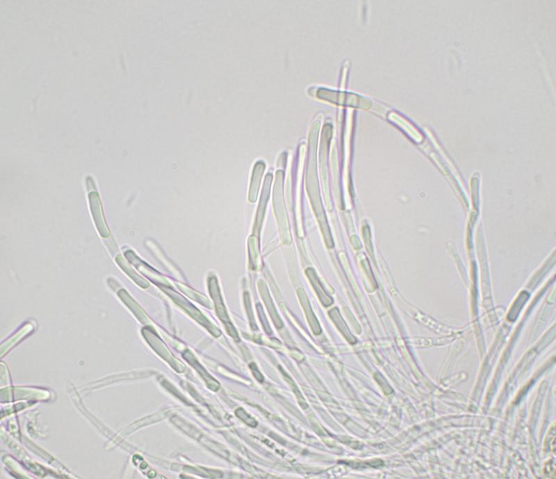
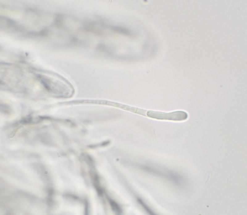
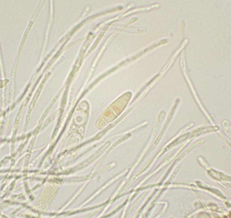
Paraphyses narrowly cylindrical (2-2.5 µm wide), sometimes swollen at apex; sometimes branched; cylindrical refractive VB in upper part.
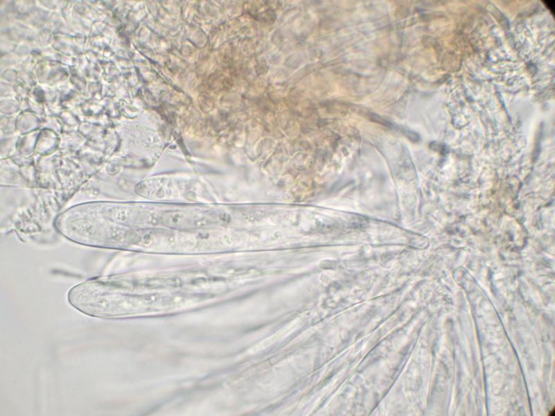
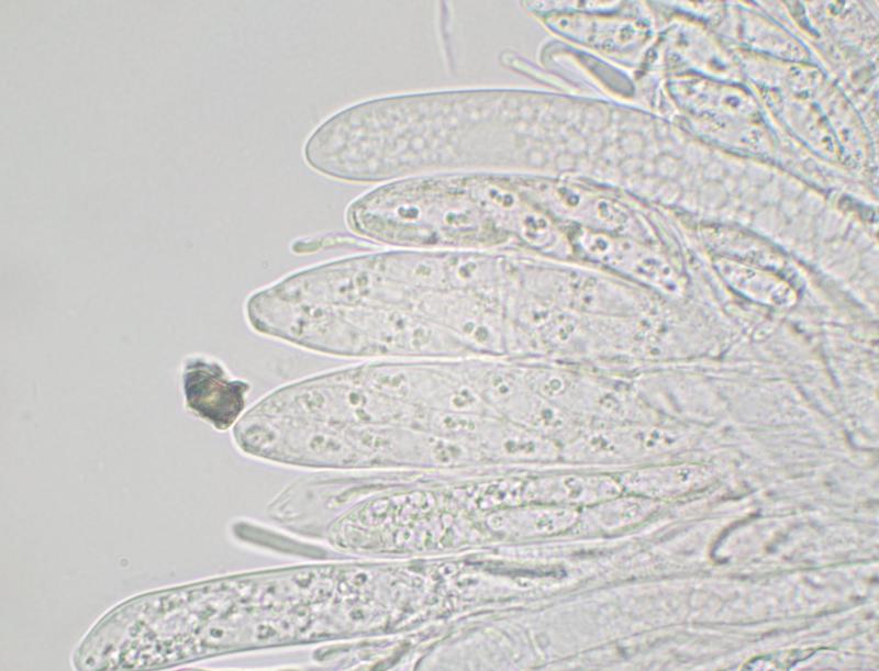
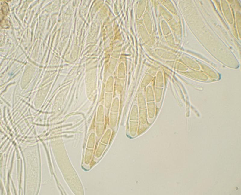
Asci clavate; ca 100-110 x 13-16 µm; 8-spored (biseriate); IKI+ blue; with shallow apical ring.
Ascospores fusiform; hyaline; free spores 21-23 x 6 µm; mostly 1-septate, but free spores sometimes 2-septate; scattered small OBs, mainly near ends of spore.
This seems to resemble Nimbomollisia (Niptera) eriophori. I didn't notice gelatinous sheaths on the spores when examining the specimen but the image of spores in the ascus in MLZ seems to show some sort of gelatinous structure at the ends of the spores. Some of the paraphyses have swollen apices but this feature isn't as well developed as I would have expected in Nimbomollisia.
I'd be grateful for a second opinion.
Thanks
Marcus
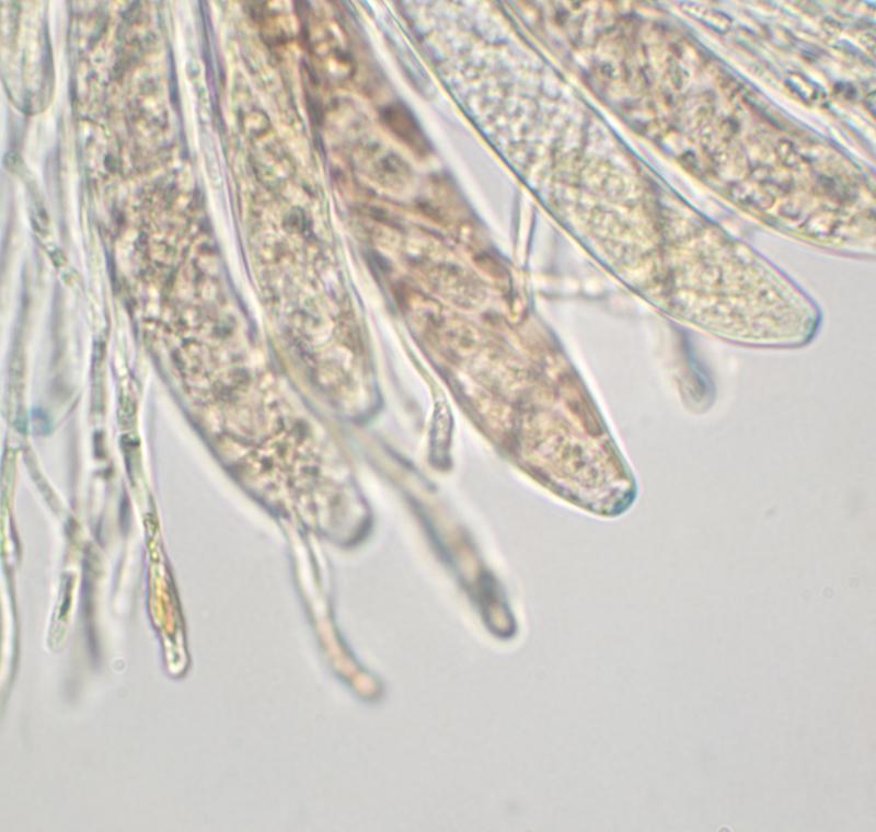
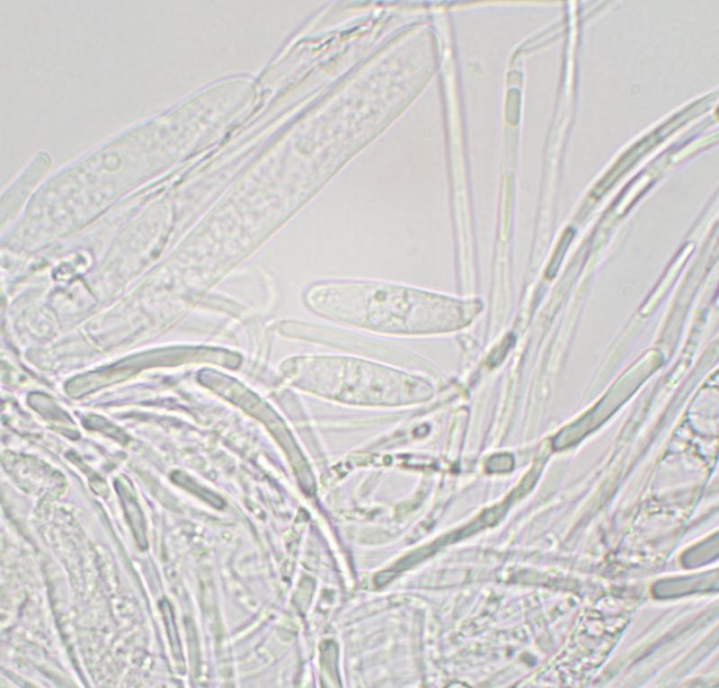

There were very few free spores. Spores in the asci were difficult to see clearly (see images) but I couldn't see any obvious caps or sheaths.