
12-04-2026 17:56
 Hardware Tony
Found on dead stems in February earlier this year
12-04-2026 15:52
Gernot FriebesHi,I'm looking for help with this anamorph collect
12-04-2026 12:22
 William Slosse
In a dune grassland in Oostduinkerke (Belgium), on
11-04-2026 15:45
 Zuzana Sochorová (Egertová)
Please, could anyone send me this paper?Moyne G.,
11-04-2026 13:34
Artem PtukhaHello, I am seeking assistance with the identific

11-04-2026 10:19
 Michel Hairaud
Chers amis d'Ascofrance , voici une très bonne no
11-04-2026 10:10
Michel Hairaud
Dear Ascofrance members, here is some very good ne
10-04-2026 23:22
Gernot FriebesHi,ascospores are 1- to 3-septate, approximately
10-04-2026 15:51
William Slosse
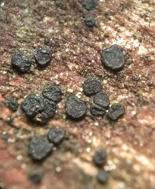
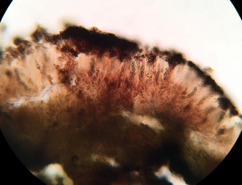
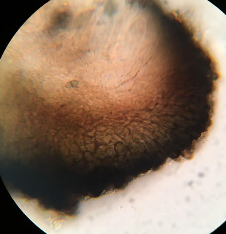
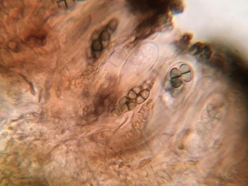
Hello everyone, On 08/04/26, I found a growth sit
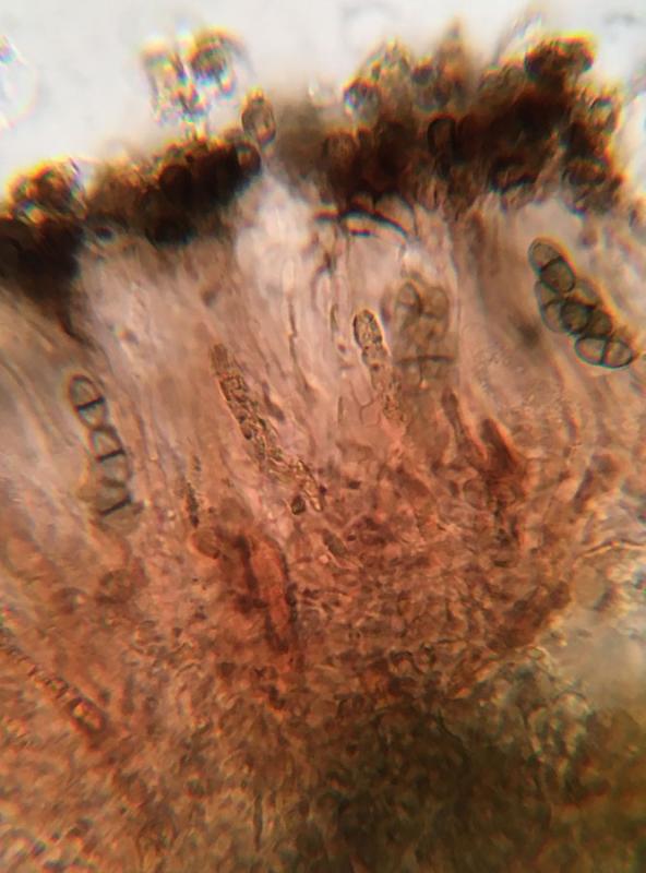
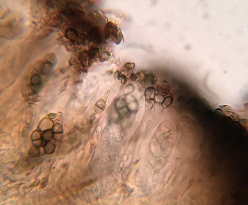
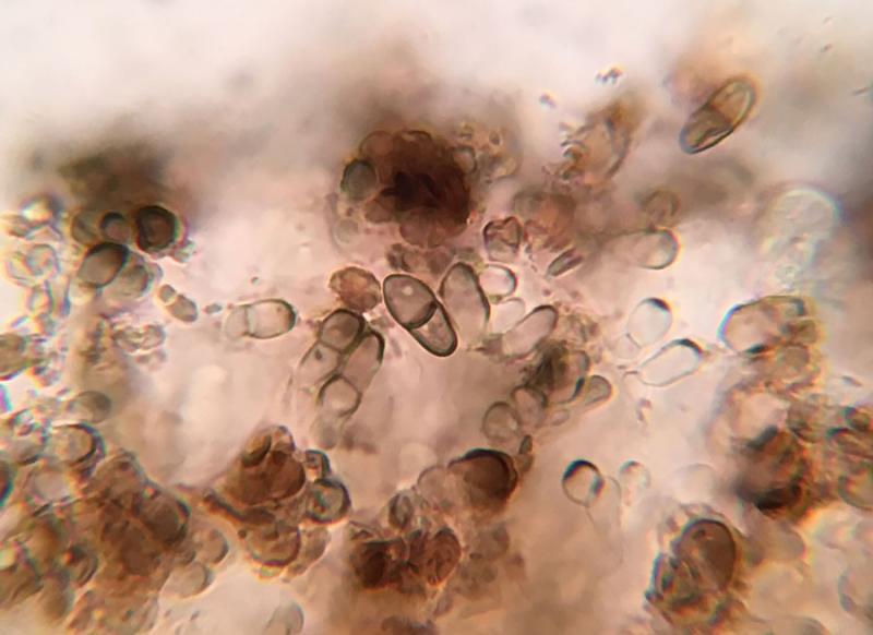
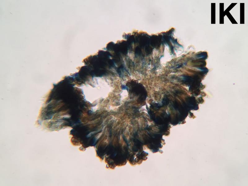
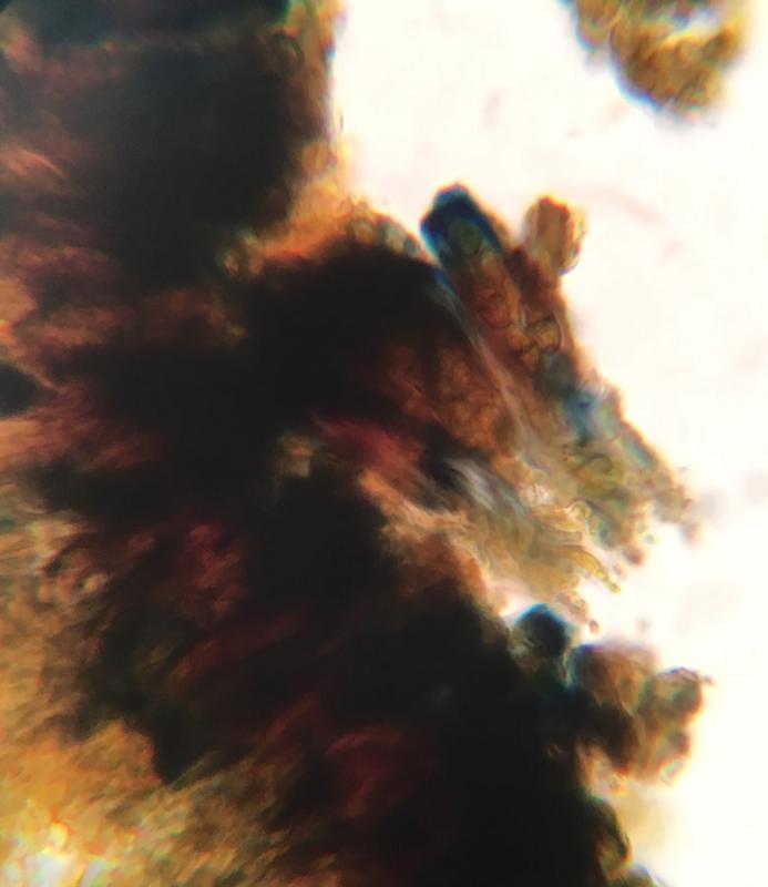
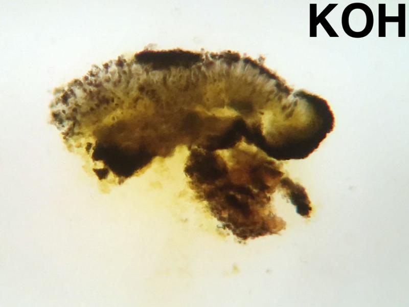
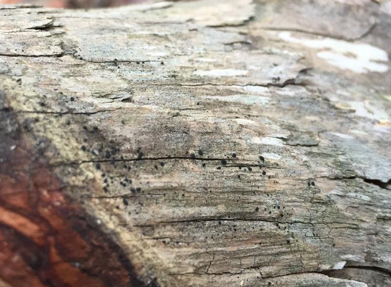
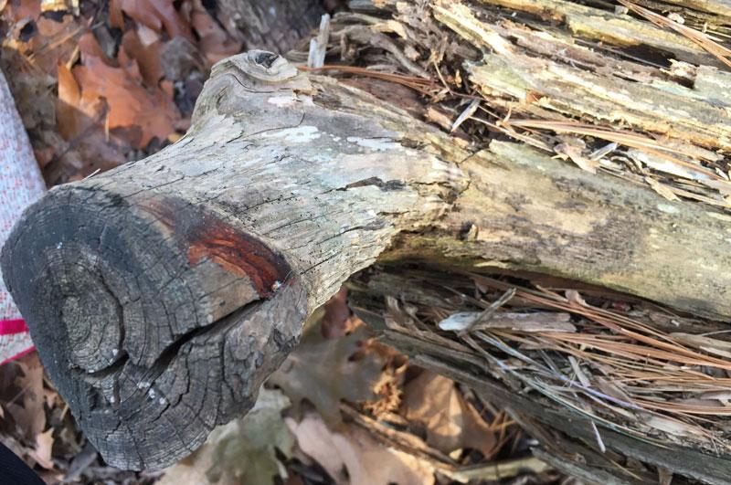
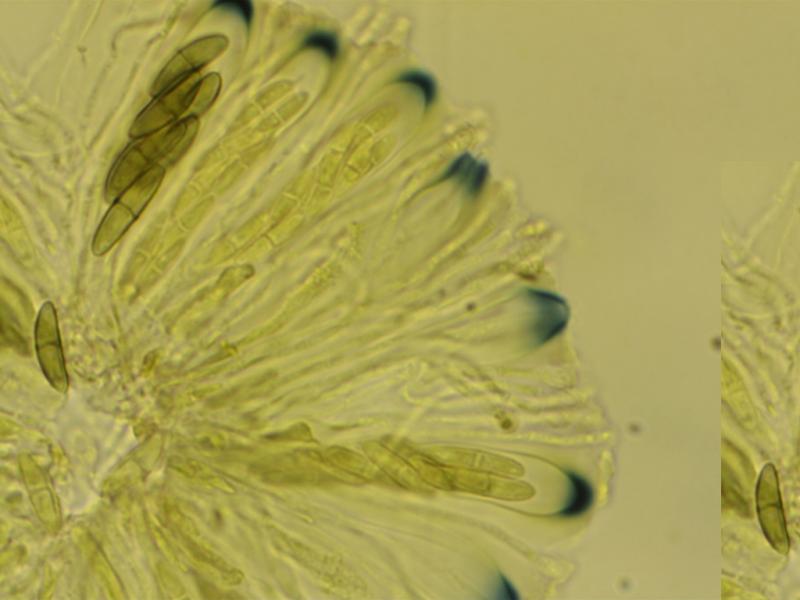
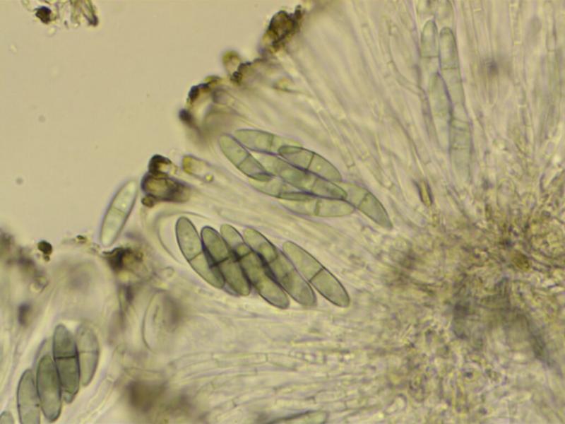
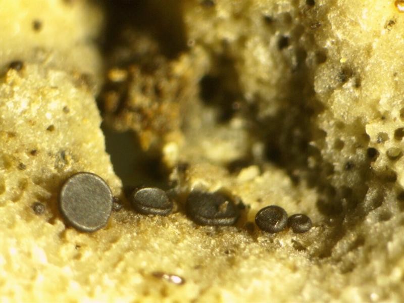
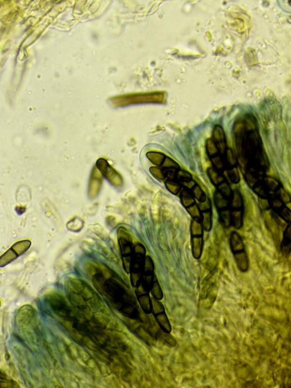
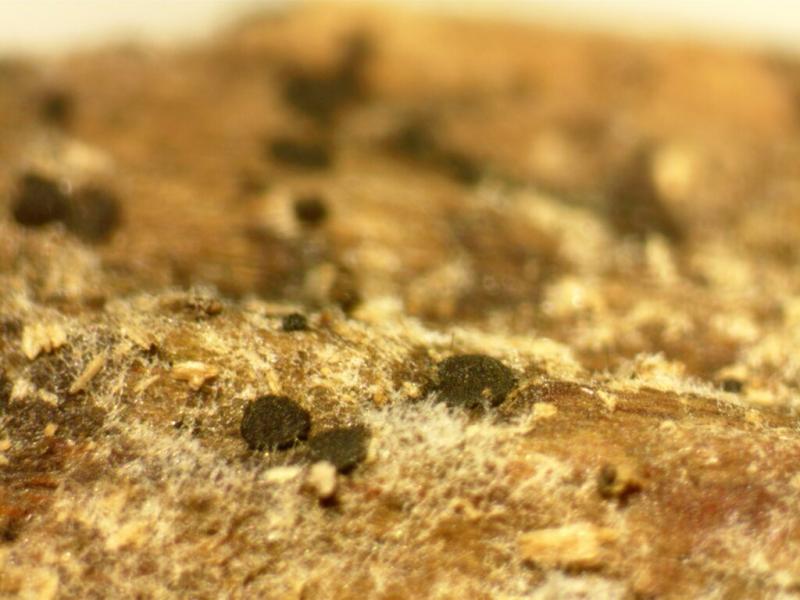
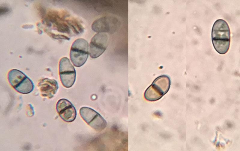
Found yesterday in a New York City park on decorticated pine (Pinus strobus?). There are red pigments in the excipulum. Reddish brown pigments in the epihymenium. These seem to dissolve in KOH and produce in a yellow liquid. The asci are broadly clavate -- almost saccate. A large portion of the ascus stains blue in Lugol's, not just the tip. Spore are brown, one septate 8-11 x 4-5µm. Paraphyses are septate ending in broad cells. I had a thought that this might be a non-lichenicolous Dactylospora, but I can't find anything that fits. Can anyone point me in the right direction?
Thanks,
Ethan

Hi Ethan,
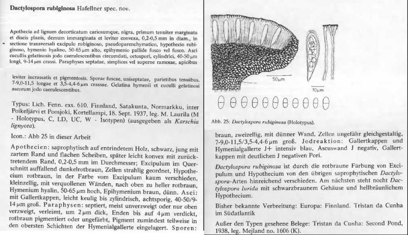
What about Rhizodiscina? It doesn't look right for R. lignyota (spores not slender enough) but might there be other species in the genus?
Cheers,
Charles.
Hi again Ethan,
I was able to compare Dactylospora stygia with Rhizodiscina lignyota as I found both species in limestone woodland on Anglesey where I live. Macroscopically and microscopically they are quite similar but R. lignyota has a rougher appearance. Both species have asci with quite a spectacular Melzer's reaction but it seems more diffuse in R. lignyota. The shape of the asci differ and there are slight differences in the ascospores. The first three photos are D. stygia.
Cheers,
Charles.
IMO, this is a very close relative of a species that I often find on Quercus (robur and petraea). Because of the ascus type it clearly belongs to the Sclerococcomycetidae (the earlier Dactylosporaceae) but differs morphologically and genetically from the classic Dactylospora species.
In the next few days I will upload a sequence from my "Dactylospora flavescens" to GenBank. If you can barcode your find, I would like to ask you to let me know the sequence.
If not, I'd like to sequence your collection.
An ML tree based on Gen 5.8S is attached here.
Best regards,
Guy
 Sclerococcomycetidae-1985650-ML-tree-rep1000-from-5-8S-0001.pdf
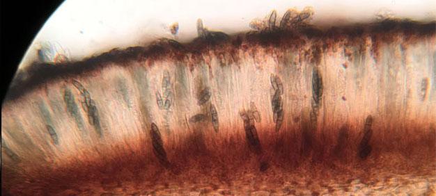
Yes, these Sclerococcomycetideae are a polyphyletic group of sometimes very adventurous genera. A good morphological feature to distinguish Dactylospora from the other genera are the two-colored gel caps of the asci with IKI-negative apical apparatus but hyaline pore after the addition of IKI (and not pretreated with KOH). In Dactylospora (sensu Hafellner) the gel surrounding the ascus is always hemiamyloid towards the ascus base, first blue, then red-brown (see: Baral, H.O. (1987). Lugol's solution/IKI versus Melzer's reagent: hemiamyloidity, a universal feature of the ascus wall. – Mycotaxon 29: 399–450.) and up around the porus, with a sufficiently high iodine concentration the gel stains blue/blue. All Dactylospora species known to me have +/- red-brown apothecia and are probably all mycoparasites. Various species not known to me are lichen parasites.
In two species, which I also have provisionally classified as Dactylospora, the gel at the ascus base turns blue to dark blue and turns black-blue around the porus. Both species also a hyaline porus. One of these species ("D. grisea") parasitizes Hymenochaetopsis tabacina and the other species is "Dactylospora flavescens" although it is not certain whether it is a mycoparasite or growing on wood only. In the real Dactylospora species the spores actually develop from hyaline to gray to a beautiful blue to finally blue-brown to dark brown with age (in dead Asci). The spores are shot off by vital asci with a blue-gray color. The spore shape is mostly symmetrical, except in muriform spores the upper cells are often slightly widened. The spore walls are usually smooth in the young stage, but can be (indistinctly visible) from smooth to longitudinally streaked or warted/punctured in age, with only few or no genetic differences between the punctured and longitudinally streaked spores. When adding KOH in microscopic sections, a yellow discoloration of the hymenium becomes visible in D. flavescens into the medium (a yellow ionomidotic reaction). In Dactylospora muriformis (nom. prov.) a rose or bright pink coloration of the exipulium can occasionally be observed, but this color does not spread into the medium, so it is not ionomidotic (sensu Korf, Baral, etc ..). And in Dactylospora grisea (nom. prov.) the excipulum sometimes turns gray to greenish or blue when KOH is added, but in most cases only brown (not ionomidotic). There are two different sequences of Rhizodiscina lignyota on GenBank which I have now integrated into an ML tree. Whereby I ascribe the New Zealand sequence to a genus unknown to me. The IKI reaction of R. lignyota is very different from that of the classic Dactylospora species. In R. lignyota, the entire ascus reacts light blue with IKI. Thick gel caps like in Dactylospora I did never noticed in Rhizodiscina (R. lignyota and R. proteae). In addition, the spores are two-celled and asymmetrical with a wider upper cell. If I ever come across Rhizodiscina lignyota, I can send it to you if you like?
Cheers,
Guy