12-04-2026 17:56
 Hardware Tony
Found on dead stems in February earlier this year
12-04-2026 15:52
Gernot FriebesHi,I'm looking for help with this anamorph collect
12-04-2026 12:22
 William Slosse
In a dune grassland in Oostduinkerke (Belgium), on
11-04-2026 15:45
 Zuzana Sochorová (Egertová)
Please, could anyone send me this paper?Moyne G.,
11-04-2026 13:34
Artem PtukhaHello, I am seeking assistance with the identific

11-04-2026 10:19
 Michel Hairaud
Chers amis d'Ascofrance , voici une très bonne no
11-04-2026 10:10
Michel Hairaud
Dear Ascofrance members, here is some very good ne
10-04-2026 23:22
Gernot FriebesHi,ascospores are 1- to 3-septate, approximately
10-04-2026 15:51
William Slosse
Hello everyone, On 08/04/26, I found a growth sit
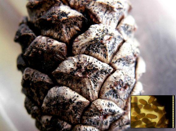
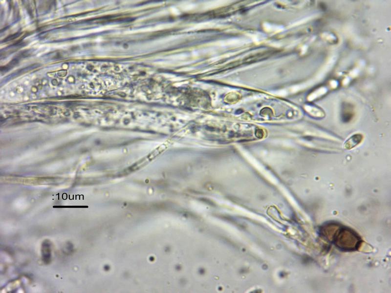
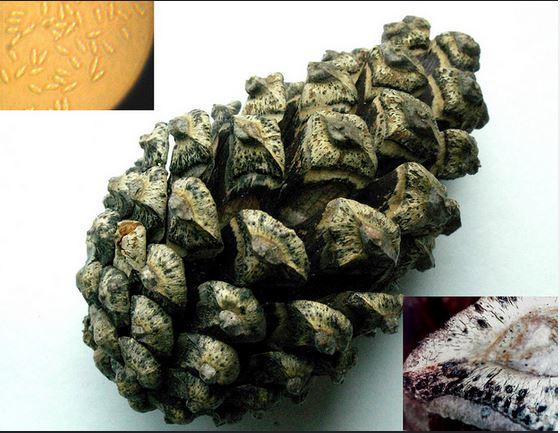
I found these very nice spores in a rather indistinct semi-immersed Coelomycete (?) on the base of a Pinus sylvestris cone at Longshaw Estate, Derbyshire, Northern England. They are somewhat similar to Hepalocystis berkeleyi (no. 840 in Ellis and Ellis) which has two septa and square appendages and is on Platanus.
The brown biseptate spores were about 12-15 long, 7-8 wide, with hyaline appendages up to 7 long.
In addition there were numerous hyaline bigutullate conidia 6.5-8 x 3-3.5.
I found some asci which were up to 12 wide.
Does anyone recognise these beautiful spores – they aren't on Pinus in Ellis and Ellis.
Regards,
Steve
Bonjours,
J' ai découvert ces très belles spores dans un coelomycètes (je crois) sur la base d'un cône de Pinus sylvestris à Longshaw Estate, Derbyshire, nord de l'Angleterre. Ils sont assez semblables à Hepalocystis berkeleyi (no. 840 dans Ellis et Ellis) qui comporte deux septa et se trouve sur Platanus.
Les spores biseptate bruns étaient environ 12-15 de long, 7-8 de large, avec des appendices hyalins jusqu'à 7 microns.
En plus il y avait des nombreuse conidies hyalins bigutullates 6.5-8 x 3-3,5.
J' ai trouvé quelques asques qui étaient jusqu'à 12 de large.
Est-ce que quelqu'un reconnaisse ces belles spores - ils ne sont pas sur Pinus à Ellis et Ellis.
Cordialement,
Steve

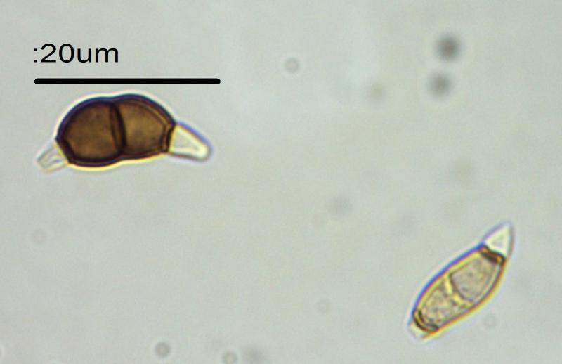
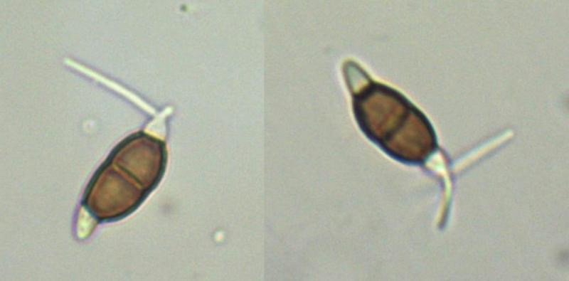
ciliate conidia could be from the genus Truncatella, cf. T. conorum-piceae.
Regards,
Ernestas
Many thanks, I have never seen these before.
I thought the cilia were germination tubes! This does like Truncatella conorum-piceae
http://www.fredis-pilzseite.de/foto-truncatella-conorum-piceae-1/
On Pinus.
Infection secondary to Sphaeropsis
http://www.forestryimages.org/browse/Taxthumb.cfm?fam=902&genus=Truncatella
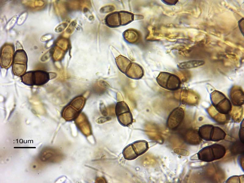
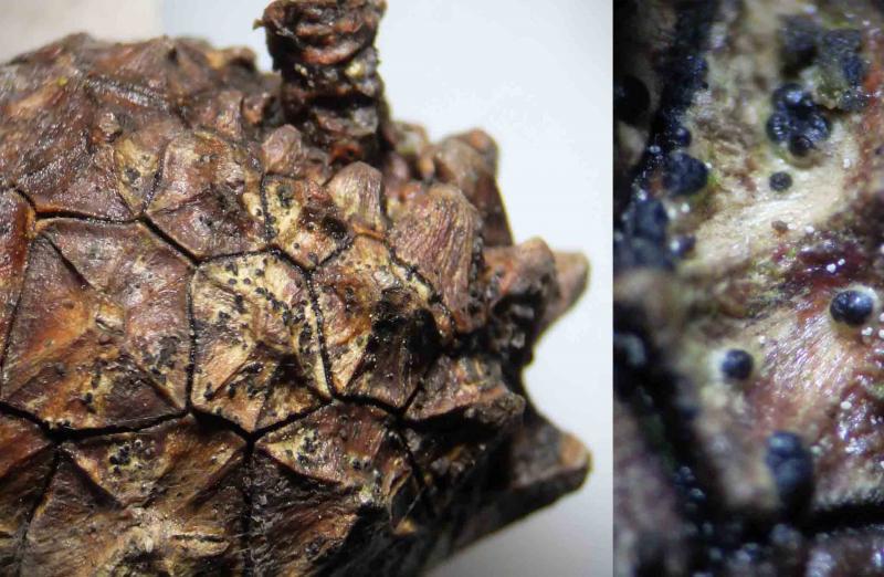
Here are a couple more ciliated spores from my specimen:
Regards,
Steve

try to find Guba's Monograph of Monochaetia and Pestalotia. Maybe this helps.
In your pictures are smaller hyalinous biguttulate spores that may correspond to the asci you saw. Did you see other fruitbodies other than those black ones?
Regards
Martin
Thanks for the help.
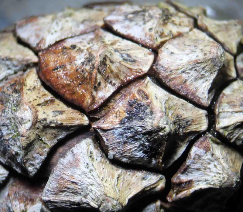
I have had an interesting morning looking at the cone in more detail.
I found 3, possibly 4 fungi, in addition to the Truncatella.
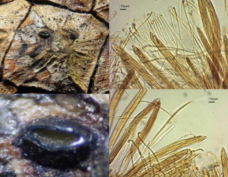
1) Lophodermium conigenum
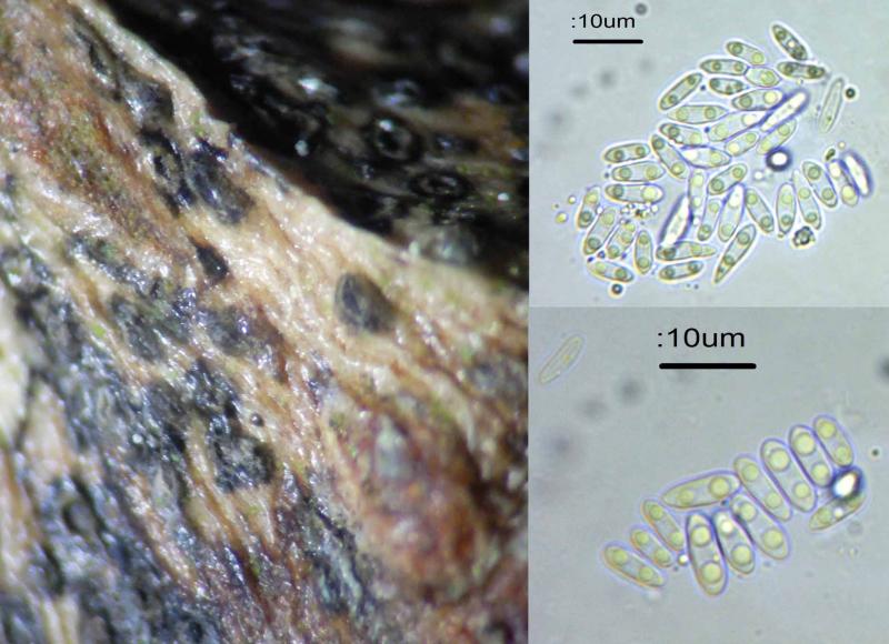
2) Sporonema diamandidis (I am less sure of this as I get my conidia slightly larger than in Ellis & Ellis) – 6-9 x 2-3 rather than 4-8 x 1.5-2. Also, the conidiomata were only up to 0.4 x 0.2 mm. My previous ID of this one in 2010 was done with less accurate equipment.
3) Tiny black rounded asco, up to 0.3mm. Associated with minute white fungi(?). Asci approx 60 x 7 reddening in Lugol, tips blueing I think.I suspect the free spores are from these – none or 1-septate, 10-13 x 3.3.5. Other asci are about 90 x 12 (containing 2 septate spores up to 17 x 6).
4) Minute white bodies associtaed with the previous. These are about 40-70 um, round to oval, with amorphous contents. Fungal? Or eggs of some kind.
Best wishes,
Steve
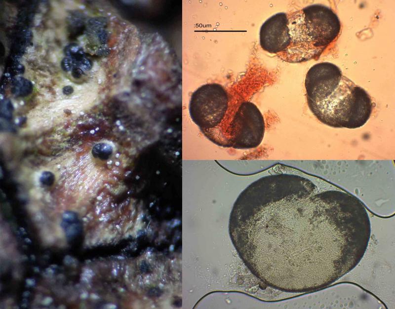
But I don't recognize a possible candidate for the biguttulate spored fungus.Please try to find it again and stain your slide with Lugol's. Your #4 are pollen of pine.
Best regards
Martin
I shall now leave it to incubate.
The monograph costs £20 from Amazon.
Thanks again for the help,
Steve

moi pour ma part, et sur la base des photos présentées, je propose Sphaeropsis sapinea (Diplodia pinea). les deux extrémités hyalines seraient des tubes germinatifs de la spore brune bicellulaire.les jeunes spores encore hyalines qu'on aperçoit à l'arrière de la photo, ne présentent pas d'appendices à leurs extrémitées. d'autres prélevements et d'autres photos détaillées des frutifications et des spores seront d'une grande utilité.
I for my part, and on the basis of the presented photos, I propose Sphaeropsis sapinea (Diplodia pinea). Both hyalines extremities would be germinal tubes of the brown spore. young conidia still hyaline which we perceive behind the photo, do not present appendixes. Other detailed photos of frutifications and the spores will be of a big utility.

http://www.apsnet.org/publications/PlantDisease/BackIssues/Documents/1985Articles/PlantDisease69n10_838.PDF
Cordialement
Chris
I have recorded Sphaeropsis sapinea several times and it does have very large spores.
In the inset in the picture the scale is in 10 um intervals.
Regards,
Steve