
28-04-2026 20:33
Vitus SchäfftleinHello, I found Trochila ilicina on Ilex aquifoliu

30-04-2026 10:28
Rot BojanHello, by appearance I would say that I am dealing
27-04-2026 18:48
Tony MoverleyCollected 23rd April 2026, Norfolk, EnglandSwarms
14-04-2026 05:32
Ethan CrensonHi all, A few weeks back a friend pointed out som
27-04-2026 20:52
 Lothar Krieglsteiner
Found on hanging tiwg of Olea europaea in dried-ou
28-04-2026 22:51
 Bernard CLESSE
Bonsoir à toutes et tous,Pourriez-vous m'aider à
29-04-2026 08:01
Lothar Krieglsteiner
... on twig attached to small tree of Citrus auran
29-04-2026 10:44
Lothar Krieglsteiner
growing at moist, drying-out soil at the side of a

Fungus on Eriophorum angustifolium
Spooren Marco,
15-06-2012 21:38
 Hello.
Last week i collected a small fungus on the submersed parts of Eriophorum angustifolium.I kept the plantparts in an aquarium and for a while the thing puzzled me.It apeared not to be ripe.Yesterday I promised on this forum to make some microscans.and in the proces i had some older,or dead material.I now think that the observes characters points to Diplonaevia cf. emergens (Karst ) Hein,but i must jump in the literature and under the microscope again.
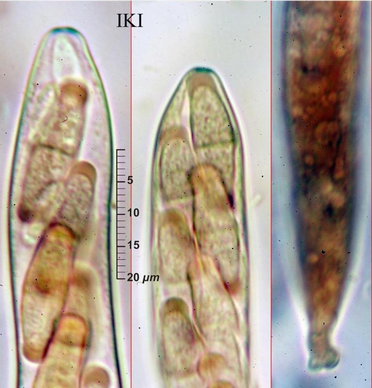
I made a few scans.The spores i measured were appox. 16x4 Mu.On the microscan you see one-septate spores and a ascusapex bleued by Lugol. The spores are reddisch brown due to the Lugol. Diplonaevia emergens is given for Juncus effusus and conglomeratus.
I hpoe I uploaded two scans.
Best regards,
Marco
Spooren Marco,
15-06-2012 21:41
Re : Fungus on Eriophorum angustifolium
It didnt work,so here the second scan,
Marco
Marco
Hans-Otto Baral,
15-06-2012 22:24

Re : Fungus on Eriophorum angustifolium
Hi Marco
Diplonaevia emergens has much smaller spores and the apothecia are erumpent below the epidermis which forms a crown-like margin around the urceolate apothecia, which look a bit like a Cryptodiscus.
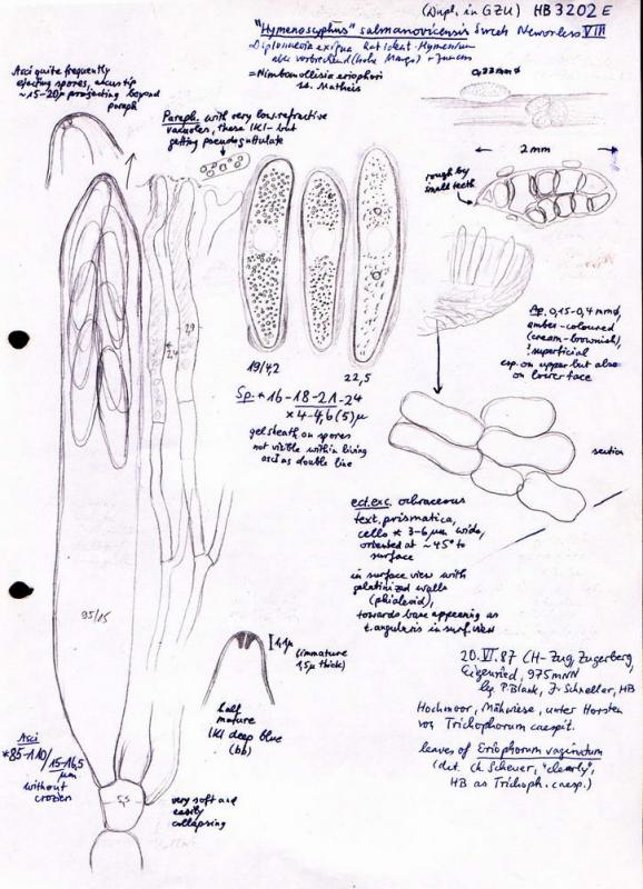
I cannot see anything on your macro, but if you have a seemingly superficial discomycete, I would suspect you have "Hymenoscyphus" salmanovicensis, which is typical on Eriophorum in bogs. The spores would fit that fungus, which is totally misplaced, and possibly belongs in the Naevioideae.
To get more certainty you should look at the ascus base which is without croziers, and if you find living spores they contain only small LBs. Here my drawing attached.
Zotto
Diplonaevia emergens has much smaller spores and the apothecia are erumpent below the epidermis which forms a crown-like margin around the urceolate apothecia, which look a bit like a Cryptodiscus.
I cannot see anything on your macro, but if you have a seemingly superficial discomycete, I would suspect you have "Hymenoscyphus" salmanovicensis, which is typical on Eriophorum in bogs. The spores would fit that fungus, which is totally misplaced, and possibly belongs in the Naevioideae.
To get more certainty you should look at the ascus base which is without croziers, and if you find living spores they contain only small LBs. Here my drawing attached.
Zotto
Hans-Otto Baral,
16-06-2012 19:07
Re : Fungus on Eriophorum angustifolium
Hi Marco
you are probably right with your doubts. Important would be to see the construction of the apothecium, best in a section. valuable would also be to know the characters of the paraphyses.
Zotto
you are probably right with your doubts. Important would be to see the construction of the apothecium, best in a section. valuable would also be to know the characters of the paraphyses.
Zotto
Spooren Marco,
16-06-2012 21:19
Re : Fungus on Eriophorum angustifolium
Hello Zotto.
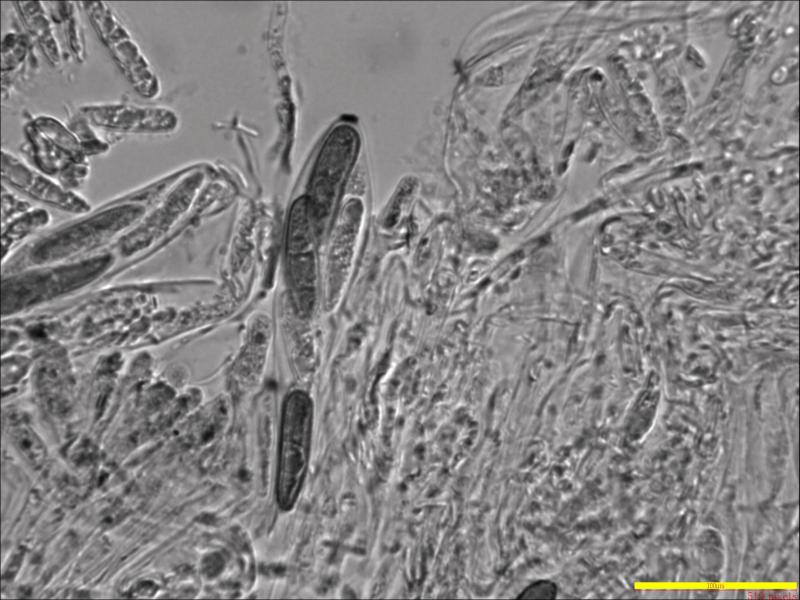
No,my fungus is not "Hymenoscyphus salmanovicensis.Svrcek depicts a fungus,and described as such,with unicellular spores.My fungus has ,see scan,one sept and i suspect a second one one the same spore in my scan.
Svrcek says about the parafyses that the are ramose at the upper part and hyaline.The parafyses of my fungus are single,not ramose,a few between the asci( a few parafyses and then an ascus etc. )and the tips of the parafyses ere hockey-stick formed and (seemingly ) twisted.the tips,(thickened ) have a brown granulation,of which i dont know if it is on the inside or the outside.At least when they are young.In the preparation of which i made my scan i didnt see that anymore.
I looked at the descriptions of the parafyses of these groups of fungi by Rehm (Rabenhorst-flora )but it didnt bring much up till now.
I shall try to make scans of the parafyses,especiallly when they are young.
A propos,I think to remember to see not only little LBs,but also bigger ones.Try to make a scan of that also.Tomorrow.
I think it is very difficult to make a section of an apothecium,they are very small and maybe a pressed one on a microscope-slde gives the same information.
Marco
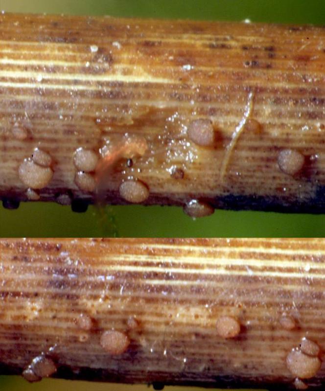
I send a scan,more light,of the apothecia in vivo,you see that they are erumpent.But doesnt look all Naevioideae superficial but are in reality erumpent?
No,my fungus is not "Hymenoscyphus salmanovicensis.Svrcek depicts a fungus,and described as such,with unicellular spores.My fungus has ,see scan,one sept and i suspect a second one one the same spore in my scan.
Svrcek says about the parafyses that the are ramose at the upper part and hyaline.The parafyses of my fungus are single,not ramose,a few between the asci( a few parafyses and then an ascus etc. )and the tips of the parafyses ere hockey-stick formed and (seemingly ) twisted.the tips,(thickened ) have a brown granulation,of which i dont know if it is on the inside or the outside.At least when they are young.In the preparation of which i made my scan i didnt see that anymore.
I looked at the descriptions of the parafyses of these groups of fungi by Rehm (Rabenhorst-flora )but it didnt bring much up till now.
I shall try to make scans of the parafyses,especiallly when they are young.
A propos,I think to remember to see not only little LBs,but also bigger ones.Try to make a scan of that also.Tomorrow.
I think it is very difficult to make a section of an apothecium,they are very small and maybe a pressed one on a microscope-slde gives the same information.
Marco
I send a scan,more light,of the apothecia in vivo,you see that they are erumpent.But doesnt look all Naevioideae superficial but are in reality erumpent?
Hans-Otto Baral,
16-06-2012 21:56
Re : Fungus on Eriophorum angustifolium
Are the apothecia moist when you photograph them or are they retracted and dry? The lower left on your photo seems to have a triangular opening by marginal lobes. This would mean that the apothecia are immersed in the host tissue and develop below the epidermis.
Your photos do not have any colour? Has the fungus a pigmented excipulum? the shape of the paraphyses sounds important, please try a photo.
Zotto
Your photos do not have any colour? Has the fungus a pigmented excipulum? the shape of the paraphyses sounds important, please try a photo.
Zotto
Spooren Marco,
16-06-2012 22:41
Re : Fungus on Eriophorum angustifolium
Hello Zotto,
I scanned the apothecia fresh out of the aquarium.My scanner doesnt see color,you must do that yourself with buttons ,but i am not such a hero with colors.
Yes,there are immersed apothecia,but also a lot seemingly superficial ones,and the thought passed that i have 2 fungi.But i interpreted it as what i saw on Naevala minutissima,: erumpent in the first but superficial at the end.The excipulum is lightly brown pigmented,nearly hyaline.
I try to make the pictures tomorrow.I also must see if the two fungi are one fungus .I have a lot to do.
Greetings,
Marco
Spooren Marco,
17-06-2012 21:35
Re : Fungus on Eriophorum angustifolium
Hello,
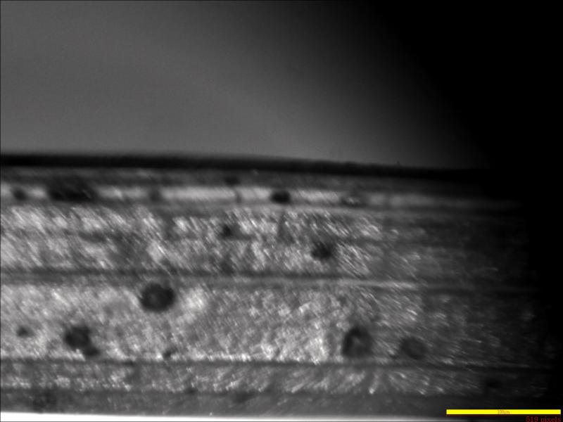
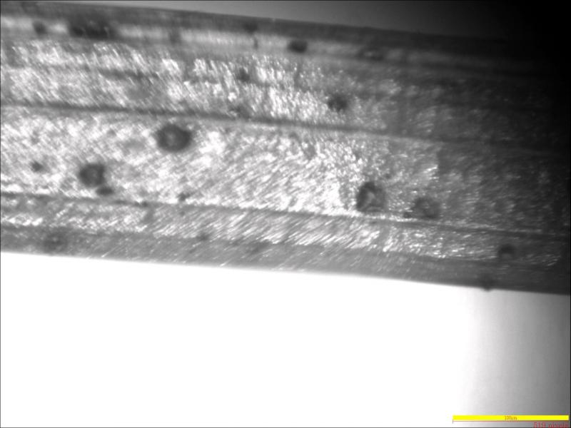
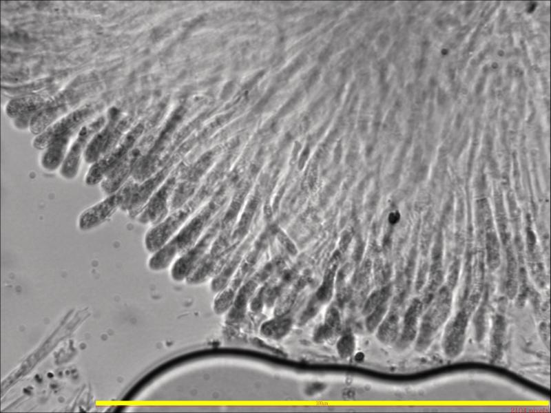
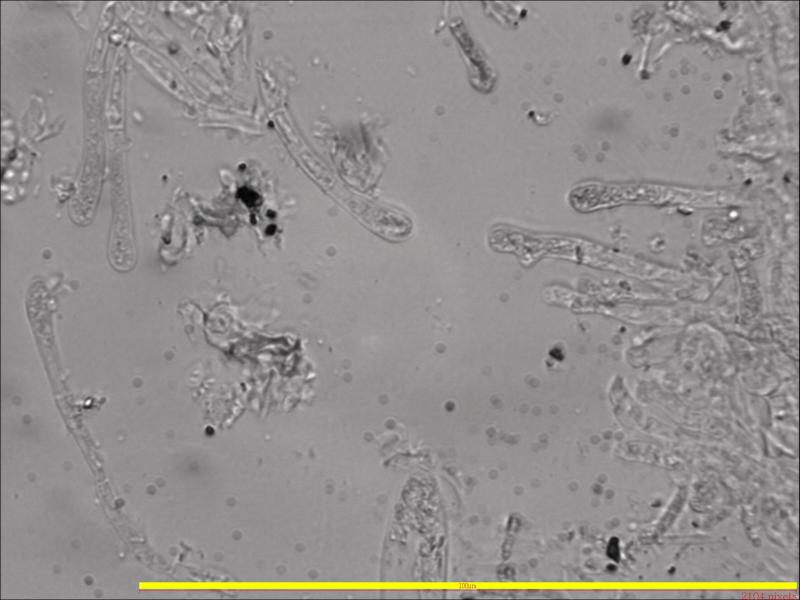
After searching a whole evening to find some goed material,I made some scans,I hope man can make something out of it.Now I am convinced that there is one fungus,immersed in the beginning and breaking through the substratum.Then they are not ripe yet.My scans are from such material.Later the ascomata become superficial and ripe.On one scan you can see the parafyses good,almost as I described them earlier.The must develop also.But man can see the granulation and slightly twisted head.Also on one scan man can see a young ascus.
Marco

Hans-Otto Baral,
17-06-2012 21:52
Re : Fungus on Eriophorum angustifolium
Hi Marco
so the paraphyses are enlarged above and a bit irregular. Regrettably all your material is dead, and the granular interior is mainly a result hereof. Maybe also that you apply too much pressure on the preparation.
Regettably, I can still not give a hint to what it might be. Is the fungus still fresh or did you dry it? I assume it will not survive drying. Vital charact5ers are very important, though they are usually not to be found in the literature.
And any information on the excipulum will also be helpful.
Zotto
Spooren Marco,
22-06-2012 22:26
Re : Fungus on Eriophorum angustifolium
Hello,
Has someone else an idea?
Marco
Has someone else an idea?
Marco
Gernot Friebes,
22-06-2012 23:32
Re : Fungus on Eriophorum angustifolium
Hi,
what about Niptera/Nimbomollisia eriophori? Ascospore size and septation would fit. The spores should have hyaline appendages though which I cannot see on your photo. Here is a description to compare with: http://www.cybertruffle.org.uk/cyberliber/59350/0075/002/0300.htm and of course also on Zotto's CD.
Best wishes,
Gernot
Hans-Otto Baral,
22-06-2012 23:42
Re : Fungus on Eriophorum angustifolium
Hi Gernot
this is quite a good idea. I am not sure whether the fungus can be erumpoent as Marco's photo suggests. But apical ring asn spore size/shape could fit. Anyhow, Marco's spores look a bit clavate.
here is N. eriophori, after photos by Enrique Rubio. The gel caps are not so easy to see in dead material
Spooren Marco,
23-06-2012 10:06
Re : Fungus on Eriophorum angustifolium
Thank you Gernot and Zotto for the ideas,
I sall look at the Nannfeldt article's.
Marco
I sall look at the Nannfeldt article's.
Marco