
28-07-2016 18:09
 Bernard CLESSE
Bonjour à tous,Hier j'ai trouvé sur tronc compl�
28-07-2016 21:11
Bernard CLESSE
Bonsoir à tous,J'imagine que vous avez déjà ré
29-06-2016 20:17
 Alvarado Cordobes Manuel
Buenos dias :Lo encontré en tallos del año anter
20-07-2016 19:02
Adam PolhorskýHello everyone, i have trouble IDing couple collec
22-07-2016 22:56
Bernard CLESSE
Voici un petit asco jaune stipité, trouvé dans u
25-07-2016 21:23
 Enrique Rubio
Hi to all Because I couldn't to obtain original d
26-07-2016 18:01
 Castillo Joseba
encontrado en excrementos de vacunoHe visto ests a
26-07-2016 16:10
BELLIDO BERMEJO FERNANDOFructificaciones de 1-2 mm de diam, pseudoestipita
Byssosphaeria ?
Ethan Crenson,
08-03-2016 08:53
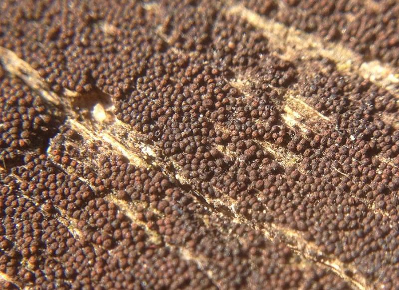
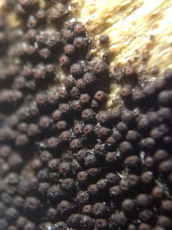
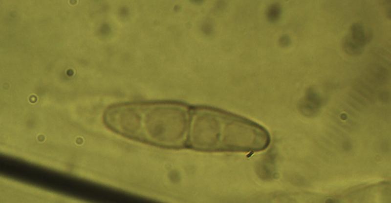
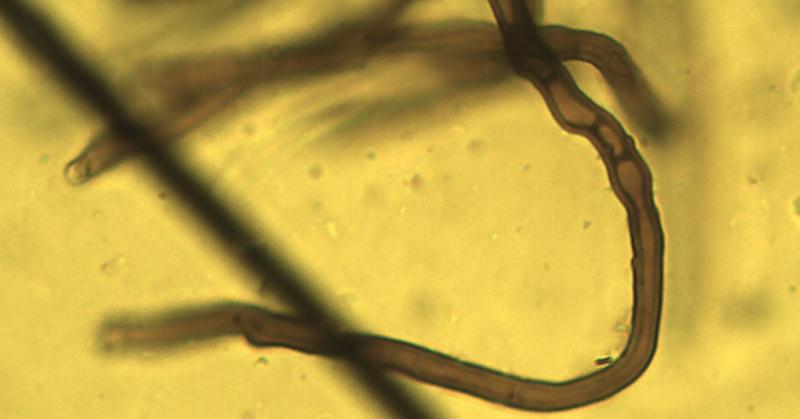
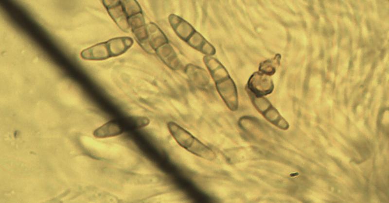
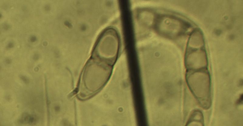
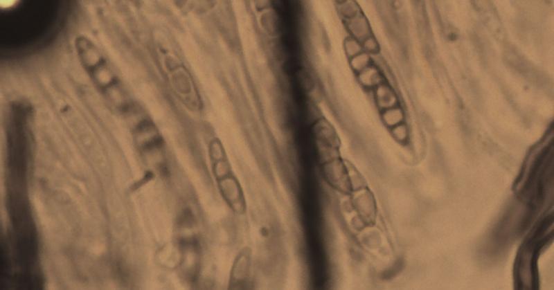
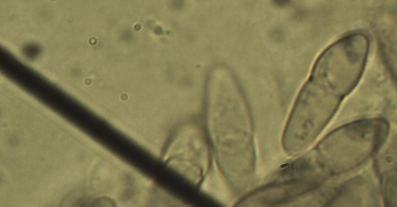
I found this tiny pyrenomycete in Brooklyn. Tiny black roughened ascomata, less than .5mm, superficial on bare wood (probably Quercus). Red around apical opening. 1, 2 & 3-septate hyaline spores measuring 16-20µm by 5-5.5µm.
Any help would be greatly appreciated.
Thanks!
Jacques Fournier,
08-03-2016 09:08

Re : Byssosphaeria ?
Hi Ethan,
your fungus seems to fit well Byssosphaeria rhodomphala. Are you on the tropical side of Brooklyn?
Cheers,
Jacques
your fungus seems to fit well Byssosphaeria rhodomphala. Are you on the tropical side of Brooklyn?
Cheers,
Jacques
Jason Karakehian,
30-07-2016 04:53

Re : Byssosphaeria ?
Hi Jacques, I was just looking at Ethan's photos and notes on this and following your lead. It really does seem to be Byssosphaeria rhodomphala! We looked at a paper Algunas especies del género Byssosphaeria (Melanommataceae, Pleosporales) de Veracruz, México by Santiago Chacón-Zapata y Fidel Tapia-Padilla. Revista Mexicana de Biodiversidad 84: 739-745, 2013. The taxon is given from Mexico and Ohio in the United States. We feel like this is a good determination given that information, but we wondered if you have much experience with the genus and might offer additional input?
Thank you!
Thank you!