31-07-2016 23:27
 Viktorie Halasu
Hello, would anyone have this paper please? Beyer
27-07-2016 21:09
 Enrique Rubio
Hi again These gregarious, small apothecia (150-
31-07-2016 21:08
Elisabeth StöckliBonsoir,Sur bois mort de Calluna vulgaris (Lande s

31-07-2016 11:27
 Angel Pintos
Ascomata on average 300 microns in diameter, immer
30-07-2016 22:23
 Bernard CLESSE
Bonsoir à tous,Cet après-midi, sur alluvions sab
26-07-2016 10:09
 Filip Fuljer
Hi guys,i need help with identification, again.I f
28-07-2016 22:06
Bernard CLESSE
Bonsoir à tous,Trouvé sur terre nue, dans une or
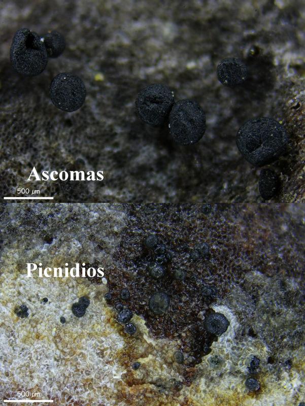
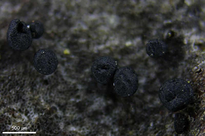
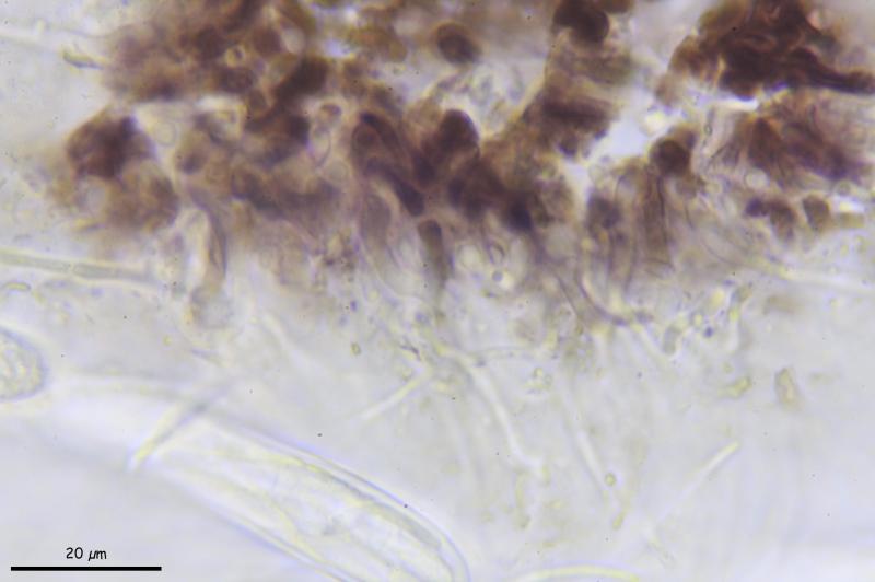
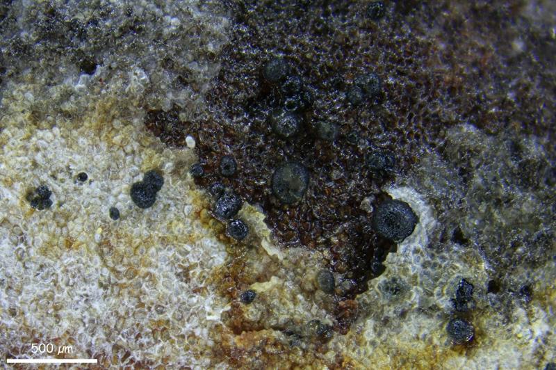
Tengo estos apotecios creciendo sobre la corteza de Pinus halepensis que pienso que puede ser Pragmopora, pero no la logro identificar.
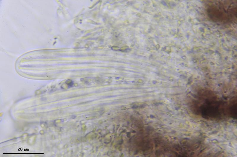
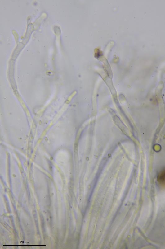
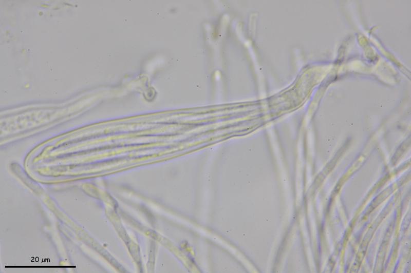
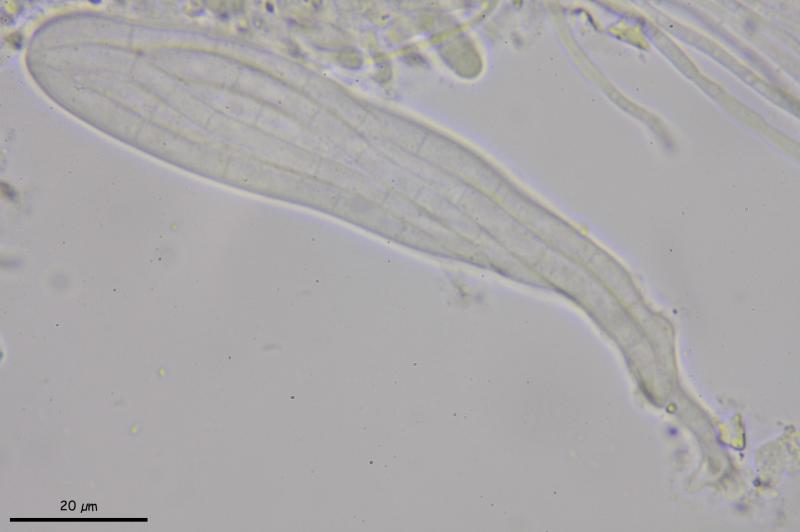
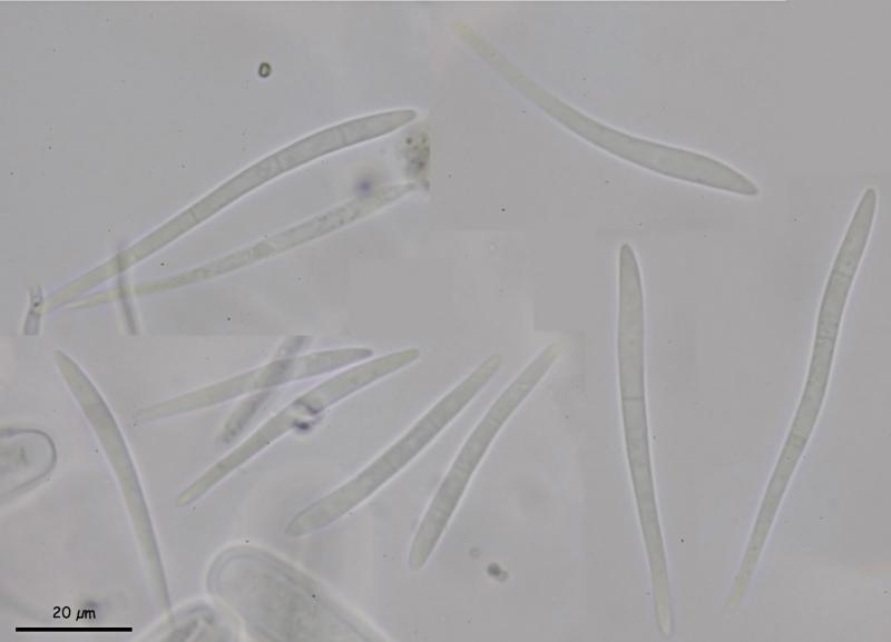
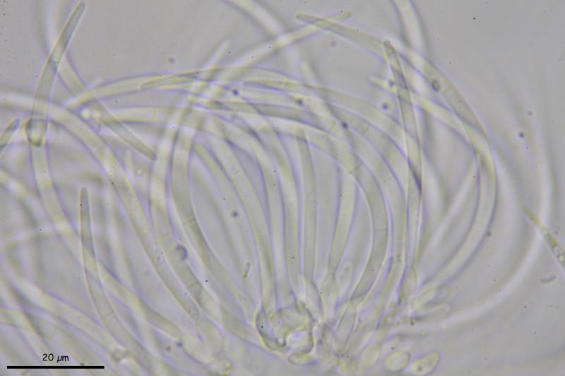
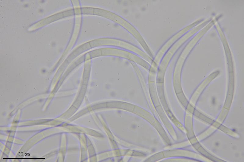
Los apotecios miden hasta 0,5 mm de diámetro y tienen ascas claviformes, octospóricas de (87-)90-113(-114) × (14-)14,3-16(-17,7) µm, Ascosporas de (43-)47-77(-78) × (3,4-)3,5-4,6(-4,7) µm, Q = (10,3-)11,5-20,3(-21,6), Me = 62,7 × 4 µm ; Qe = 15,7, con (0) 3 (-4) septos transversales (generalmente menos cuanto más inmaduras son las esporas). Paráfisis hialinas, septadas, generalmente bifurcadas o ramificadas en el ápice, donde están algo ensanchadas y cubiertas por una masa marrón.
En la misma muestra hay unos picnidios de (45,7) 70,4 - 152,7 (228,7) µm, con conidios hialinos, curvados, con 3 septos de (62-)64-81(-83) × (2,5-)3-3,4(-3,5) µm de longitud. ¿Tal vez el anamorfo?
Saludos.
Salvador
Salvador.

Durandiella would have such anamorph. Do ana- and teleomorph differ by macroscopy?
Zotto
Yo no aprecié que hubiese una capa cubriendo los apotecios, pero lo puedo revisar de nuevo.
Si que la microscopía podría encajar con Durandiella y tengo una muestra de Durandiella con el amamorfo de conidios similares, pero la descarté porque en este hongo los apotecios están solitarios y no reunidos en grupos como en Durandiella.
No entiendo (Do ana- and)
Salvador.
I wanted to ask how the conidiomata look in comparison to the apothecia.