
17-03-2018 18:57
 Blasco Rafael
Hola, he recogido una posible Orbilia en rama de E
03-03-2018 08:10
 Nivalys Sylvain
Bonjour à tous !Comme le précédent, voici un as
16-03-2018 00:47
Valencia Lopez Francisco JavierHola a todos/asEstos hongos están recolectados en
06-03-2018 21:17
Patrice TANCHAUDBonsoir, depuis plusieurs jours j'avais des souci
05-03-2018 17:15
hannie wijersGoodday,Today I found between concrete some Peziza
12-03-2018 16:13
 Vasileios Kaounas
Found in burned forest with Pinus halepensisDimens
10-03-2018 10:52
Vasileios Kaounas
Found 09-03-18, in a burned forest of Pinus halepe
16-03-2018 16:10
 Christopher Engelhardt
On Rubus, partly immersed in old twig. Ascospores
16-03-2018 01:07
 Viktorie Halasu
Hello forum,would anyone have this article please?
Orbilia en Quercus
Blasco Rafael,
17-03-2018 18:57
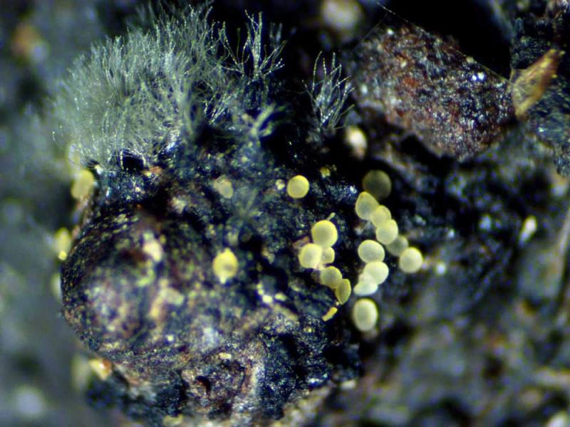
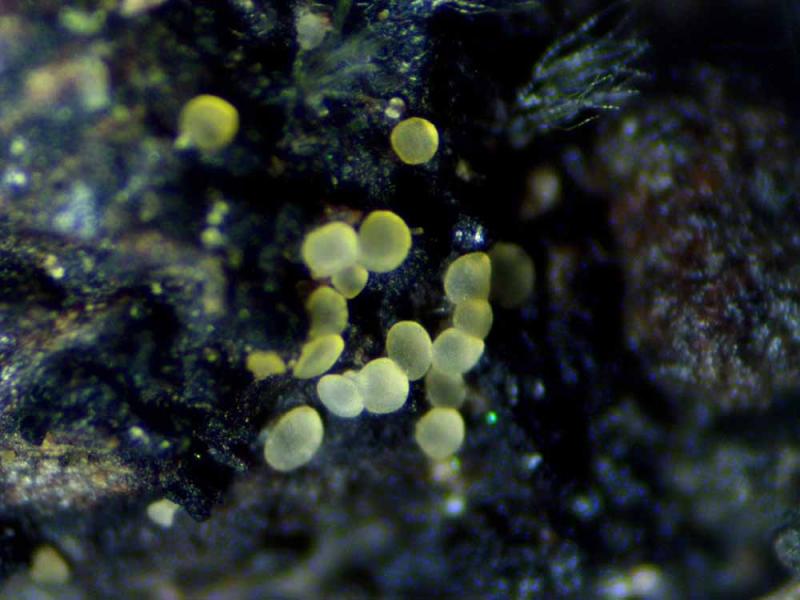
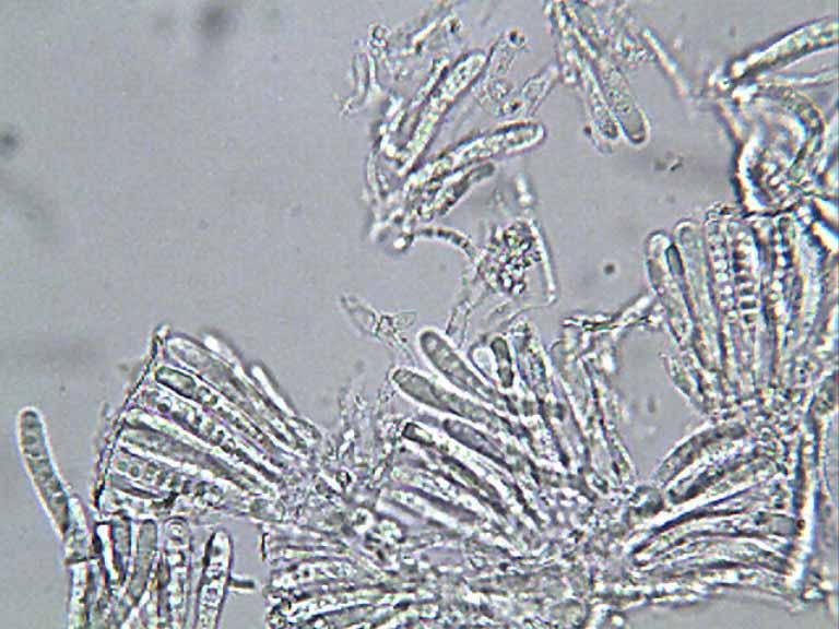
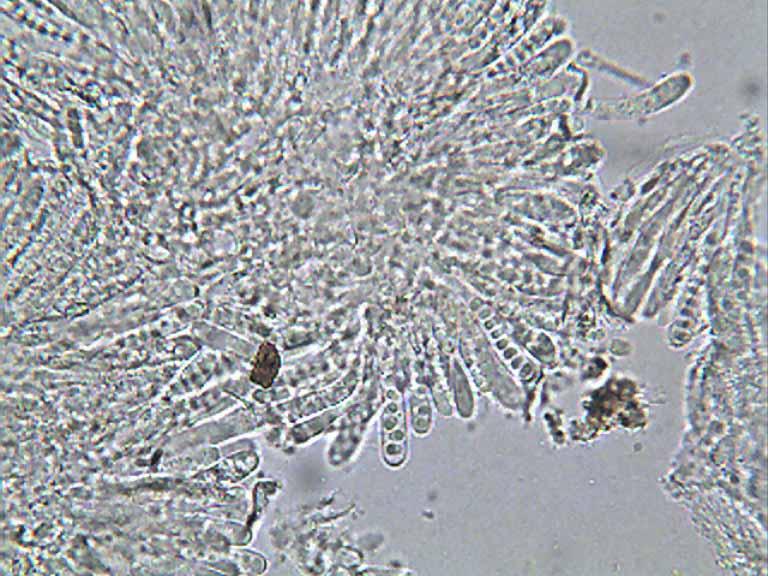
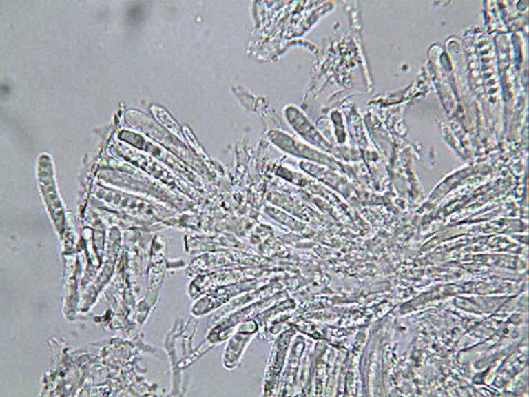
Hola, he recogido una posible Orbilia en rama de Encina de unos 8 mm, caida sobre musgos, en ella estan ungrupo de Posibles Orbilias, desconocidas a las que he visto hasta ahora.Diametro 0,15--0,22 mm
parafisis no vistas
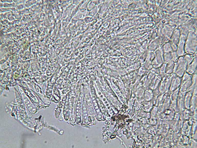
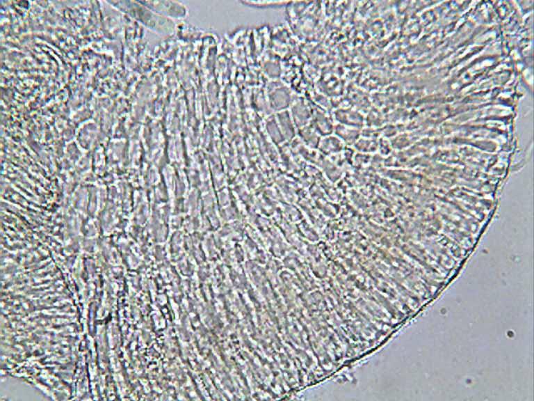
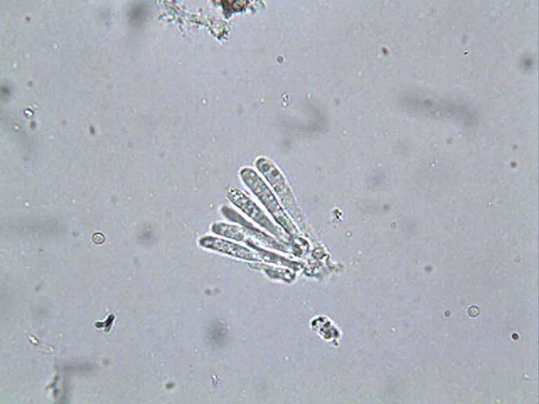
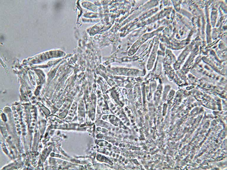
ascas 25--34 x 4,5--5
esporas solo vistas dentro de Asca, las dejare madurar unos dias para ver si esporulan
Un saludo
Rfael
Hans-Otto Baral,
17-03-2018 20:56

Re : Orbilia en Quercus
Hi Rafael
the spores are fully mature inside the asci, they have a "large" globose eccentrical spore body.
This is Hyalorbilia erythrostigma, often growing on old pyrenos.
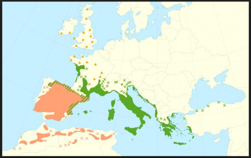
Encina is Quercus ilex - or ist it rotundifolia?
Let me please know the place and date.
Zotto
the spores are fully mature inside the asci, they have a "large" globose eccentrical spore body.
This is Hyalorbilia erythrostigma, often growing on old pyrenos.
Encina is Quercus ilex - or ist it rotundifolia?
Let me please know the place and date.
Zotto
Blasco Rafael,
17-03-2018 22:34
Re : Orbilia en Quercus
Gracias Zotto por su ayuda.
es quercus ilex
Zaragoza
Montes de zuera (cuatro caminos)
ETRS 89
Latitud 41º 54'29 , 44 "N
longitudinal 0º 55 '29, 71 " O
Altitud 539m
10-03-18
Rafael
es quercus ilex
Zaragoza
Montes de zuera (cuatro caminos)
ETRS 89
Latitud 41º 54'29 , 44 "N
longitudinal 0º 55 '29, 71 " O
Altitud 539m
10-03-18
Rafael
Hans-Otto Baral,
18-03-2018 08:16
Re : Orbilia en Quercus
Thanks! From a map that I have I guess it should be Q. rotundifolia. But on what do the apos grow, it looks like a pyreno-stroma?
The site is a S-exposed shallow slope at a road, and just at the border of the wodland. I assume it was dry on the ground, or you have just rain or snow?
A thin twig or a thick branch?
The site is a S-exposed shallow slope at a road, and just at the border of the wodland. I assume it was dry on the ground, or you have just rain or snow?
A thin twig or a thick branch?
Blasco Rafael,
18-03-2018 19:34
Re : Orbilia en Quercus
Hola Zotto
si si fallo al anotar, es Quercus rotundifolia
esta sobre viejos Pyrenos
suelo un poco humedo, pero la rama estaba entre restos de poda muy degradados que mantenian la humedad de las lluvias
son caminos entre bosques de pino/encina mezclados, muy sombreados y bastante musgo en su suelo, lo que mantiene la humedad
Rafael