09-08-2020 07:49
 Lothar Krieglsteiner
... 8.8.20, in the Eifel (Germany), on the same lo

07-08-2020 01:56
 Enrique Rubio
I'm looking for descriptions of teleomorphs (only
06-08-2020 09:59
 Hans-Otto Baral
Hello everybodyas you all know, the monograph of O

04-08-2020 22:32
 Edouard Evangelisti
Bonjour le forum, J'ai trouvé dernièrement une
04-08-2020 15:52
 Hardware Tony
Found a small group of inoperculate cup ascos unde
Camarops on Quercus
Lothar Krieglsteiner,
09-08-2020 07:49
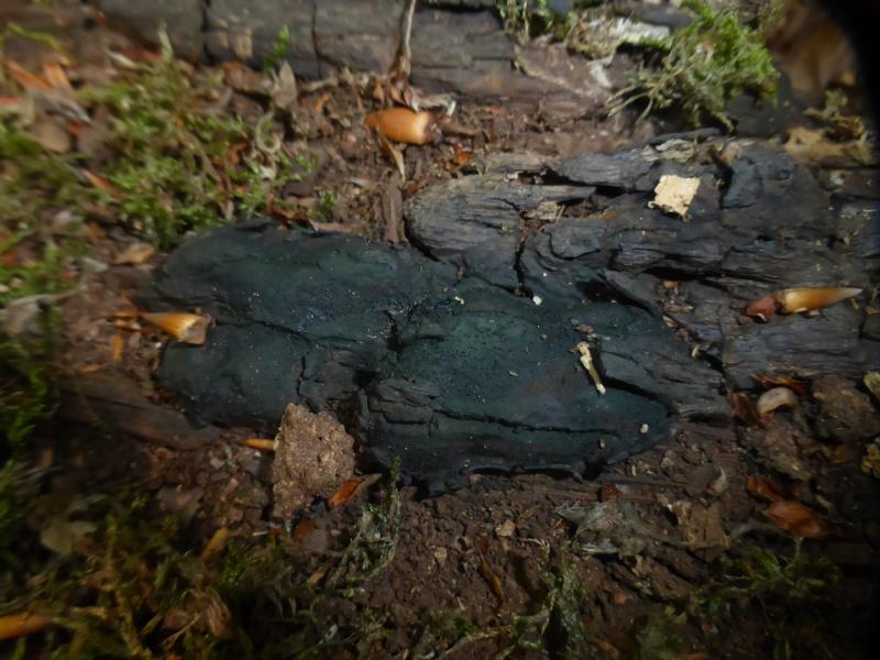
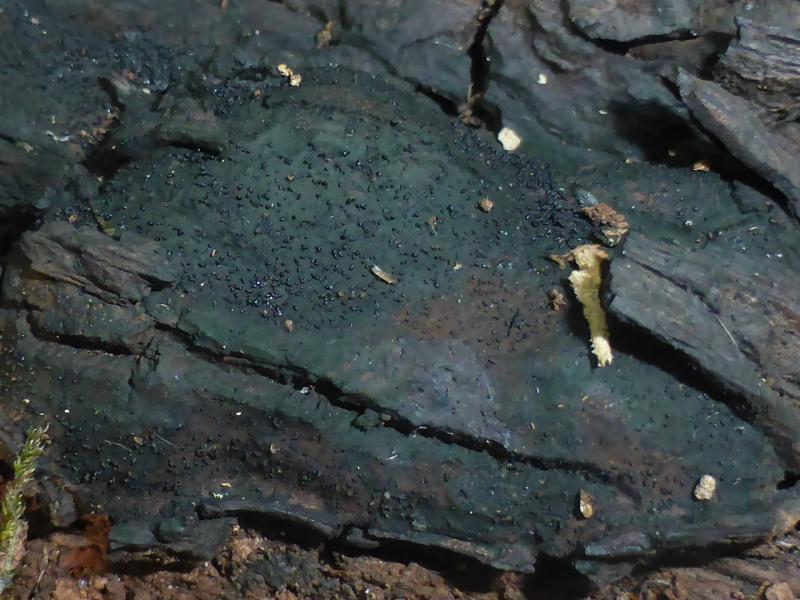
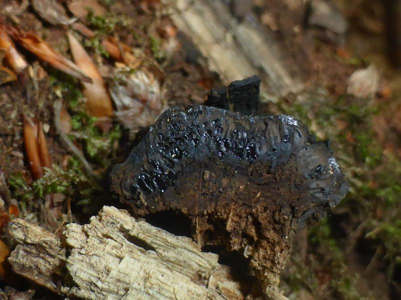
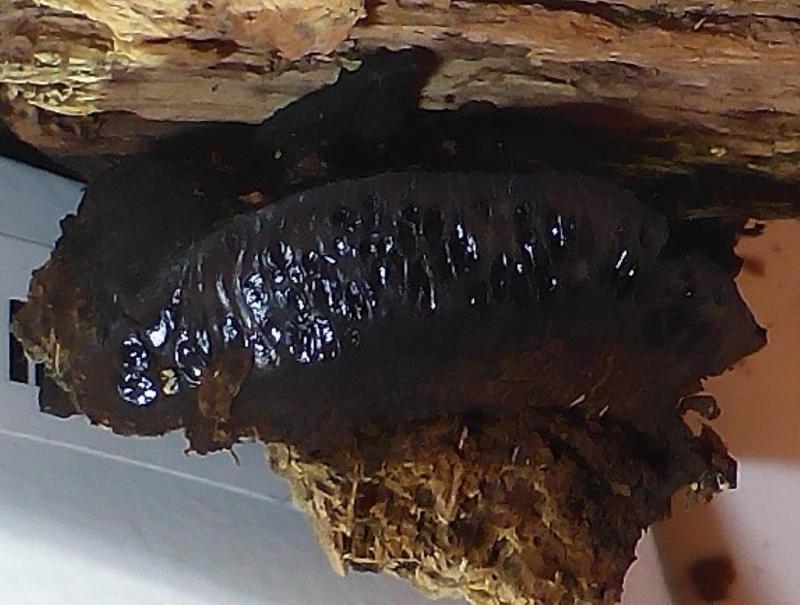
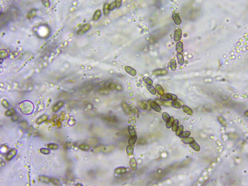
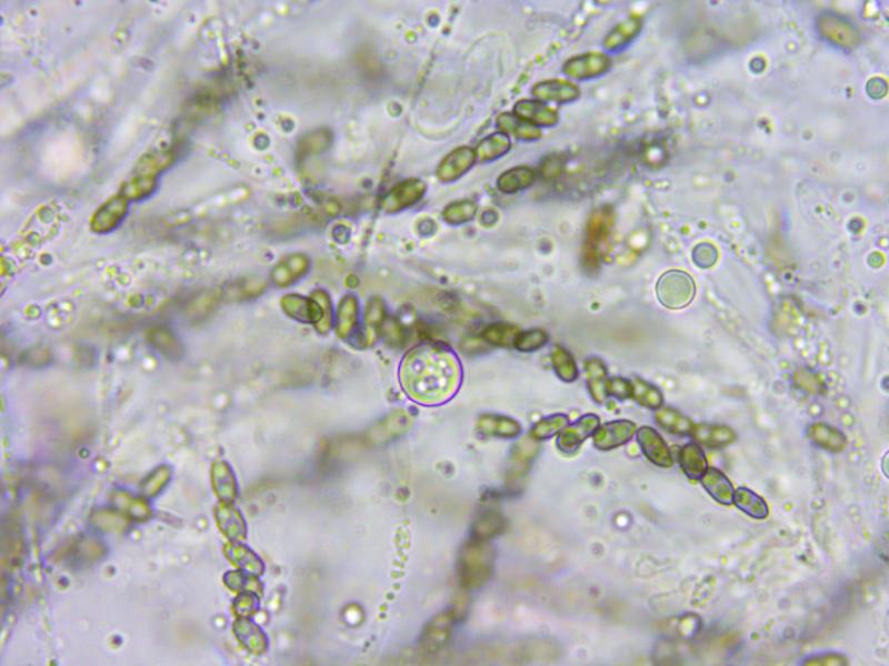
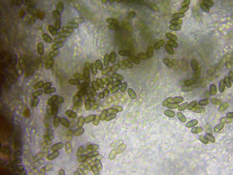
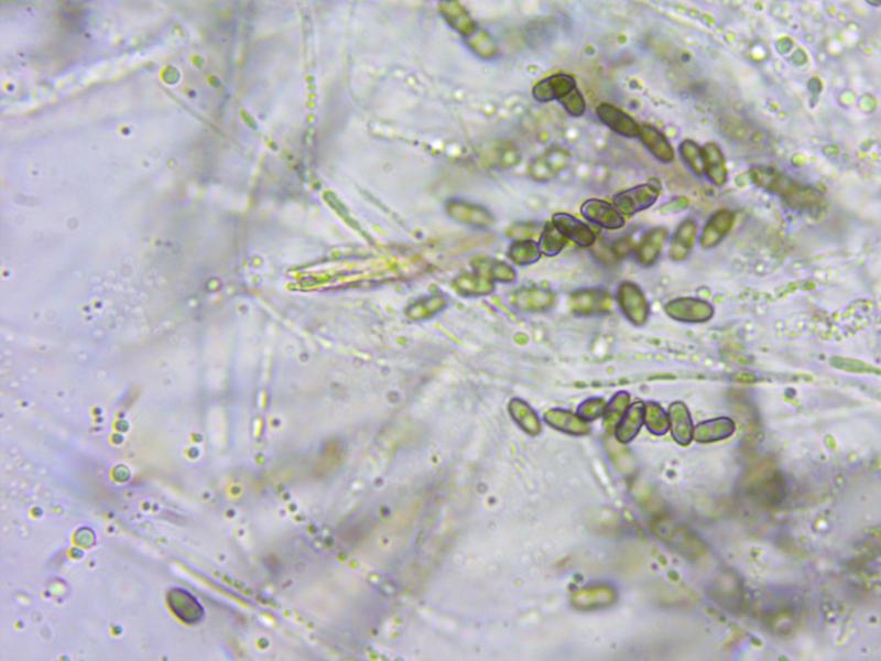
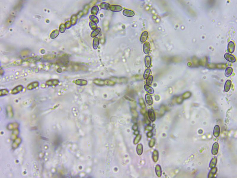
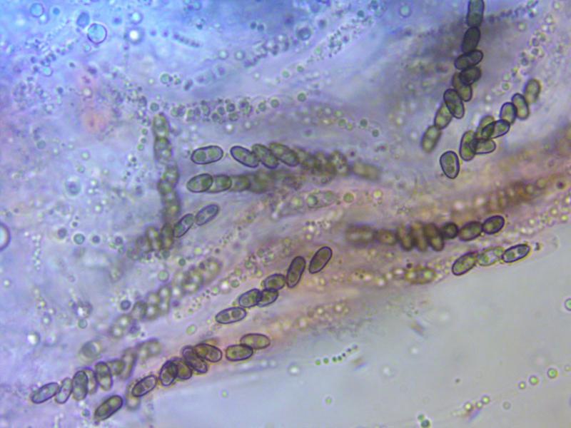
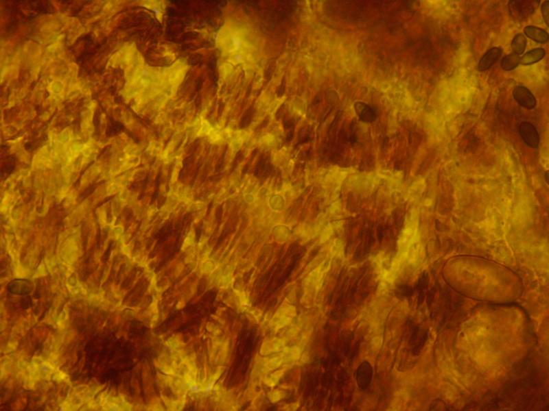
... 8.8.20, in the Eifel (Germany), on the same log with Daedalea quercina.I would have thought this to be C. polysperma, but Quercus would be an unusual substrate. C. petersii is said to grow on Quercus. What are clear differences? I cannot find a good key including all species. Who can help - what have I to do?
The spores are mostly about 6-7/2,5-3,5 µm large. 5 % KOH (I do not have 10 % with me) dissolves a weak greyish (lilaceous?) pigment.
Best, Lothar