
22-04-2026 01:06
 Richard VALERI
Bonjour à tous.Je vous présente cette Nectria s.
21-04-2026 22:14
Margot en Geert VullingsThis cup fungus was found on April 10, 2026, on lo
21-04-2026 13:36
Gernot FriebesHi,I am out of ideas for this one. I collected Sal
21-04-2026 13:19
Gernot FriebesHi,this Lophodermium on Typha has ascospores measu
21-04-2026 13:05
Gernot FriebesHi,this hyphomycete feels familiar but I was not a
20-04-2026 22:00
 Malcolm Greaves
These pale yellow, hairy ascos were growing on cul
19-04-2026 21:23
Steve ClementsBonjour, I found this anamorphic fungus on old pl
19-04-2026 20:46
Steve Clements1 mm diameter approx spherical conidiophores on pl
12-04-2026 17:56
 Hardware Tony
Found on dead stems in February earlier this year
Ostropales indet. 2
Hans-Otto Baral,
03-10-2009 18:22
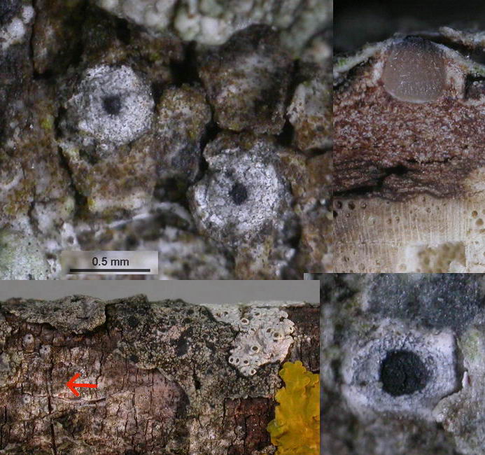
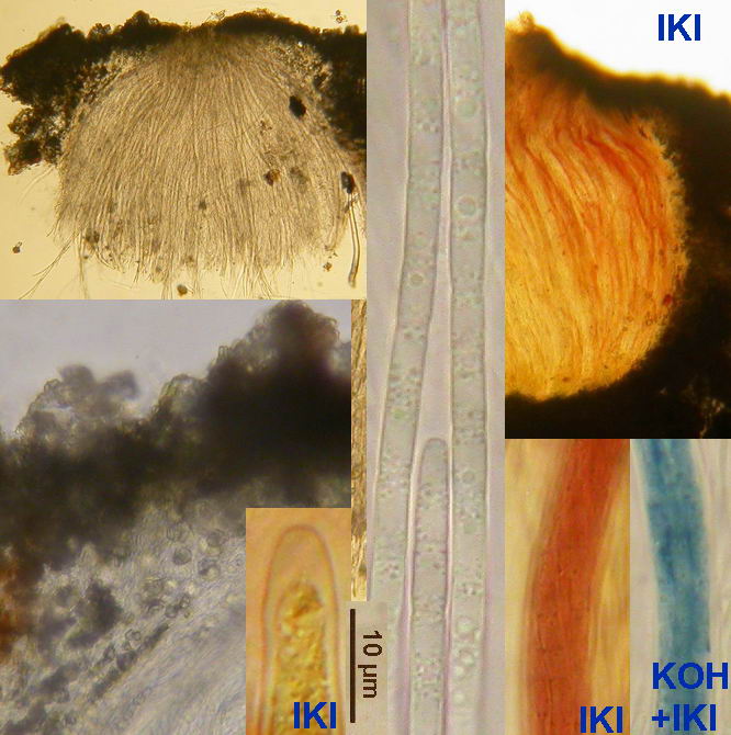
 Here is the second one. This has an inamyloid hymenium (like Ostropa and Robergea), but the spore sheath is very distinctly hemiamyloid!
N of Digne, Quercus pubescens branch 10 mm thick. Sp. ca. 300 µm long, *2.5-3.2 µm wide, cells 8-8 µm long, lipid content 1.5-2.5. Asci and whole hymenium inamyloid, but spores in dead state (sometimes also living?) IKI 2rr, after shortly boiling IKI bright blue.
Zotto
Hans-Otto Baral,
03-10-2009 18:25
Re:Ostropales indet. 2
The ascomata are almost perithecioid here.
Gernot Friebes,
04-10-2009 14:48
Re:Ostropales indet. 2
Hi Zotto,
could it be Schizoxylon albo-atrum? At least this is my outcome with the key of Schizoxylon by Martha Sherwood.
Best wishes,
Gernot
could it be Schizoxylon albo-atrum? At least this is my outcome with the key of Schizoxylon by Martha Sherwood.
Best wishes,
Gernot
Hans-Otto Baral,
04-10-2009 23:15
Re:Ostropales indet. 2
Hi Gernot
thanks, that's a good idea! Sherwoods illustration on p. 112 fits quite well. The ascospore cells she gave as 4-5 x 2 µm, while I measured 5-8 x 2.5-3.2 µm in the living state (sorry for my error above). It is a pity that we do not know whether the spores are also hemiamyloid in Sherwood's material, especially Rehm's type. Sherwood says for the paraphyses J- or faintly J+ blue, but we must know that she used Melzer, and a hemiamyloid hymenium like in my Ostropales indet. 1 would be in Melzer just like that, J- or faintly blue. In one of her material of alboatrum (from Oregon) she reported a strongly amyloid epithecium. And I do not understand why she says "apparently common" but cites only 7 collections.
Zotto