
12-06-2026 14:50
 François Freléchoux
Bonjour, Voici la brève description d'une Mollis
10-06-2026 21:16
François Freléchoux
Bonsoir,Le dernier du jour, en attendant votre avi
11-06-2026 19:01
 William Slosse
Hello all,In an attempt to make a culture of a sus
11-06-2026 19:03
 Nicolas VAN VOOREN
Chers membres d'Ascofrance,Le site sera placé en

09-06-2026 18:32
Camille MertensSur morceau de roseau immergé 0,5 - 0,7 mm de dia
10-06-2026 12:54
Steve ClementsBonjour encore, Pouvez-vous m'aider, s'il vous pl
10-06-2026 21:07
François Freléchoux
Toutes les tiges de gentianes jaunes de l'an pass�
10-06-2026 13:41
François Freléchoux
Bonjour à nouveau, Voici une trouvaille d'hier.
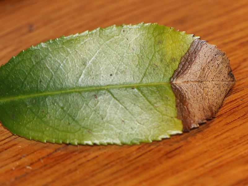
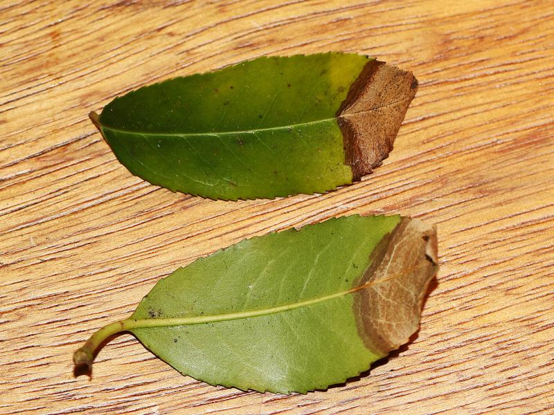
 On 6 february 2025 i stumbled upon a leafspot on Prunus lusitanica in Aerdenhout (The Netherlands). Can someone confirm that it is indeed Coleophoma prunicola? or maybe something completly different?
 For more photo's see: https://waarneming.nl/observation/338565267/
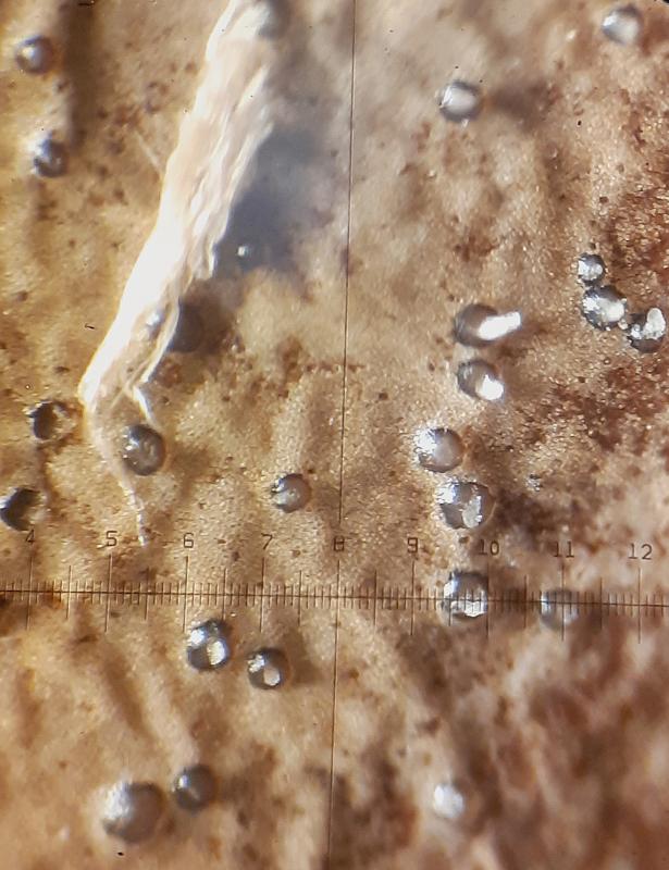
Conidiomate pycnidia, ca. 150- 200 µm diameter (N=10).
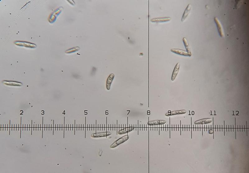
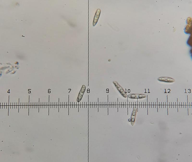
Conidia holoblastic, hyaline, aseptate, cylindrical, apex obtuse, base truncate, thin-walled, smooth, guttulate with several large guttules.
Conidia 20,8 - 23,4 - 26 ?m × 4-5,2 ?m (N=25)
400 X mafnification: 1 div. = 2,6 ?m
Following the Key for Coleophoma in the article: 'Reinstatement of Coleonaema for Coleophoma oleae and notes on Coleophoma' it should be Coleophoma prunicola
(Duan, J.X., Wu, W.P. and Liu, X.Z. (2007). Reinstatement of Coleonaema for Coleophoma oleae and notes on Coleophoma. Fungal Diversity 26: 187-204.) https://www.researchgate.net/publication/237440307_Reinstatement_of_Coleonaema_for_Coleophoma_oleae_and_notes_on_Coleophoma

To know this you should see the conidiogenous cells and compare them with those in the article.
Best wishes
Angel
Thanks for your comment. I can try another time to see the conidiogenous cells and compare them with those in the article. I already tried once but failed to see the conidiogenous cells sadly.
Kind regards,
Jorian