08-06-2026 17:00
François BartholomeeusenGood day everyone, On June 5 2026, I collected de
08-06-2026 10:16
 Spooren Marco
I don`t have a clou about this fungus,it is not in
07-06-2026 15:10
 William Slosse
Hello everyone,On 05-06-26, I found following asco
05-06-2026 11:02
Thomas Læssøehttps://svampe.databasen.org/observations/10596691
07-06-2026 12:09
 François Freléchoux
Bonjour, Voici une brève description de ce qui m
07-06-2026 12:43
Steve ClementsBojour. This was a strange find on a stick on my
12-07-2015 00:05
 Nedim Jukic
This one from the same locality as the previous on
06-06-2026 17:44
Steve ClementsBonjour, This disco was on planed wood 3 x 1.5 cm
14-08-2016 23:15
 Alex Akulov
Dear friendsCan you help me to find the descriptio
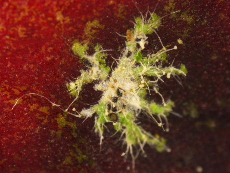
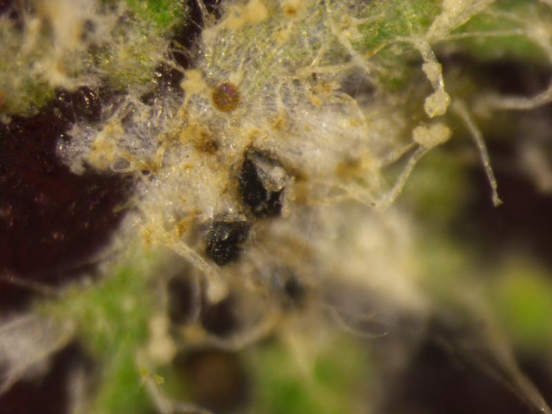
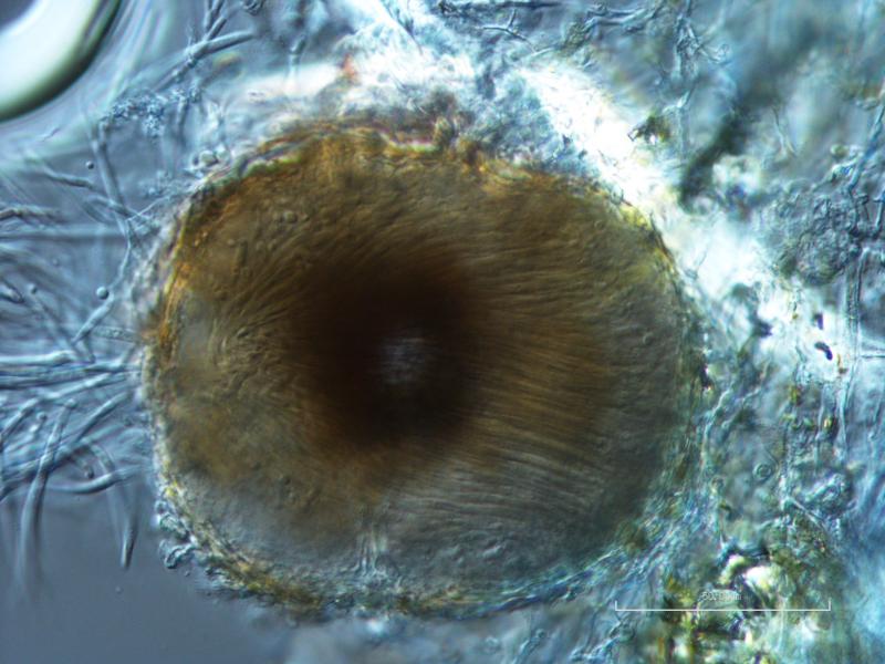
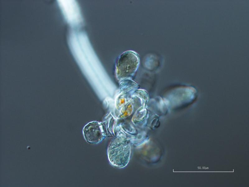
 Hello! I wanted to share a fascinating thing I saw this week. This is the coelomycete anamorph state of pyrenolichen Phyllocharis orbicularis (=Strigula orbicularis), which is apparently not illustrated anywhere. I was lucky enough to be able to ask Robert Lücking for help, and he ID'd it right away, having seen it before; when I asked where I could find an illustration, he admitted that he didn't think one existed, only the ascomata and the macromorphology of the thallus. The thallus of this particular example is somewhat poorly lichenized, and looks more like the photobiont (Cephaleuros virescens) than the typical thallus, but the conidia are the really fun part anyway.
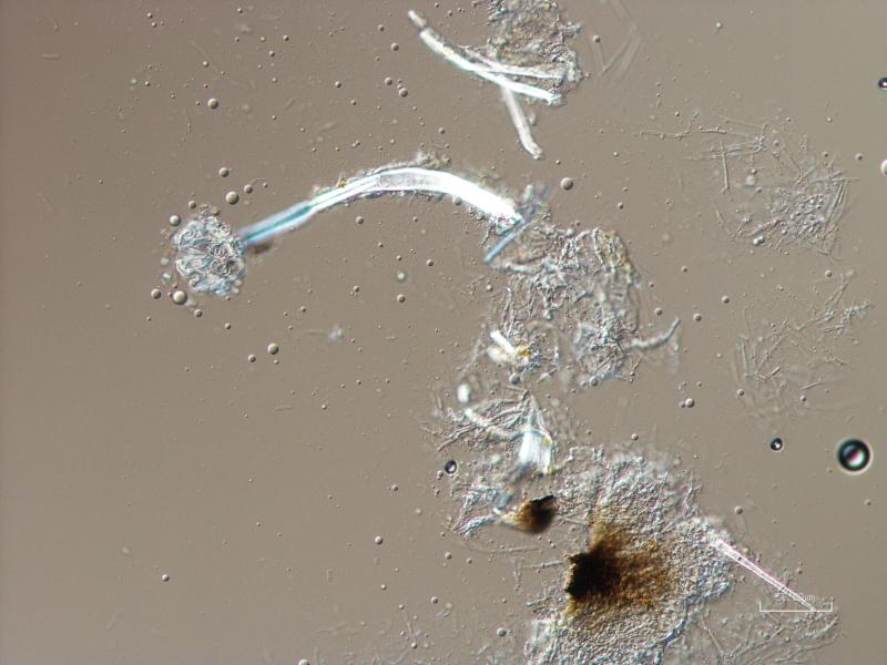
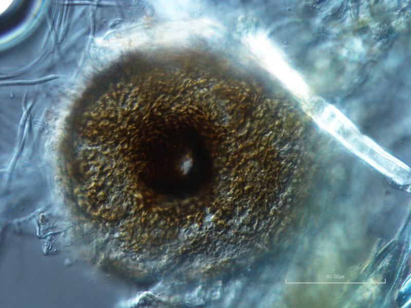
The conidia are hyaline, 4- to 6-septate, 40-45 x 2-3.5 ?m excluding the appendages, with a non-cellular, mucoid appendage at each end, which are quite variable in length, but generally less than 10 ?m, and often curving into a hook. Conidiophores are small, lageniform, reduced to conidiogenous cells, and integrated into the inner wall of the pycnidium.
I really wanted to put these photos out there, so that if anyone else is struggling to identify this beautiful and distinctive anamorph they'll be able to find some reference images! I can't thank Dr. Lücking enough for his kind help in the identification of this fungus.