08-06-2026 17:00
François BartholomeeusenGood day everyone, On June 5 2026, I collected de
08-06-2026 10:16
 Spooren Marco
I don`t have a clou about this fungus,it is not in
07-06-2026 15:10
 William Slosse
Hello everyone,On 05-06-26, I found following asco
05-06-2026 11:02
Thomas Læssøehttps://svampe.databasen.org/observations/10596691
07-06-2026 12:09
 François Freléchoux
Bonjour, Voici une brève description de ce qui m
07-06-2026 12:43
Steve ClementsBojour. This was a strange find on a stick on my
12-07-2015 00:05
 Nedim Jukic
This one from the same locality as the previous on
06-06-2026 17:44
Steve ClementsBonjour, This disco was on planed wood 3 x 1.5 cm
14-08-2016 23:15
 Alex Akulov
Dear friendsCan you help me to find the descriptio
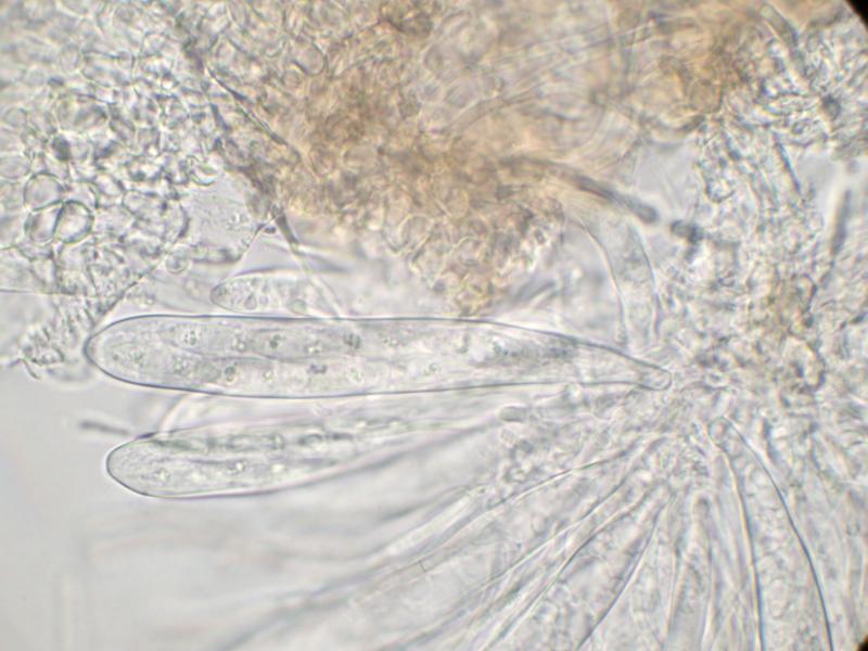
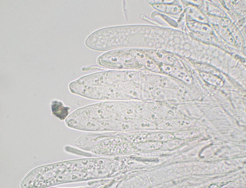
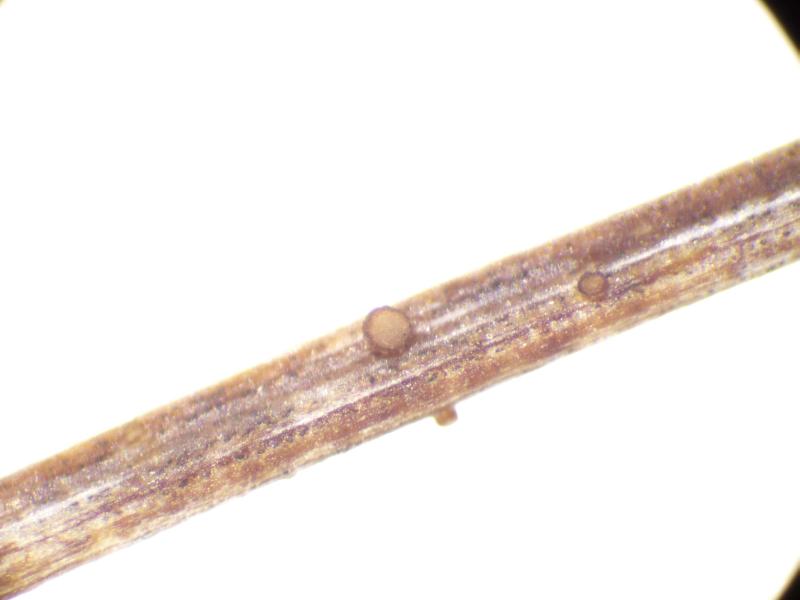
Apothecia sessile; cup-shaped; ca 300 µm diam.; hymenium pale grey; exterior brownish orange.
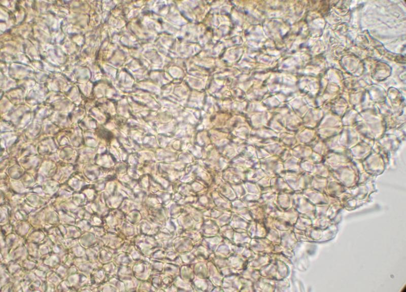
Excipulum brown textura globulosa.
Paraphyses narrowly cylindrical (2-2.5 µm wide), sometimes swollen at apex; sometimes branched; cylindrical refractive VB in upper part.
Asci clavate; ca 100-110 x 13-16 µm; 8-spored (biseriate); IKI+ blue; with shallow apical ring.
Ascospores fusiform; hyaline; free spores 21-23 x 6 µm; mostly 1-septate, but free spores sometimes 2-septate; scattered small OBs, mainly near ends of spore.
This seems to resemble Nimbomollisia (Niptera) eriophori. I didn't notice gelatinous sheaths on the spores when examining the specimen but the image of spores in the ascus in MLZ seems to show some sort of gelatinous structure at the ends of the spores. Some of the paraphyses have swollen apices but this feature isn't as well developed as I would have expected in Nimbomollisia.
I'd be grateful for a second opinion.
Thanks
Marcus
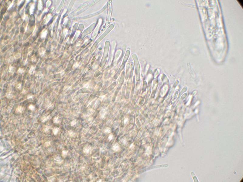
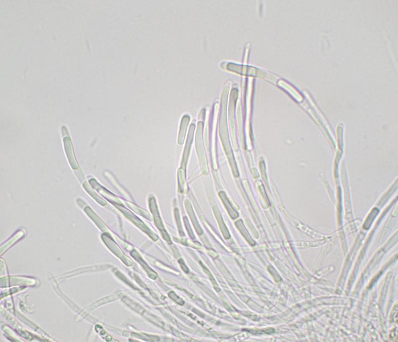
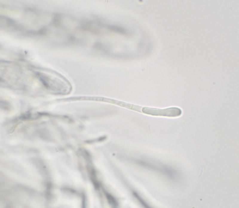
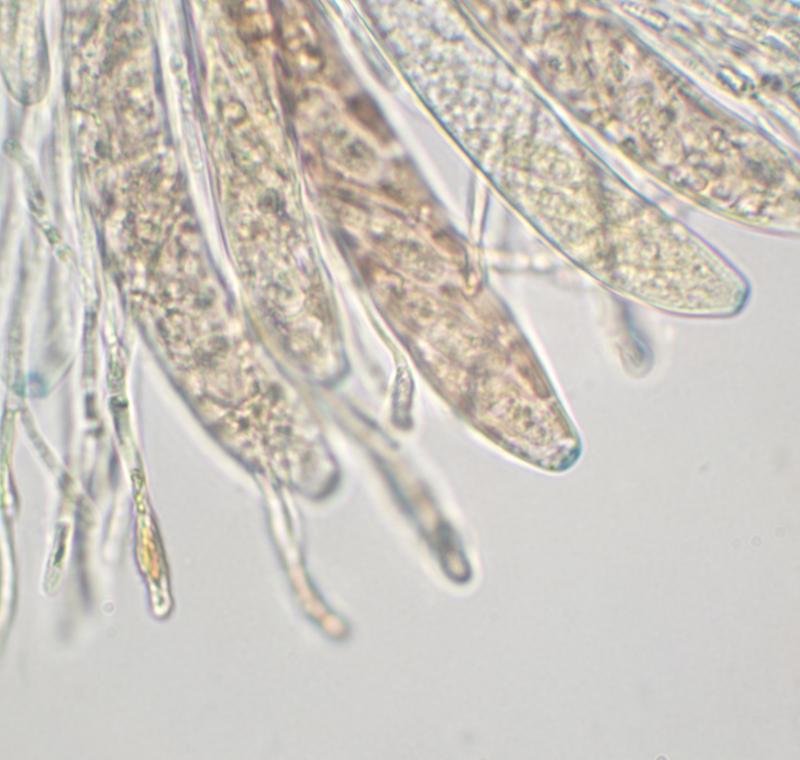
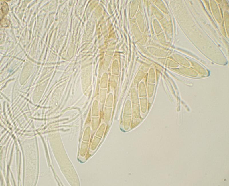
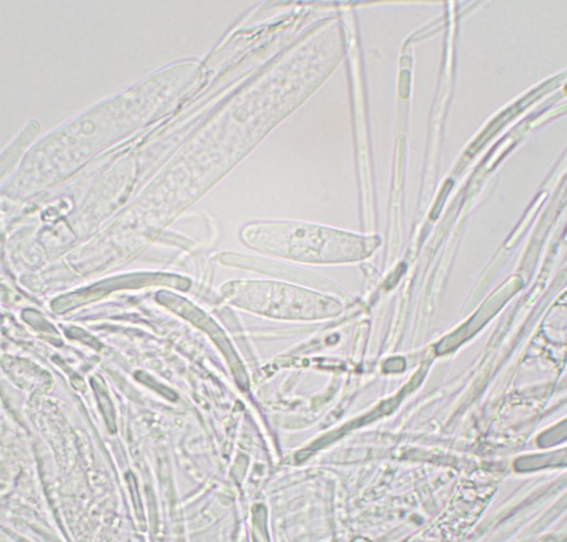
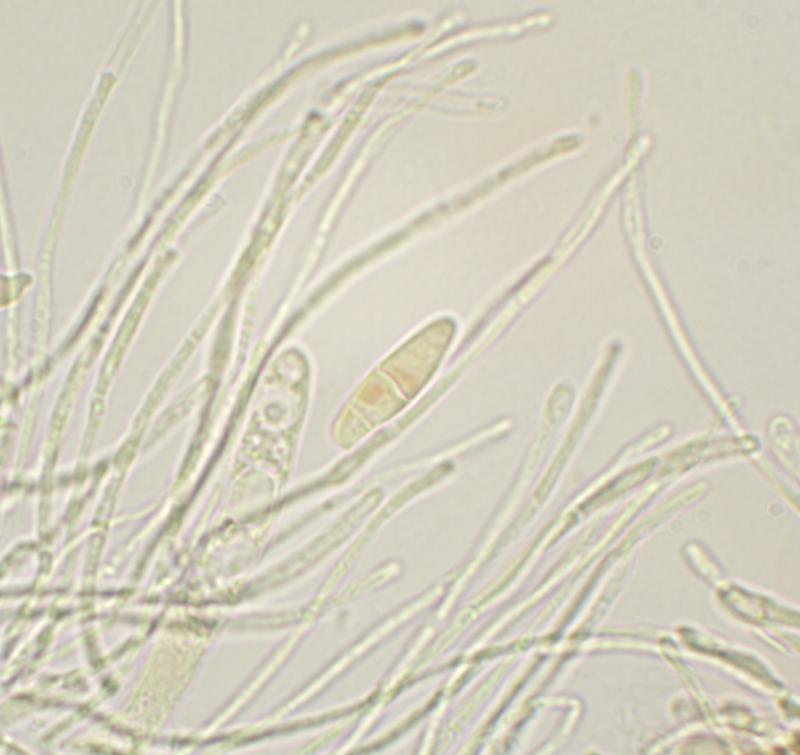

There were very few free spores. Spores in the asci were difficult to see clearly (see images) but I couldn't see any obvious caps or sheaths.