
08-06-2026 17:00
François BartholomeeusenGood day everyone, On June 5 2026, I collected de
08-06-2026 10:16
 Spooren Marco
I don`t have a clou about this fungus,it is not in
07-06-2026 15:10
 William Slosse
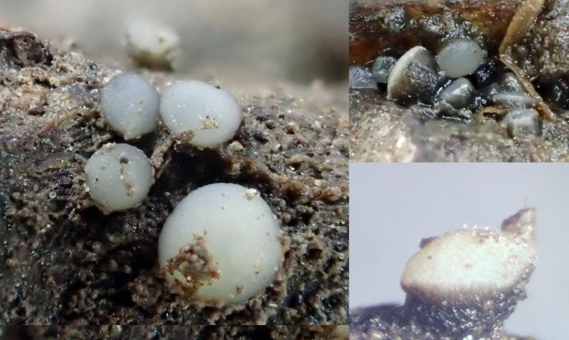
Hello everyone,On 05-06-26, I found following asco
05-06-2026 11:02
Thomas Læssøehttps://svampe.databasen.org/observations/10596691
07-06-2026 12:09
 François Freléchoux
Bonjour, Voici une brève description de ce qui m
07-06-2026 12:43
Steve ClementsBojour. This was a strange find on a stick on my
12-07-2015 00:05
 Nedim Jukic
This one from the same locality as the previous on
06-06-2026 17:44
Steve ClementsBonjour, This disco was on planed wood 3 x 1.5 cm
14-08-2016 23:15
 Alex Akulov
Dear friendsCan you help me to find the descriptio
Vibrissea flavovirens?
Stefan Jakobsson,
08-07-2022 02:34
Can it be confirmed that this is V. flavovirens?
Hans-Otto Baral,
08-07-2022 08:39

Re : Vibrissea flavovirens?
I think the colour is not so important. Since I do not have an overniew on available measurements, I cannot easily say if such small measurements ever occurred, but I do not know a further species with these characters.
Stefan Jakobsson,
08-07-2022 11:49
Re : Vibrissea flavovirens?
Thank you!
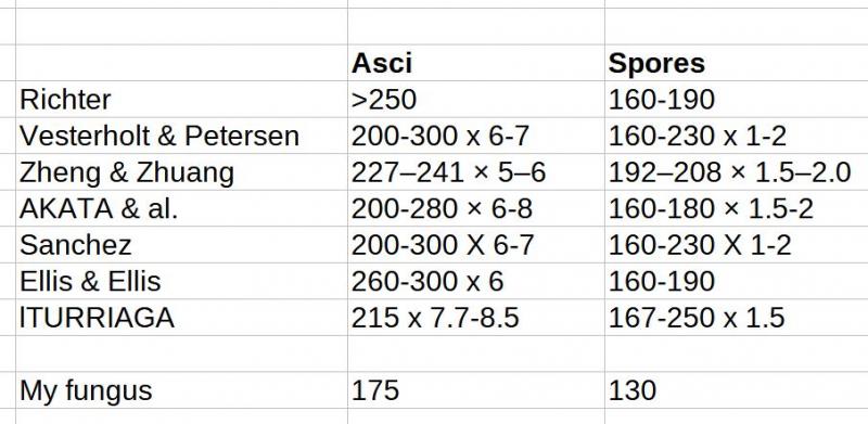
I looked up the asci and spore size in a few sources.
Hans-Otto Baral,
09-07-2022 09:42
Re : Vibrissea flavovirens?
Thanks for this survey. In my key I wrote for flavovirens:
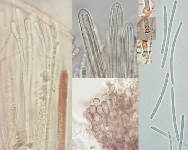
asci *260-343 x 6.5-8.8 µm
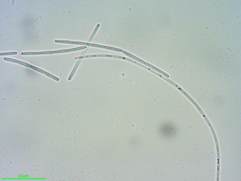
spores *125-195 x 1.2-1.8 µm
spore fragments *(27–)30-42(–51) µm
Ascus measurements in your table are probably mostly in dead state, but the differences to living asci are apparently not very high.
In the type of V. minima Velen. on Salix, which I restudied and considered a synonym, I found asci +154 x 4.8-5.3 and spore fragments 27-48 µm. So your spore fragments are a bit shorter than usual.
In a collection from Sheffield (HB 9520) I measured spores *125-134 µm long (like yours), breaking into 4 part spores of *27-38 x 1.3-1.6 µm, 4-celled (ascus length not measured).
In the case there is a continuum of measurements among collections, I suspect that living asci much shorter than 260 µm also occur.
Identities in the literature are perhaps not certain. E.g. Zheng & Zhuang 2017 do not mention the number of spore cells and fragments, but the spore photo suggests flavovirens indeed.