
08-06-2026 17:00
François BartholomeeusenGood day everyone, On June 5 2026, I collected de
08-06-2026 10:16
 Spooren Marco
I don`t have a clou about this fungus,it is not in
07-06-2026 15:10
 William Slosse
Hello everyone,On 05-06-26, I found following asco
05-06-2026 11:02
Thomas Læssøehttps://svampe.databasen.org/observations/10596691
07-06-2026 12:09
 François Freléchoux
Bonjour, Voici une brève description de ce qui m
07-06-2026 12:43
Steve ClementsBojour. This was a strange find on a stick on my
12-07-2015 00:05
 Nedim Jukic
This one from the same locality as the previous on
06-06-2026 17:44
Steve ClementsBonjour, This disco was on planed wood 3 x 1.5 cm
14-08-2016 23:15
 Alex Akulov
Dear friendsCan you help me to find the descriptio
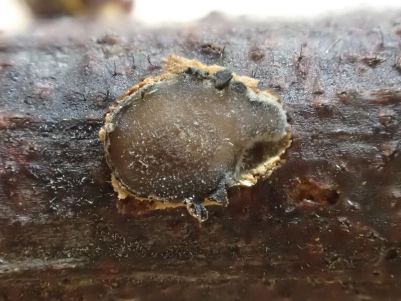
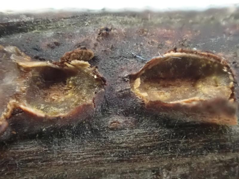
Erumpent on Wattle Part 2
Zuidland Peter,
25-06-2022 07:08
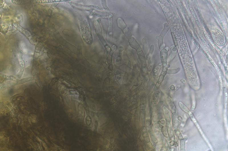
I view of possible confusion by poor images I representwhat I think is an erumpent I have found on Wattle. I have used Spooner's Heliotales of australia for years and I find nothing to help me in there, there is no budding like this on Heliotales according to him.
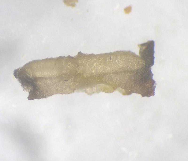
This sample forms under the bark, breaks through as it gets bigger and spreads across the wood surface. One image shows old craters with remnant excipulm around the edges under the bark.
Fresh images from today, all microscopy in water; sample is being kept moist and cool for further work if needed.
SE Victoria Australia
Cheers
Pete
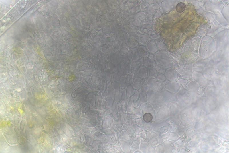
Hans-Otto Baral,
25-06-2022 09:23

Re : Erumpent on Wattle Part 2
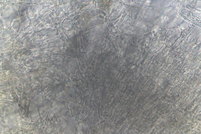
Helpful would be a closeup of the marginal lobes in section (last pic), if there are periphysoids. It could be an ostropalean fungus. Of course, free spores would be good and their measurements. Did you test IKI? MLZ is unsuitable, unless you pretreat with KOH.
Zuidland Peter,
25-06-2022 09:51
Re : Erumpent on Wattle Part 2
Thanks heaps Hans Otto,
I did check for IKI with Lugol's and it is the first micro image in my previous post on this( see below again); I have two more samples that I can try to check what you have asked for.
Take care
Pete
I did check for IKI with Lugol's and it is the first micro image in my previous post on this( see below again); I have two more samples that I can try to check what you have asked for.
Take care
Pete
Zuidland Peter,
26-06-2022 04:21
Re : Erumpent on Wattle Part 2
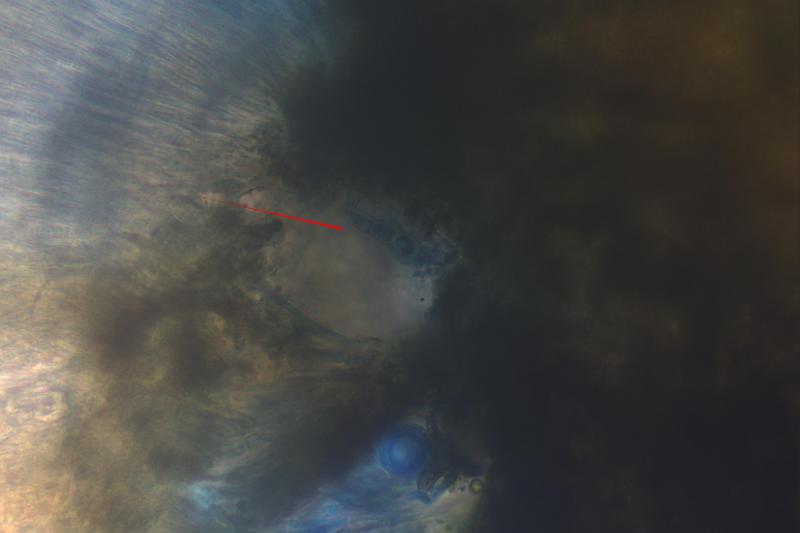
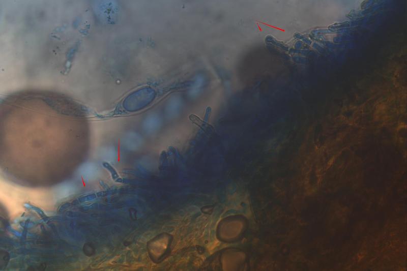
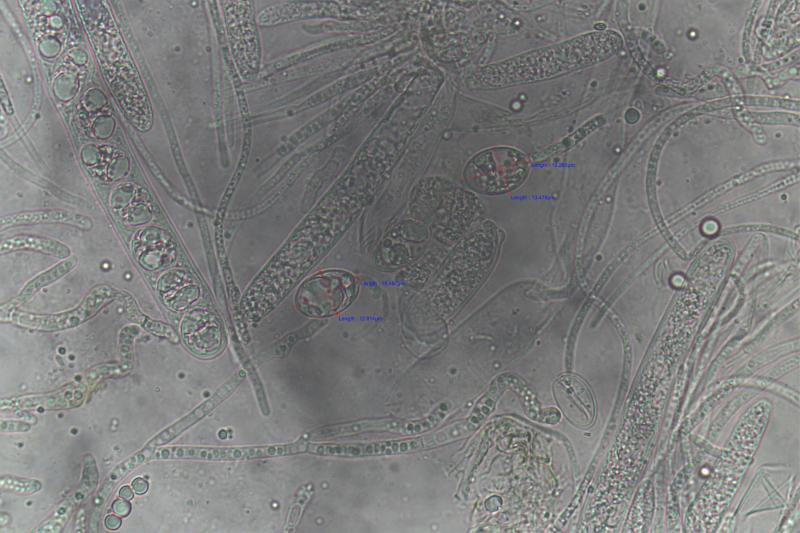
I think these are the periphysoids with arrows.
Pete
Pete
Zuidland Peter,
26-06-2022 04:24
Re : Erumpent on Wattle Part 2
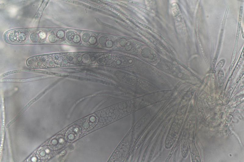
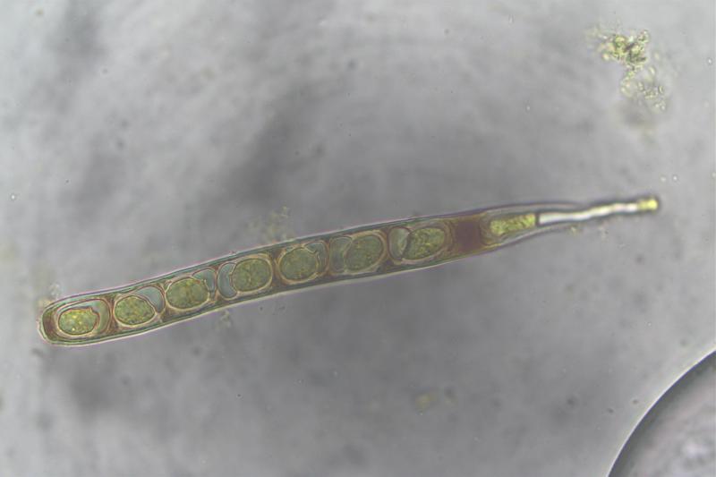
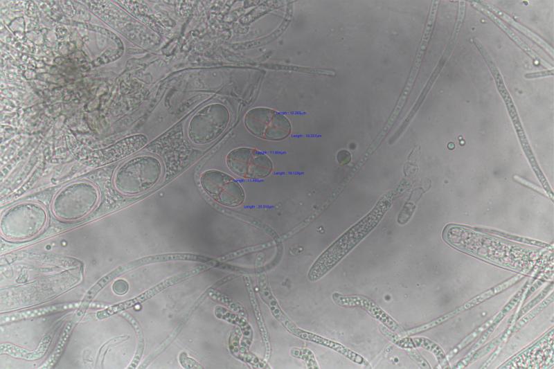
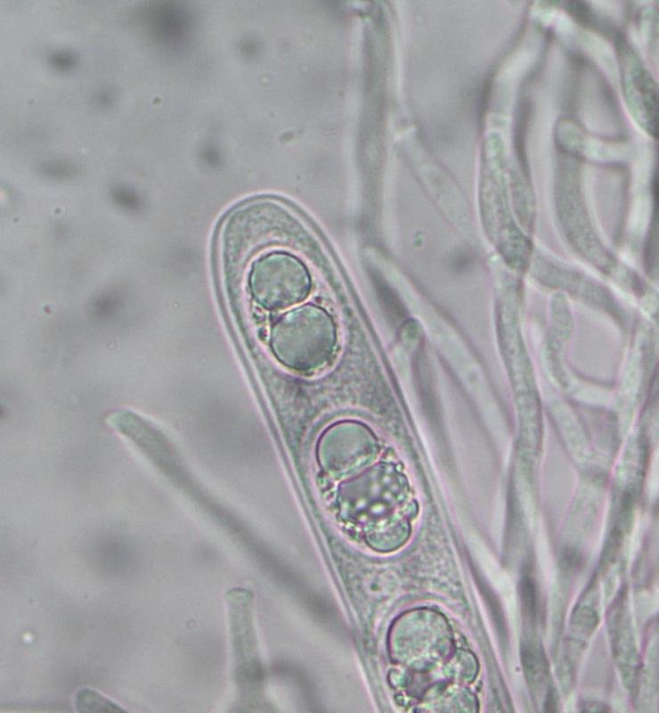
Free spores in H2O measured: 16.4 - 20.3 x 10.2 - 13.5um
many thanks
Pete
many thanks
Pete
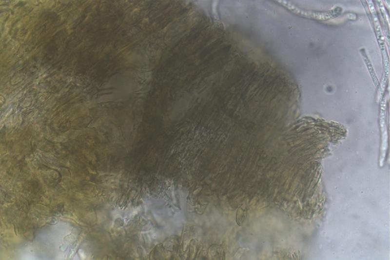
Hans-Otto Baral,
26-06-2022 18:00
Re : Erumpent on Wattle Part 2
O.k., I am now a bit confused, do you think your wattle 1 and 2 are the same species?
I saw the IKI-ascus photo in 1 and wondered if it is a hemiamyloid apical ring or only plasma. It could be a ring, and then you should either show it in larger magnification or try KOH-pretreatment to see if the ring then stains blue in IKI.
Periphysoids: I am not sure. They often form a compact layer of short parallel hyphae embedded in some gel.
Zuidland Peter,
27-06-2022 07:42
Re : Erumpent on Wattle Part 2
I am sorry that my poor skills as a non trained mycologist are causing you some grief or confusion, I don't mean to.
The specimen is the same in both series, I have no other asco near it nor with these spores.
Here is sample with just Lugol's again, from today and zoomed.
Pete
The specimen is the same in both series, I have no other asco near it nor with these spores.
Here is sample with just Lugol's again, from today and zoomed.
Pete
Zuidland Peter,
27-06-2022 07:45
Re : Erumpent on Wattle Part 2
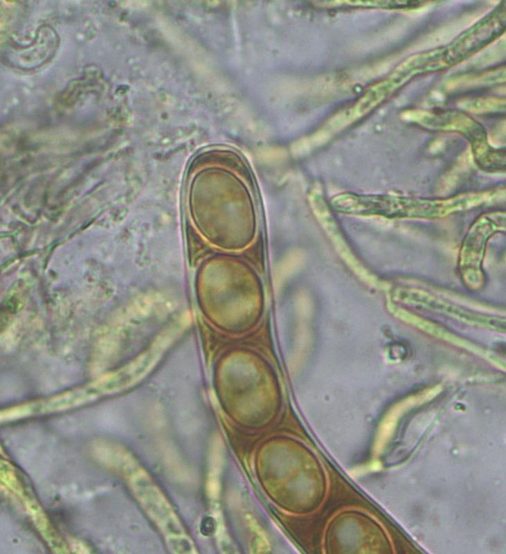
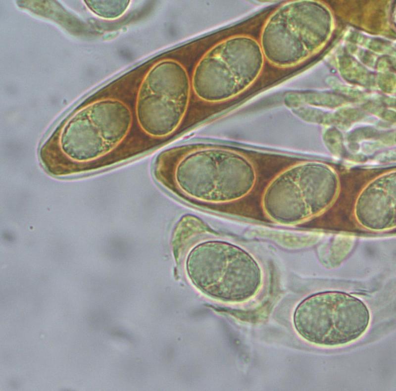
Here is the sample heat treated with 4%KOH, cooled, remove most KOH apply Lugol's.
This leaves a clear looking asci with no stain.
This leaves a clear looking asci with no stain.
Zuidland Peter,
27-06-2022 07:47
Re : Erumpent on Wattle Part 2
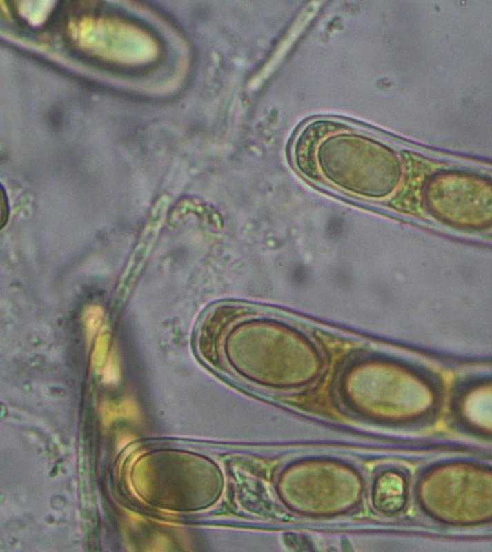
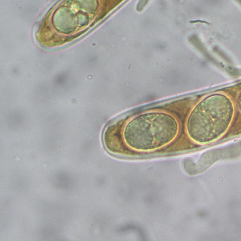
Heat treat with KOH, cool, add Lugol's, remove Lugol's then add Lugols again.
This left the asci stained.
This left the asci stained.
Zuidland Peter,
27-06-2022 07:49
Re : Erumpent on Wattle Part 2
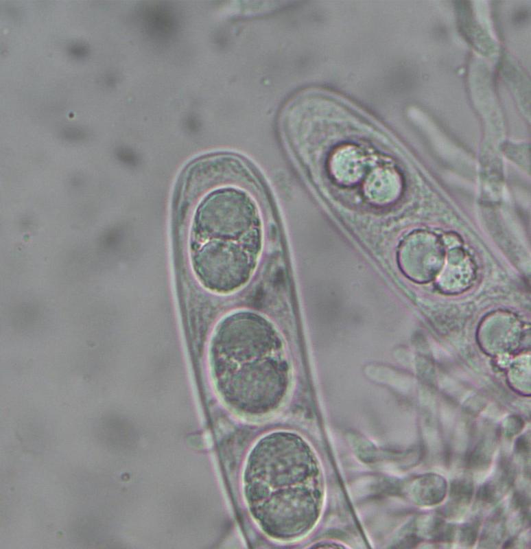
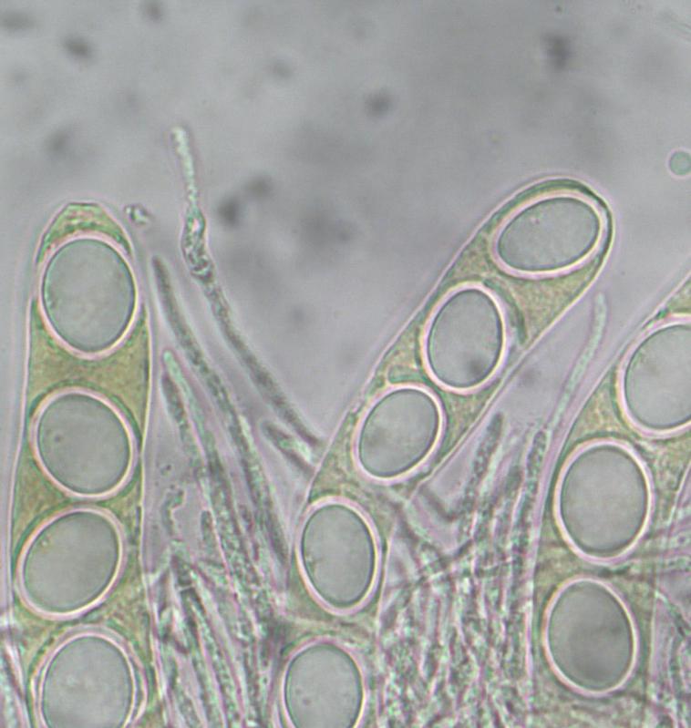
For the excercise I did it with MLZ too.
Heat with KOH, remove then add MLZ
One image looks to show a channel near the tip
Heat with KOH, remove then add MLZ
One image looks to show a channel near the tip
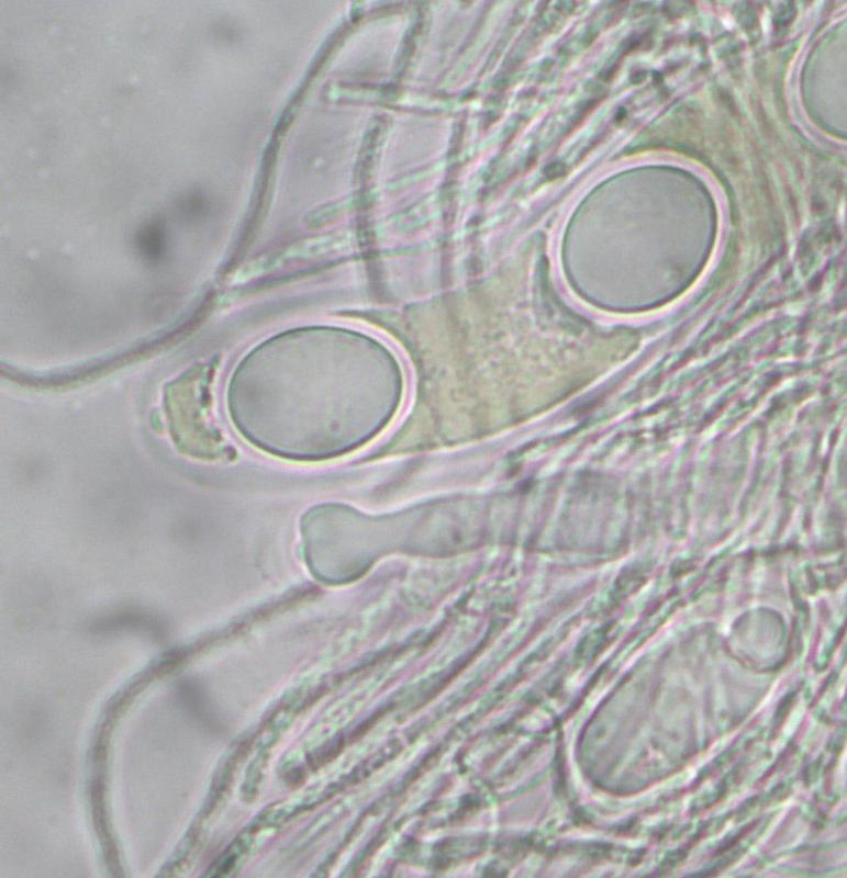
Hans-Otto Baral,
27-06-2022 08:21
Re : Erumpent on Wattle Part 2
Splendid and much better resolution! Now it is clear, the asci are inamyloid. The "channel" is an ocular chamber, maybe it is more typical of immature asci without spores or with beginning spore formation. In such asci it my be that the lateral ascus wall is thickened.