
07-06-2026 12:43
Steve ClementsBojour. This was a strange find on a stick on my
07-06-2026 12:09
 François Freléchoux
Bonjour, Voici une brève description de ce qui m
12-07-2015 00:05
 Nedim Jukic
This one from the same locality as the previous on
06-06-2026 17:44
Steve ClementsBonjour, This disco was on planed wood 3 x 1.5 cm
14-08-2016 23:15
 Alex Akulov
Dear friendsCan you help me to find the descriptio

05-06-2026 11:02
Thomas Læssøehttps://svampe.databasen.org/observations/10596691
04-06-2026 11:36
Gernot FriebesHi,found on Vaccinium myrtillus.Asci: IKI –, 8-s
05-06-2026 12:10
François Freléchoux
Capitotricha sp. sur Lonicea caerulea Caractères
Sclerotinia Sclerotiorum spores
Ale Ale,
11-09-2021 20:09
Hello,
Is there any reference in which I can find the approximate dimension of Sclerotinia Sclerotiorum spores?.
I am more interested in knowing the thickness of the cell wall and membrane. I assume the spores have a cell wall. Any help will be highly appreciate it.
I also know they are 2 nuclei
Thank you very much
Hans-Otto Baral,
11-09-2021 21:20

Re : Sclerotinia Sclerotiorum spores
Every fungal spore has a spore wall, whether conidial (mitosporic) or sexual.
Spore size depends on the living vs. dead state.
Spore sizes are not fix but underlie some variation among the populations. In my sclerotiorum folder (www.in-vivo-veritas.de) you can see my drawings with spore size and illustration of the two nuclei.
Spore size may be e.g.
*(10)11-13(14) x 5.5-6.2 µm (HB 6102)
*11.7-14.7(15.7) x 5.5-6.6 µm (HB 4412)
*11-14.5(15.5) x 6-7 (HB 9480)
or in dead state
+9-11(12 x 4.5-5(-5.5) µm (HB 238)
Spore wall thickness can only be estimated at maybe 0.2 µm.
May I ask why you need these data?
Zotto
Ale Ale,
11-09-2021 21:47
Re : Sclerotinia Sclerotiorum spores
Hello Zotto,
Thank you very much for your reply. First I should mention that I am an electrical engineer, so I have virtual no training in the biology of spores.
Im working with microdevices that can capture and detect these spores in solution, for potential applications in crop protection.
My current goal is to determine the dielectric properties of S.S spores, that is, to determine the conductivity and permittivity of the external wall (cell wall + membrane) as well as the permittivity and conductivity of the internal spore (I would assume an uniform cytoplasm).
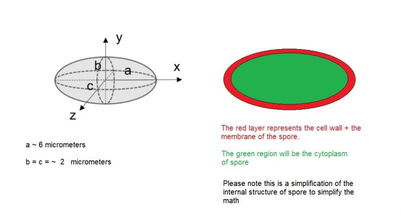
We do have spores in our lab, and the size match what you informed me. However, I could not find any info about the thickness of the spore wall. I was thinking to model them as being 80 nanometers. I attached an schematic of my spore model. What do you think?
Thank you again
Hans-Otto Baral,
11-09-2021 22:17
Re : Sclerotinia Sclerotiorum spores
Not sure what you mean with a b c, but shouldn't be b and c = 3 µm?
0.08 µm is well possible. I assume there exist TM photos of the spore wall. The fungus seems well-explored in regard to physiology and DNA, but morphology?
Ale Ale,
11-09-2021 22:45
Re : Sclerotinia Sclerotiorum spores
It is quite confusing, you are right.
a, b and c are the radius along the axis x, y and z, respectively.
Perhaps a value for b and c equal to 2.5 µm or 3 um is more accurate, based on the average size of the spores we observed (populations from praire regions of Canada).
It seems that a spore wall thickness in the range of [70nm - 150nm] will be a plausible estimation then, as there is no reports on this, at least I could not find them.
I would post here the results once I finished the experiments.
Thank you Zotto
Ale Ale,
13-10-2021 19:55
Re : Sclerotinia Sclerotiorum spores
Hi Otto,
Whats the best way to test spore viability?
Hans-Otto Baral,
13-10-2021 20:51
Re : Sclerotinia Sclerotiorum spores
Quite simple. In a water mount you must photograph them with oil immersion under bright field (aperture slightly closed, but no phase contrast or else). Then I will explain you the criteria.
Ale Ale,
13-10-2021 21:34
Re : Sclerotinia Sclerotiorum spores
Thanks, Could I use trypan blue too?
Hans-Otto Baral,
13-10-2021 21:43
Re : Sclerotinia Sclerotiorum spores
I would not apply any staining agent. It is possible to use aqueous basic dyes, they can help in vitality recognition but they are toxic when exposed for too long.