11-05-2026 12:32
 Bernard CLESSE
Pourriez-vous m'aider à identifier cette héloti
13-05-2026 15:26
 François Freléchoux
Bonjour,Voici une récolte faite il y a quelques j
12-05-2026 15:41
 Nicolas VAN VOOREN
Dear Ascolovers, especially interested in Pezizale
13-05-2026 12:05
 Thierry Blondelle
Bonjour à tous,J'aimerais avoir confirmation de c
10-05-2026 23:17
 Andreas Gminder
Hello,today we found in a moist steep decidous for
28-04-2026 20:07
 Lothar Krieglsteiner
... on twig in the air at standing Ceratonia siliq
27-04-2026 20:52
Lothar Krieglsteiner
Found on hanging tiwg of Olea europaea in dried-ou
11-05-2026 20:22
Lothar Krieglsteiner
on attached twig of standing Ficus caricaquite uns
29-04-2026 10:44
Lothar Krieglsteiner
growing at moist, drying-out soil at the side of a
Aspergillus sect. fumigati
Stephen Martin Mifsud,
26-12-2019 16:05
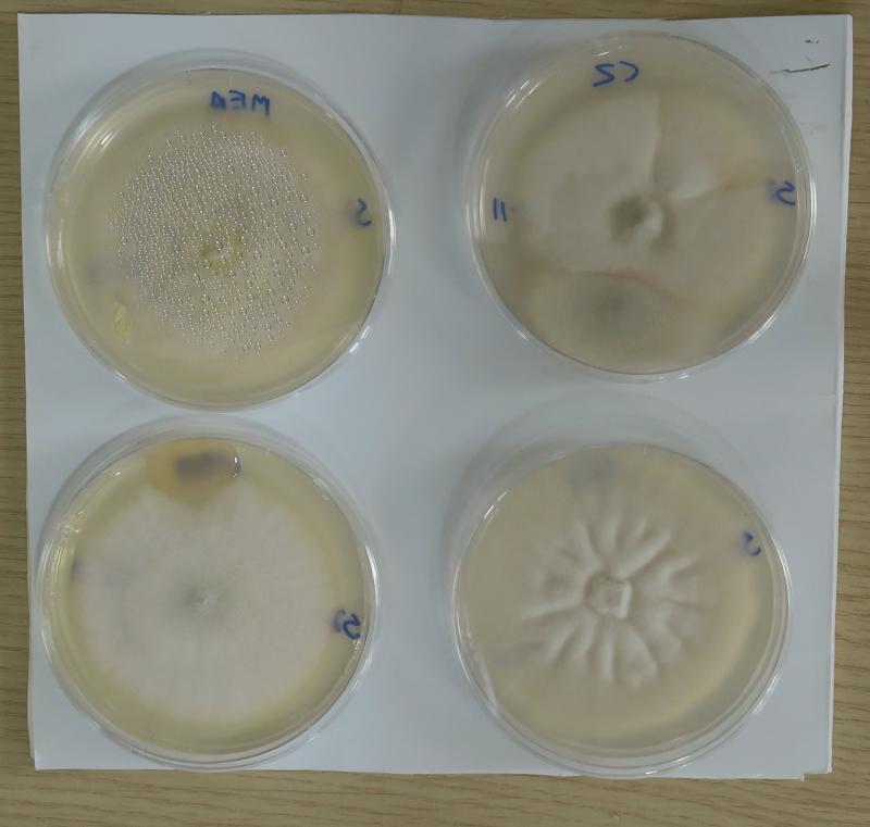
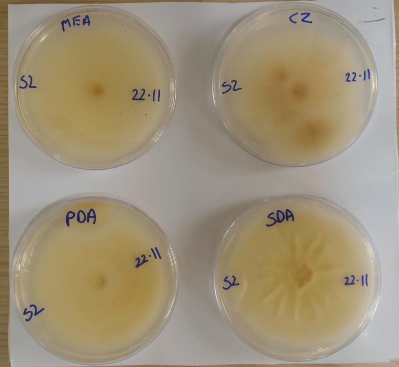
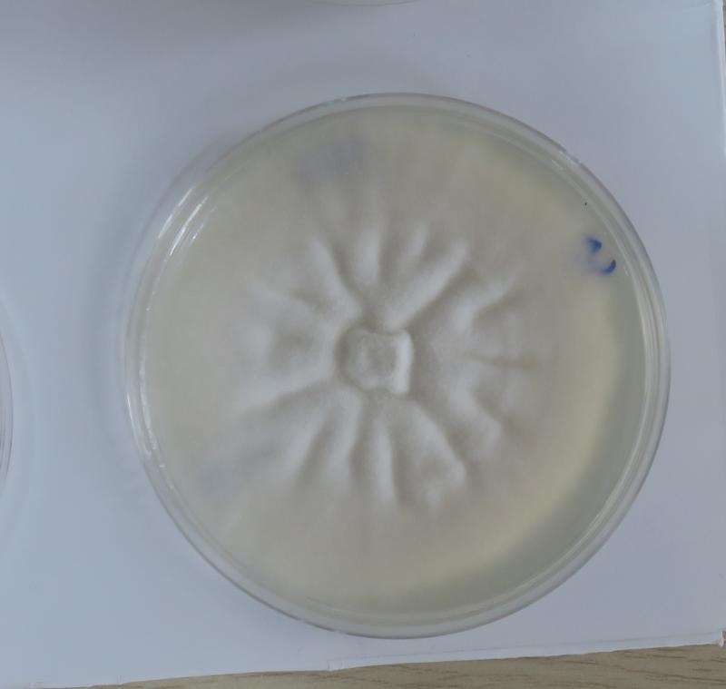
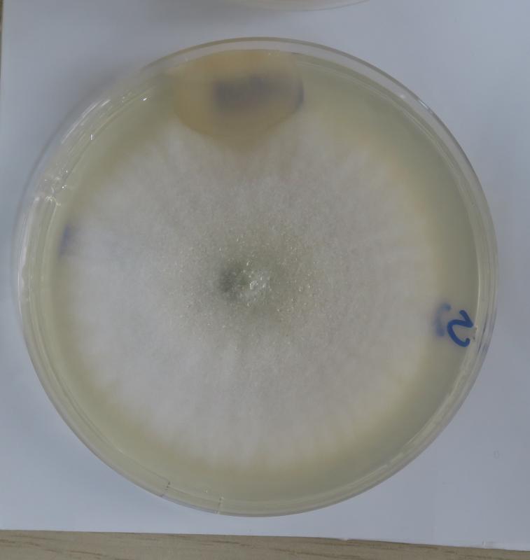
 I was trying to recultivate a species of Talaromyces growing on Washingtonia seeds fallen on the soil. The seeds and specimen was dried at room temp. Months later, I tried to recultivate the Talaromyces from the seed husks, kernels, etc and I got instead a range of interesting microfungi including three Aspergillus sp.
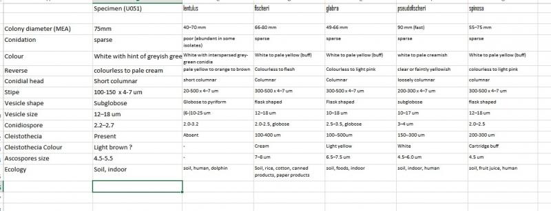
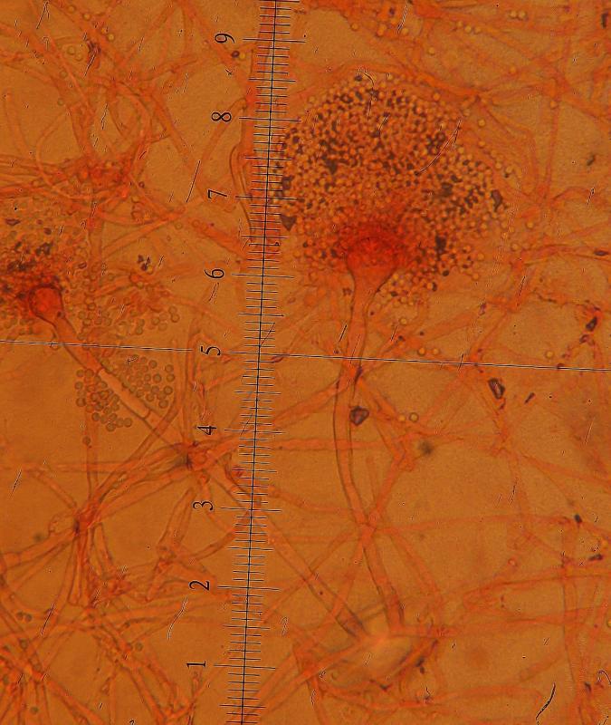
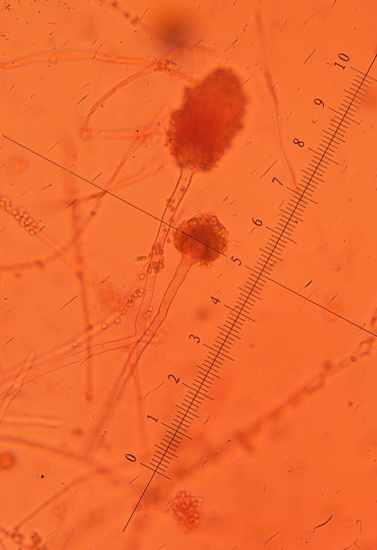
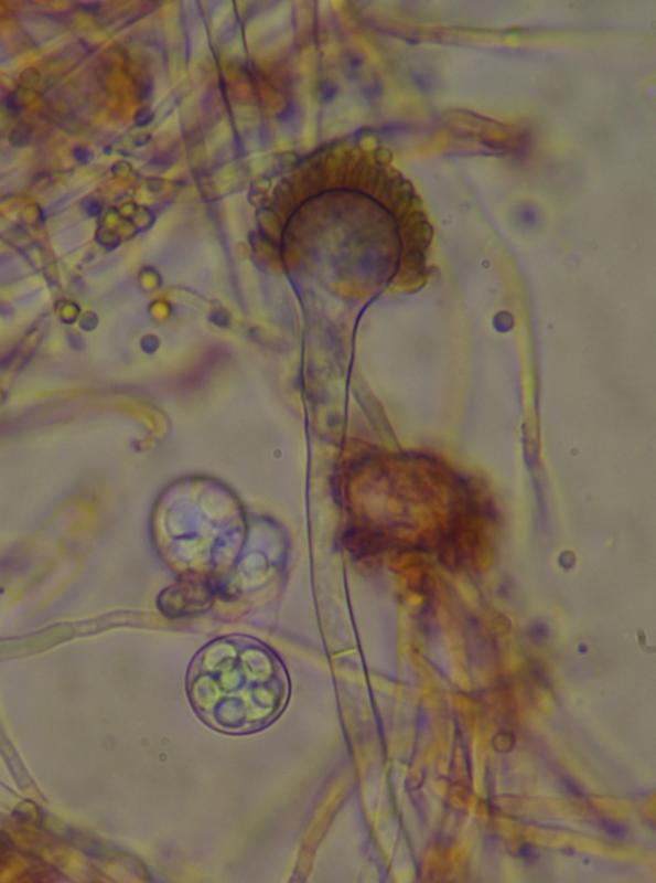
One of them formed white colonies, fast growing at 24C but on a closer look they had few grey-green conidiophores with shortly columnar conidial heads. The Vesicles were subglobular, 10-18um wide, with one series phialides about 6um long, emerging from about 2/3 of the vesicle (not fully radiating). Conidia in dense inticated chains forming 50-80um columnar or subglobose heads, each conidiospore 2.5um, sphereical, greyish-green. Conidiophores smooth, 60-120um long, 5-7um wide, somewhat expanding below the vesicle (but not always),
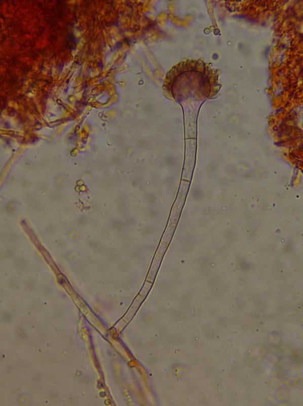
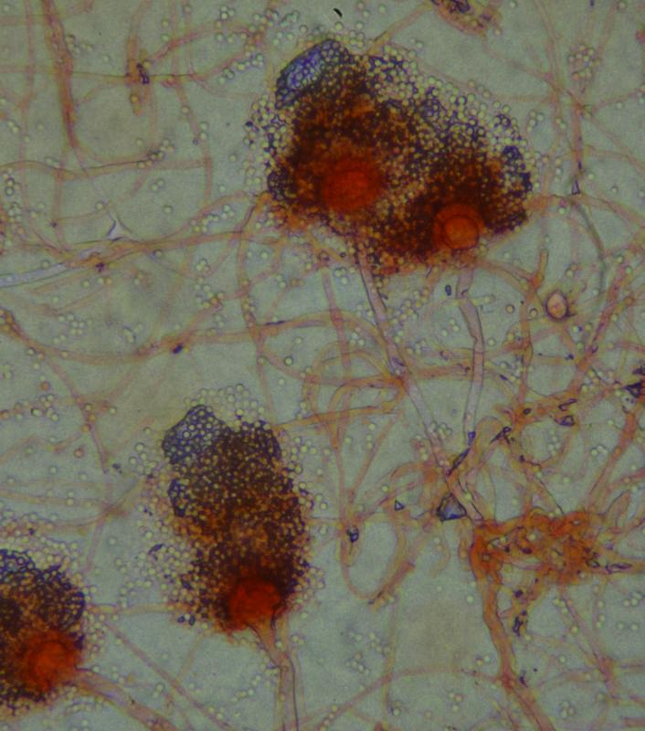
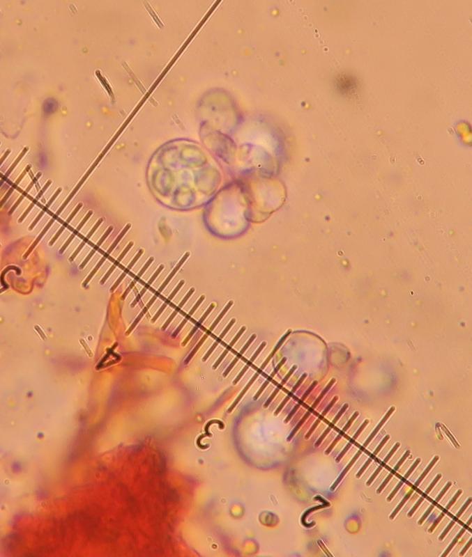
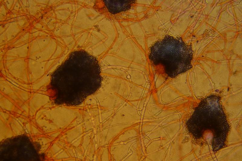
Also present where clesitotheca producing hundreds of globulae asci holding 8 ascospores. There where about 5-6um wide with their characteristical two radial wings and what I think to have seen under x1000 oil immersion, spines or warts on the dorsal/ventral side.
With the data available, I have thought the species is Aspergillus sect. fumigati, and despite not having an electron microcope to see the ornamentation of the ascospores, I think the warted surfaces and other characters collectively lead to the species pseudofisheri.
The colonies on OAT, PDA, CZ and MEA where overall white (much more green in A. fischeri)