
22-04-2026 20:17
 Marian Jagers
Is anyone familiar with the Hyphomycetes genus Pse
22-04-2026 20:54
 Enrique Rubio
Hi to everybody.This Pyrenopeziza grew in moist le
22-04-2026 01:06
 Richard VALERI
Bonjour à tous.Je vous présente cette Nectria s.
21-04-2026 13:36
Gernot FriebesHi,I am out of ideas for this one. I collected Sal
21-04-2026 13:19
Gernot FriebesHi,this Lophodermium on Typha has ascospores measu
21-04-2026 13:05
Gernot FriebesHi,this hyphomycete feels familiar but I was not a
20-04-2026 22:00
 Malcolm Greaves
These pale yellow, hairy ascos were growing on cul
Disc on fagus
Rasmus Riis-Hansen,
27-11-2019 16:54
 Hi,
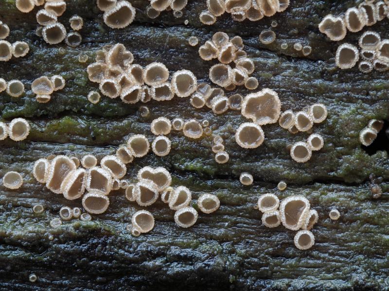
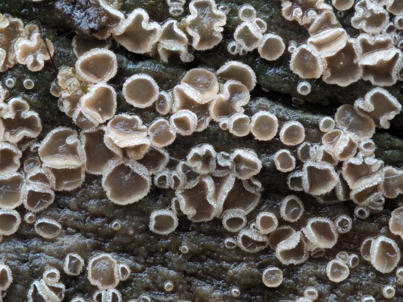
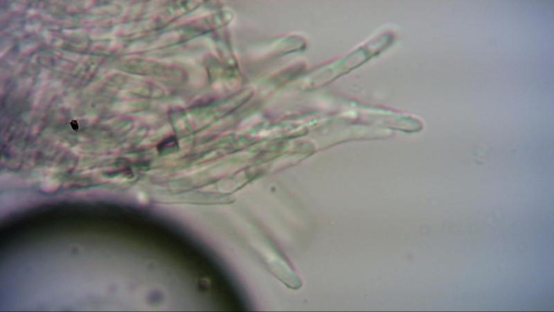
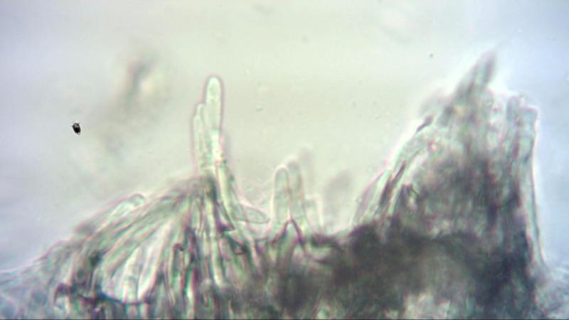
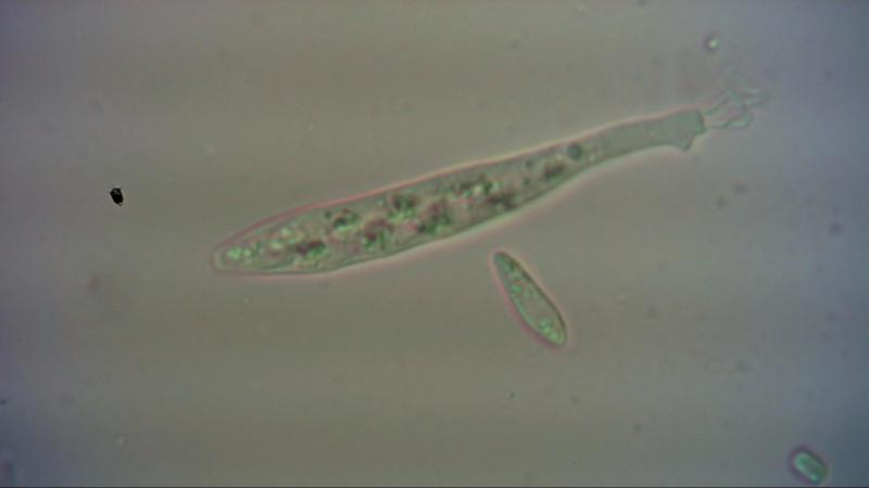
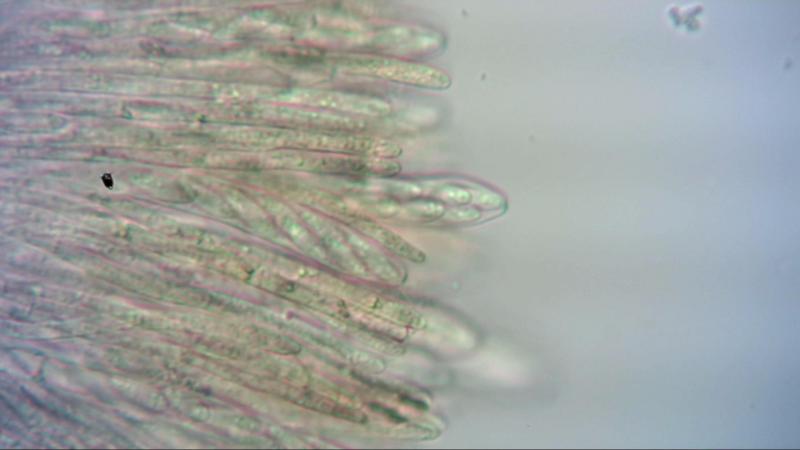
Hi,I hope you can help med with this disc found a few days ago on a medium decayed Fagus sylvatica log.
Ap. up to 1,5 mm. Hym light brown with a rosa hint. Several hundred fruitbodies.
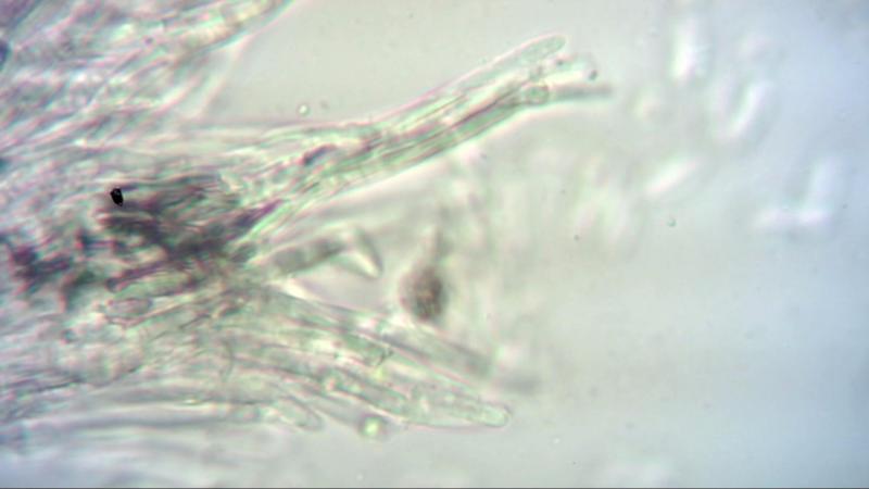
Spores with several small drops, 11-12,5 x 3 µm. Asci J+, 8-spored, 47-62 x 6,2-7,8 µm, with croziers (I think). Paraphyses cylindrical with septa, aprox. 3 µm wide, Hairs in bundles. Each hair is cylindrical and smooth, around 3 µm wide. Lengt of the hairs are around 50 µm.
/Rasmus
Quijada Luis,
27-11-2019 17:00
Re : Disc on fagus
It looks like the genus Cistella
Hans-Otto Baral,
27-11-2019 17:28

Re : Disc on fagus
Hmmm, the hairs look not really warted. What about Psilocistella quercina/Protounguicularia variepilosa? Spores perhaps too large?
Rasmus Riis-Hansen,
27-11-2019 19:52
Re : Disc on fagus
Thanks for the input Hans-Otto. As you point out the hairs are not warted - some of them have a few "spots" on them.
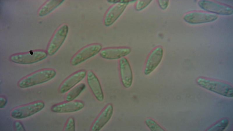
The spores are indeed to large. I measured them again to be sure. This time the range was 9-14 x 3 µm. I can see in your folder "Psilocistella quercina (cf. variepiosa)" sporemeasurement up to 10,8 µm long, but the 14 µm I have measured is still quite far from that. Otherwise ít fits nicely with the microscopical pictures and illustrations in the folder.
Psilocistella quercina/protounguicularia variepilosa is a good option, but I am not sure whether themuch larger spores is a deal breaker?
Hans-Otto Baral,
27-11-2019 20:47
Re : Disc on fagus
In my literature database P. variepilosa spores are 4-7 x 1.8-2, so very small (probably always dead).
P. quercina is 6-12.5 x 1.5-3, partly alive. Also in Quijada et al. 2014 who gave a detailed description and plates, P. quercina is *5.5-10 x 2-3.2. Spore size seems to vary rather strongly.
P. quercina is 6-12.5 x 1.5-3, partly alive. Also in Quijada et al. 2014 who gave a detailed description and plates, P. quercina is *5.5-10 x 2-3.2. Spore size seems to vary rather strongly.
Kosonen Timo,
28-11-2019 07:32

Re : Disc on fagus
Hi,
We have been working a bit with this particular "group" lately. I'd be really interested in the sample! To do sequencing and all...
How about monoseptata? That would "explain" the spores. The translucent brownish substance inside the hairs is characteristic to variepilosa/monoseptata. But maybe it exists in P. quercina as well?
bw
Timo
Hans-Otto Baral,
28-11-2019 09:28
Re : Disc on fagus
About spore size, do you have a scale to your spore photo? On my screen the spores measure 20-24 x 6-6.5(-7) mm, which might correspond to 10-12 x 3-3.3(3.5) µm.
Rasmus Riis-Hansen,
28-11-2019 19:40
Re : Disc on fagus
Timo you are very welcome to the material. I can dry it up and send it to you. Just send me an email with the info.
Rasmus Riis-Hansen,
28-11-2019 19:44
Re : Disc on fagus
Hans-Otto I do not have a scale for the spore photo, but I am sure about my measurements. I calibrated it not so long ago. I use a simple eye piece camera which goes in my trino on the microscope. I don't know the sensor size.
Hans-Otto Baral,
28-11-2019 20:17
Re : Disc on fagus
o.k, then the spores must probably be min. 3 µm wide.
Kosonen Timo,
28-11-2019 20:27
Re : Disc on fagus
all right. If it is not all moldy and wet, you could try sending a small part of the sample fresh. I'll try to make culture out of it. I'll send you the info.
Timo
Kosonen Timo,
05-12-2019 16:19
Re : Disc on fagus
I got the sample today. When studied in water there no resinous substance in- or outside the hairs. The excipulum is brownish, also in the less mature fruitbodies. It appeared more like even pigment rather than something outside the excipular cells. Ascus apex is blue in MLZ, otherwise no amyloid reactions whatsoever, no nodules or anything. The spores measure regularly 10-12 in length (free, in water) and there are septate spores inside the asci as well. I'll continue with it, but right now I don't have good suggestions. The material in Zottos folder "Psilocistella quercina - variepilosa" has atleast superficially similar material. But in my mind the P.variepilosa/monoseptata sensu Raitviir/Huhtinen are something else. It's not this anyway.
Timo
Kosonen Timo,
06-12-2019 10:41
Re : Disc on fagus
oh, one more. The excipulum (not the hairs, paraphyses or asci) appeared to take a lot of MLZ staining it brightly yellow. Right, things look yellow when in MLZ, but this appeared to be something else. Maybe it relates to the brownish excipulum. I'd be happy to hear of similar reaction in other groups. Or am I chasing an artefact here?
Timo
Hans-Otto Baral,
06-12-2019 10:46
Re : Disc on fagus
Hi Timo
you should present a photo of this reaction. I am not used to test MLZ, did the yellow appear intracellular or were the walls stained?
Zotto
Kosonen Timo,
06-12-2019 10:48
Re : Disc on fagus
The walls stained yellow. I'll come up with more photos later today.
T
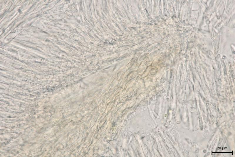
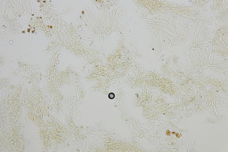
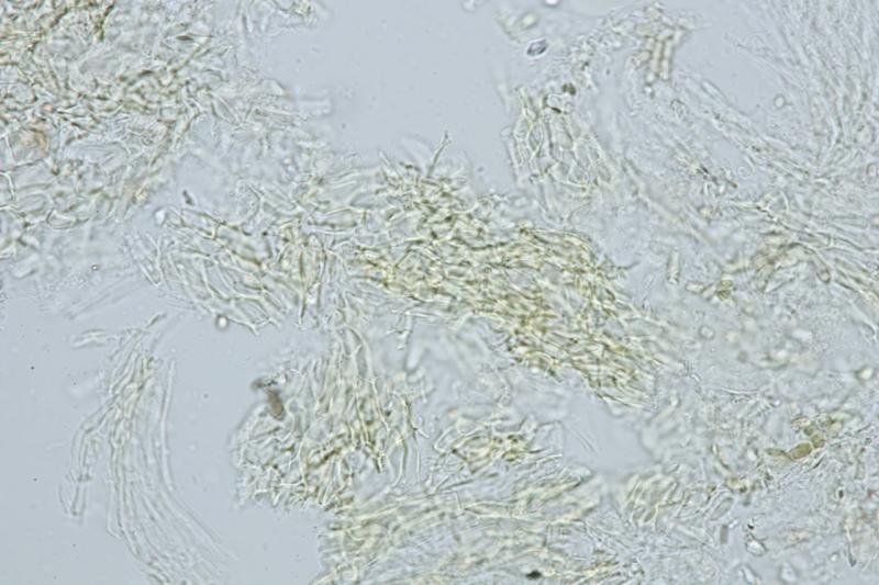
Kosonen Timo,
06-12-2019 16:39
Re : Disc on fagus
I attach three pics. My conclusion is that the reddish-brownish pigment is the substance that "turns" yellow. So, MLZ doesn't penetrate the cell wall, but the narrow space between cells goes yellow because the pigmented substance incorporates the MLZ somehow. But maybe not that big deal in the end.
Timo
Hans-Otto Baral,
06-12-2019 16:45
Re : Disc on fagus
I think also this is the natural colour of exudate between cells. But: MLZ surely penetrates the cell wall, as you can see when it stains glycogen in the cells, e.g. the spores.
Kosonen Timo,
09-03-2020 13:56
Re : Disc on fagus
Seems Luis had a hunch :-)
Got some sequence data now. It seems to belong to the "Cistella&Urceolella" clade (or Vandijckellaceae it should be called perhaps). It's a bit preliminary and also I don't have a clear overall picture of this clade, but it doesn't seem to be very closely related to any sequenced Urceolella or Cistella although member of this clade. So, not really suggesting any particular genus yet.
Timo