05-05-2026 22:40
Gernot FriebesHi,I believe this is a Plagiostoma growing on a Sa
04-05-2026 18:13
 Stephen Martin Mifsud
ID request for what seems to be a true aquatic fun
04-05-2026 16:39
Stephen Martin Mifsud
ID request: This specimen was collected in Malta o
28-07-2011 18:31
 Alex Akulov
Dear FriendsToday I made the pdf file of Velenovsk
28-04-2026 20:07
 Lothar Krieglsteiner
... on twig in the air at standing Ceratonia siliq
04-05-2026 09:50
 Castillo Joseba
Me mandan el material seco de Galicia,(España) re
02-05-2026 12:42
Alain BRISSARDBonjour à tousJeuidi 30 avril dernier on m'a remi
02-05-2026 13:06
Pauline. PennaBonjour Please can someone help me with this id
01-05-2026 22:45
 Thierry Blondelle
Bonjour à tous, Une récolte sur bouse séchée d
Schizothecium tetrasporum
Joop van der Lee,
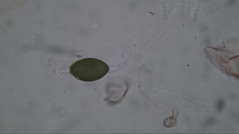
07-04-2019 10:18
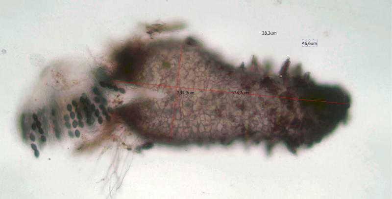
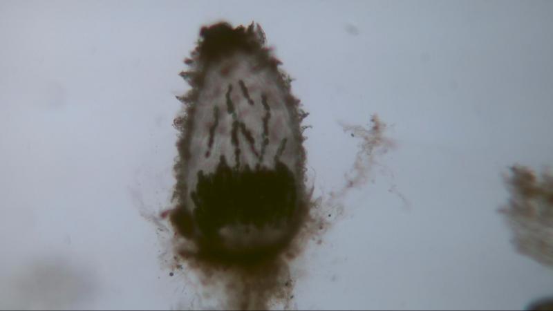
 Found on deer dung,
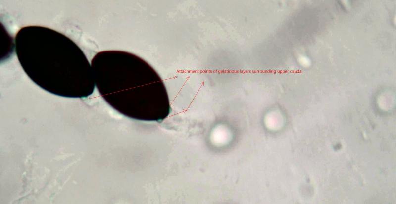
The fact that pedicel and upper cauda are covered with a gelatinous layer does not heve much attantion in documentation. In my opinion it is best described in "Coplrophilous fungi in New Zealand. I. Podospora species with swollen agglutinated perithecial hairs" Mycologia 87(3) 1995 pp. 375-396. Under Podospora tetraspora page 393.
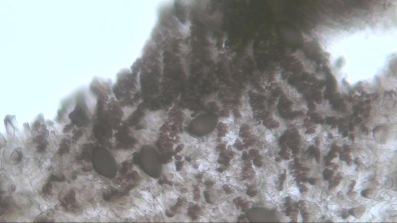
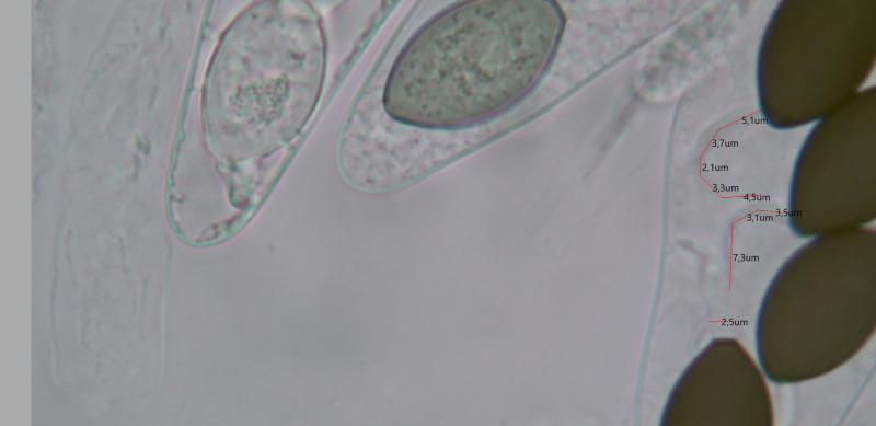
Perithecia: 574x237 um; neck and area just below the neck covered width short hairs; one third of the body covered with agglutinated hairs 38-46 um.
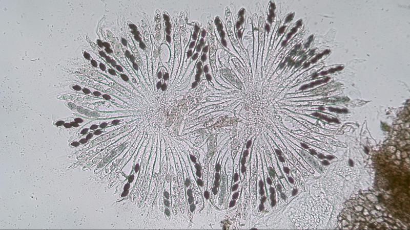
Asci: 81-spored; 196-204x22-24 um
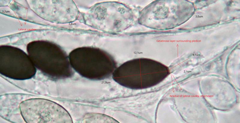
Spores: 21.8x12.1 um; pedicel 8.2-9.1x2.1-2.5 um, at least two lateral cauda at the base of the pedicel, pedicel covered with a gelatinous layer; upper cauda 13-15x1.1-1.6 um, cauda covered with gelatinous layer originating on both sides of the germ pore.
Residue of lateral caudea on base of the pedicel is visible by means of black or lighted spots.
The same is visible with the gelatinous layer around the upper cauda originating just beside the germ pore.
It is exeptional to see that the width of the pedicel is greater with immature spores than with mature spores. 3.2 um against 2.3 um.
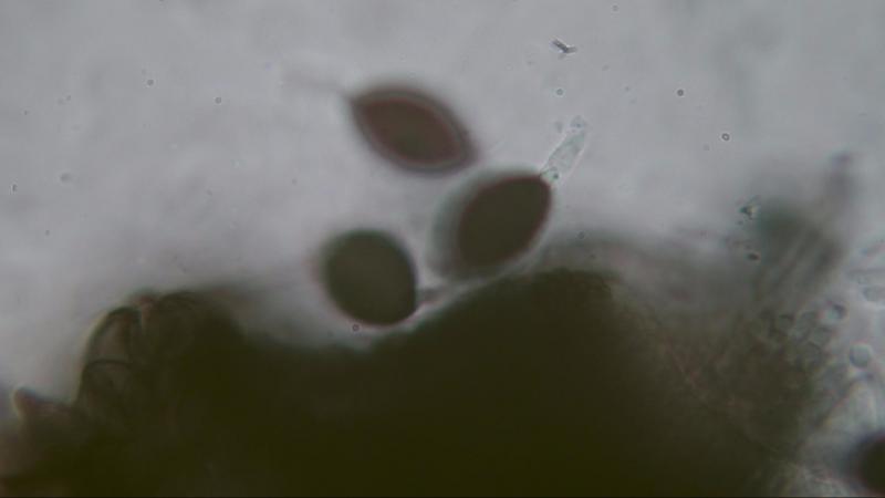
Photos 7-9 are from a S. tetrasporum with a smaller spore size 15.3-18x8.2-9.2 um and found on rabbit dung.
Perithecia: 398x215 um.
Photo 8 shows the gelatinous layer around the pedicel.
Photo 9 shows the gelatinous layer around the upper cauda originating on both sides of the germ pore.
Joop