
21-04-2026 13:36
Gernot FriebesHi,I am out of ideas for this one. I collected Sal
21-04-2026 13:19
Gernot FriebesHi,this Lophodermium on Typha has ascospores measu
21-04-2026 13:05
Gernot FriebesHi,this hyphomycete feels familiar but I was not a
20-04-2026 22:00
 Malcolm Greaves
These pale yellow, hairy ascos were growing on cul
19-04-2026 21:23
Steve ClementsBonjour, I found this anamorphic fungus on old pl
19-04-2026 20:46
Steve Clements1 mm diameter approx spherical conidiophores on pl
12-04-2026 17:56
 Hardware Tony
Found on dead stems in February earlier this year
17-04-2026 19:16
 Enrique Rubio
Hi to everybodyI would appreciate any assistance r
14-04-2026 05:32
Ethan CrensonHi all, A few weeks back a friend pointed out som
17-04-2026 15:14
 Bruno Coué
Bonjour.Récoltes du 16/04/2026, sur feuilles mort
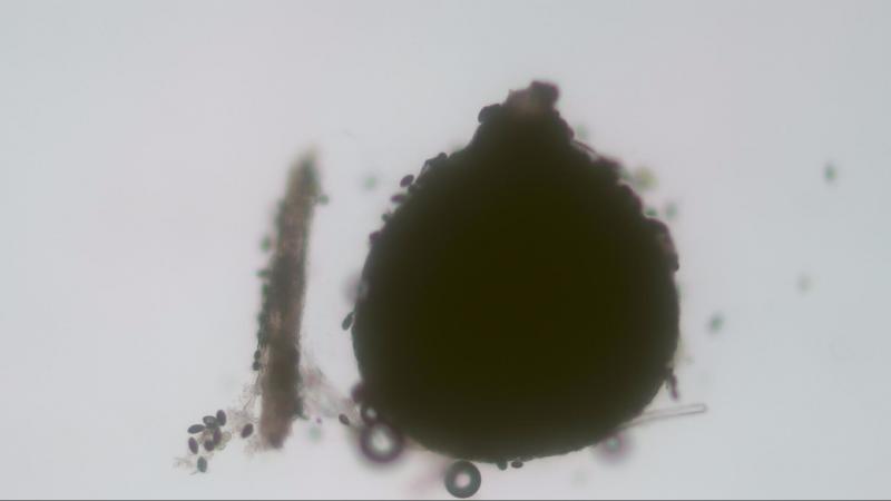
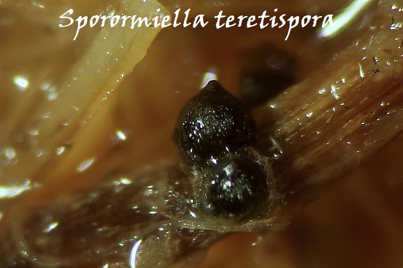
Sporormiella teretispora
Joop van der Lee,
28-03-2019 19:12
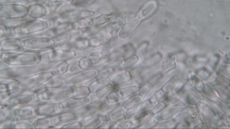
 Found on horse dung.
Perithecia: 370x343 um (Perithecia was covered with spores form Conichaeta)
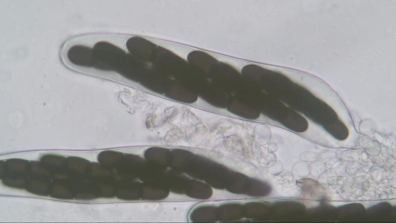
Asci: 8-spores; 195.6-196.8x33-34.7 um
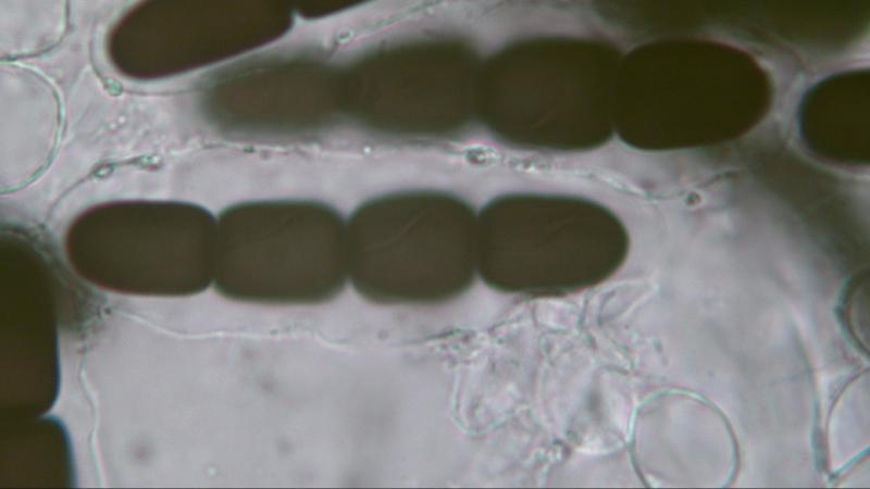
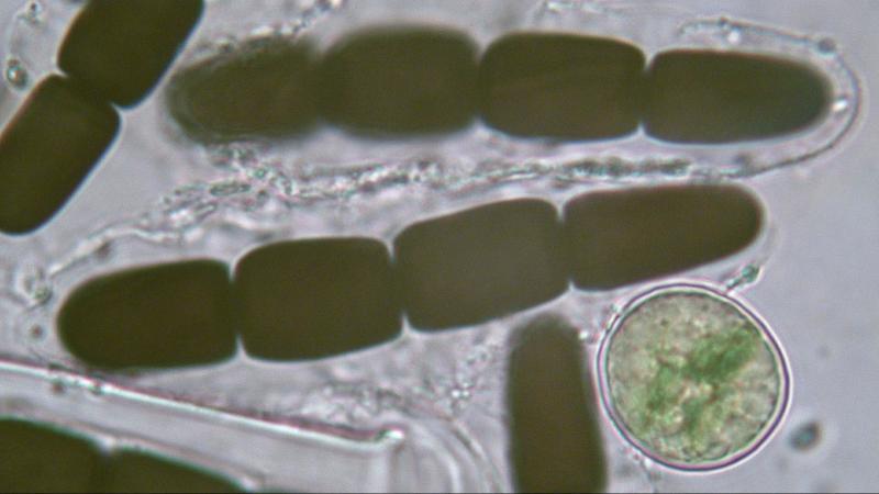
Spores: biseriate; 59.75-74.1x10.35-12.85; germ slit sligthly oblique.
Paraphyses: septated; 3.45-5.2 um
Yulia Lytvynenko,
29-03-2019 06:10

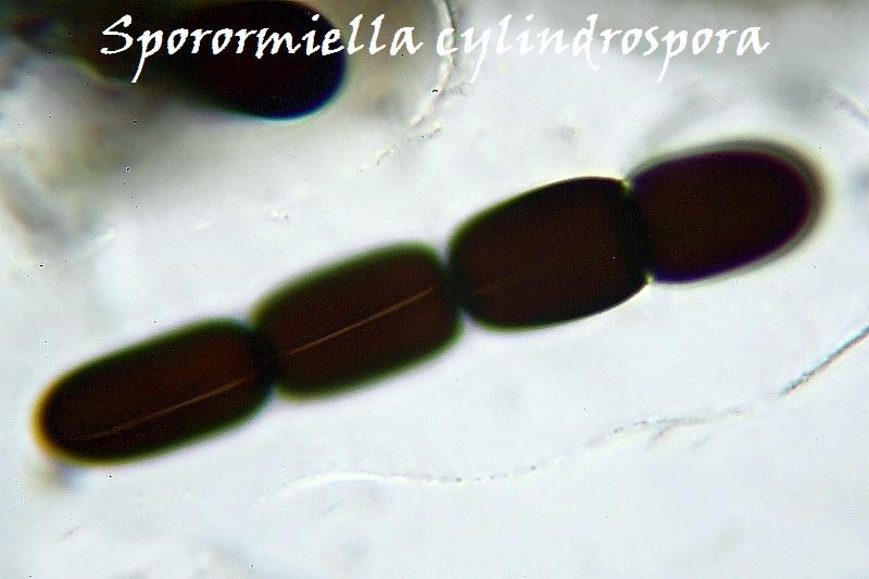
Re : Sporormiella cylindrospora?
Dear Joop!
Sporormiella cylindrospora has spores larger in size ((67)70-80x13-16). And why, for example, is not S. teretispora (ascospores 60-66x10-13)? What confuses you?
Sporormiella cylindrospora has spores larger in size ((67)70-80x13-16). And why, for example, is not S. teretispora (ascospores 60-66x10-13)? What confuses you?
Michel Delpont,
29-03-2019 07:11

Re : Sporormiella cylindrospora?
I think it is indeed S. teretispora; S. cylindrospora spores have different germinal slits.
Michel.
Peter Püwert,
29-03-2019 12:15

Re : Sporormiella cylindrospora?
Hi all,
here two pictures and two links about Sp. teretispora and cylindrospora.
Greetings Peter.
Joop van der Lee,
29-03-2019 14:17
Re : Sporormiella cylindrospora?
Vielen dank Peter.
Joop
Joop
Yulia Lytvynenko,
30-03-2019 09:09
Re : Sporormiella cylindrospora?
Dear Joop.
Regarding the size of the spores of Sporormiella teretispora.
According to different authors, it is quite different. 60-66 X 10-13 (Ahmed&Cain), 55-67 X 11-14 (Bell), (61.7-) 63.6-68.4 (-69.3) x 01.4-) 11.8-12.3 (-12.8) (Doveri). Mungai et al. give size 66–73.5 × 11–15.5 (as. S. aff. teretispora). Last size is similar to your measurements.
The morphology of the germ slits is also given different.
Ahmed&Cain: germ slit nearly parallel, in some collections the germ slit is curved at each end;
Doveri: germ slit an imperceptibly oblique to almost parallel, often flexuous; and besides, indicates that germ slits almost parallel. rather than oblique, germ slits.
Mungai et al.: germ slit diagonal, sigmoid to oblique.
In this regard, I would like to ask a question both to you and all my colleagues in this forum.
What do you think, is the morphology of germ slits in some species of Sporormiella a reliable diagnostic sign? How important should it be considered in the identification Sporormiella species?
Joop van der Lee,
01-04-2019 20:07
Re : Sporormiella cylindrospora?
Check your mail Yulia.
Joop
Joop