
05-05-2026 22:40
Gernot FriebesHi,I believe this is a Plagiostoma growing on a Sa
04-05-2026 18:13
 Stephen Martin Mifsud
ID request for what seems to be a true aquatic fun
04-05-2026 16:39
Stephen Martin Mifsud
ID request: This specimen was collected in Malta o
28-07-2011 18:31
 Alex Akulov
Dear FriendsToday I made the pdf file of Velenovsk
28-04-2026 20:07
 Lothar Krieglsteiner
... on twig in the air at standing Ceratonia siliq
04-05-2026 09:50
 Castillo Joseba
Me mandan el material seco de Galicia,(España) re
02-05-2026 12:42
Alain BRISSARDBonjour à tousJeuidi 30 avril dernier on m'a remi
02-05-2026 13:06
Pauline. PennaBonjour Please can someone help me with this id
01-05-2026 22:45
 Thierry Blondelle
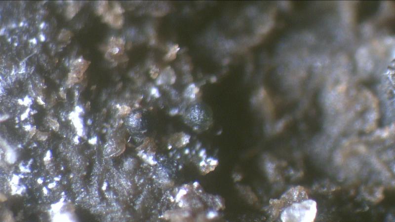
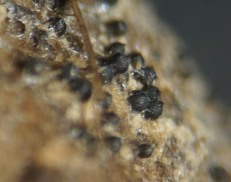
Bonjour à tous, Une récolte sur bouse séchée d
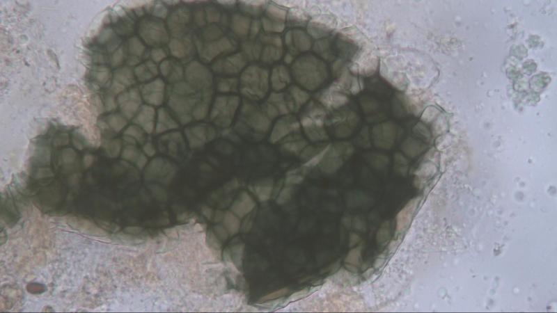
 Found on cow dung, most of the time in the visinity of Schizothecium species.
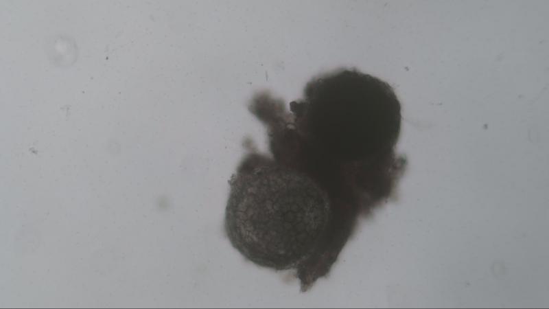
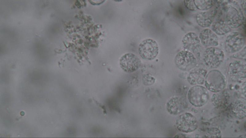
Fruitbody: Round 170,2-173 um in diameter, surrounded by a gelatinous layer approx. 60 um thick, dark green in colour.
Spores: Round and/or pointed 6.8-7.3 um covered with round warts 2.2-2.5 um.
The first time spores were measured when in water but missing the warts so the second time measurement was performed in Melzer.


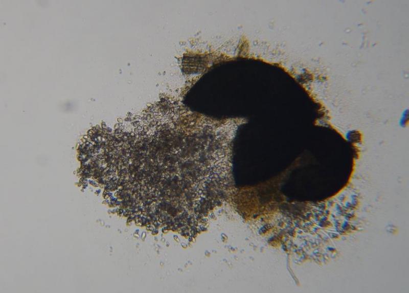
That is a very interesting fungus. Do the fruiting bodies have an opening of some kind? The round asci look like the kind you find in a cleistothecium but your pictures suggest that maybe there is an ostiole. Also, do the asci contain 8 ascospores or are there more than that?
Regards,
David

What you show on all your photos are in my opinion asci and it is very difficult to see the spores alone. Are there any hairs? Afraid to be could you look for the genera Lophotrichus, Kernia. There is also the genus Orbicula but in the latter the asci are cylindrical.
Michel.

Can you provide me the following article.
Pithoascus nidicola (Massee & E.S. Salmon) Arx, Proceedings van de Koninklijke Nederlandse Akademie van Wetenschappen Section C 76 (3): 292 (1973) [MB#320551]
Regards,
Joop

Hello Sven,
I will try to get the article in the library of Naturalis when I can find the time to do so.
If it is succesful I will send you a copy.
Joop
Joop
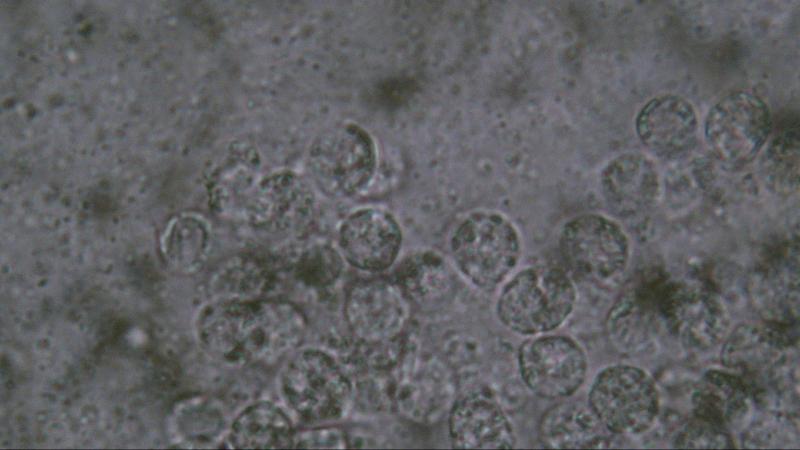
In my opinion it is a single ascus species like Thelebolus stercoreus containing hundreds of spores.
I did not find any ostiole but maybe that is possible when these species are ripe.
It is typical that these species were found together with Schizothecium conicum species.
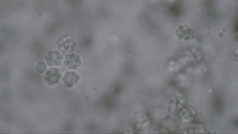
When putting pressure on the cover glass the species bursts open (photo #4).
Joop
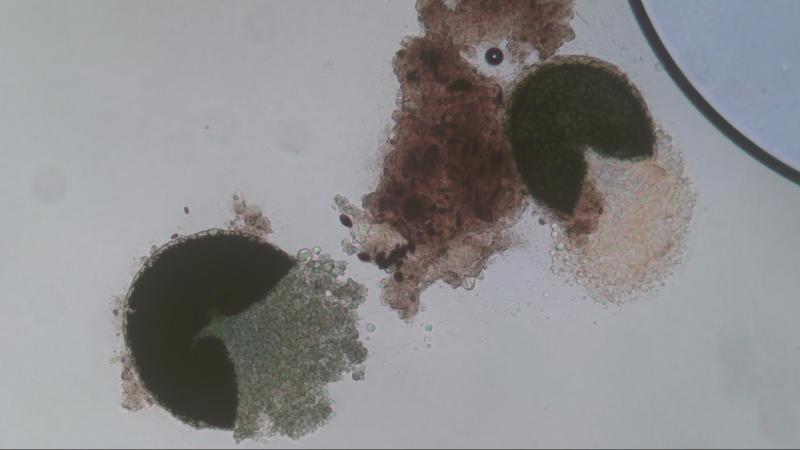
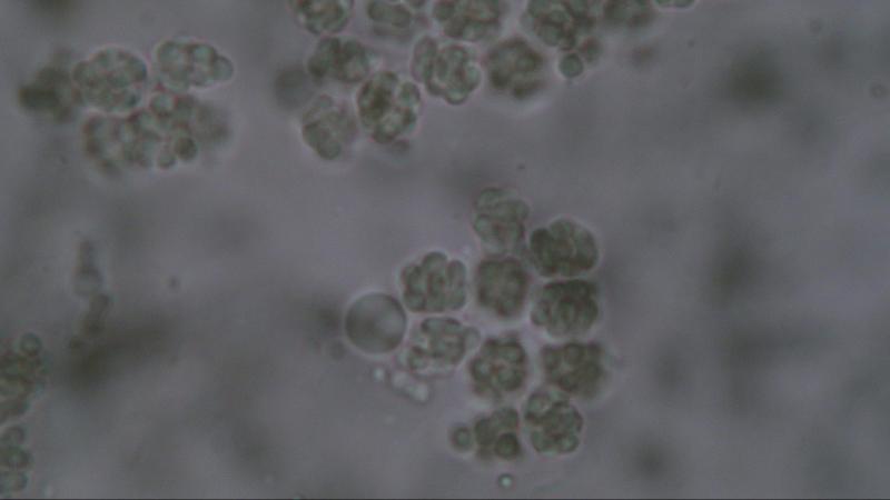
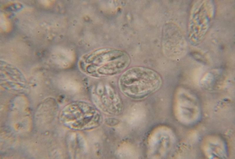
I collected some more information about this species and it will present different shapes of spores when using different fluids.
Photo #1 when using water.
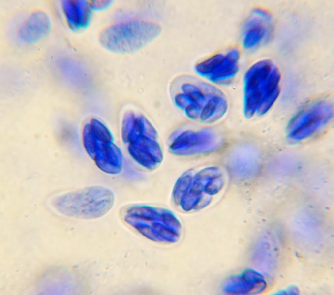
Photo #2 when using Melzer
Photo #3&4 when using Congo Red.
It seems to me that the presentation on #2 show the spores as seen in #3&4 clinging to each other. Whereby at first I thought that they were warts.
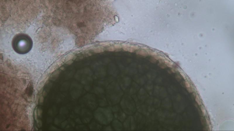
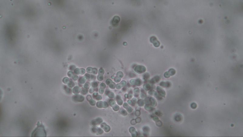
The spores consist of 2 cells, each cell measures 2.45 um in diameter, the total of 2 cells combined is 4.9 um.
Each cell is filled with a "the bary bubble".
When measuring the cells as presented in #2 (or other photos I made) the result will be the same as in #3&4 namely 2.45 um.
Greetings,
Joop
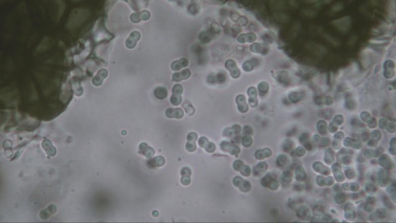
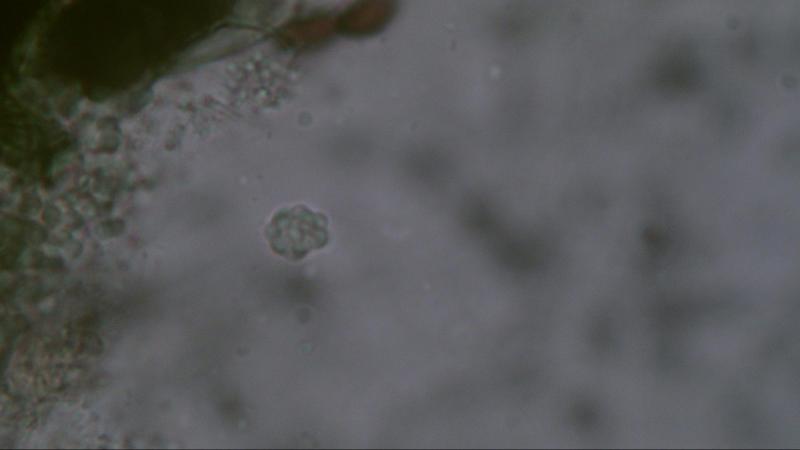
I believe this fungus is Mycoarachis inversa, a species characterized by two-celled peanut-shaped spores and a cleistothecial peridium with the hyaline layers on the outside (hence "inversa").
David
Joop
