
06-05-2026 11:25
 Castillo Joseba
Me mandan el material seco de Galicia (España) re
28-04-2026 20:07
 Lothar Krieglsteiner
... on twig in the air at standing Ceratonia siliq
05-05-2026 22:40
Gernot FriebesHi,I believe this is a Plagiostoma growing on a Sa
04-05-2026 18:13
 Stephen Martin Mifsud
ID request for what seems to be a true aquatic fun
04-05-2026 16:39
Stephen Martin Mifsud
ID request: This specimen was collected in Malta o
28-07-2011 18:31
 Alex Akulov
Dear FriendsToday I made the pdf file of Velenovsk
04-05-2026 09:50
Castillo Joseba
Me mandan el material seco de Galicia,(España) re
02-05-2026 12:42
Alain BRISSARDBonjour à tousJeuidi 30 avril dernier on m'a remi
02-05-2026 13:06
Pauline. PennaBonjour Please can someone help me with this id
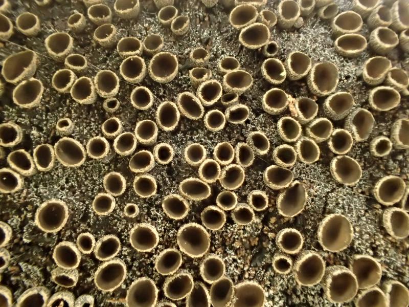
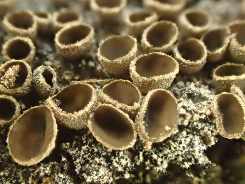
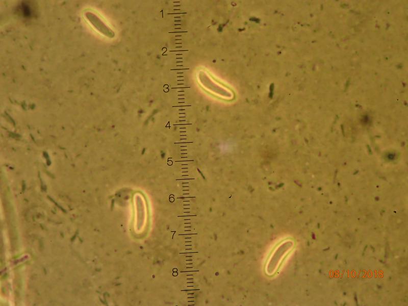
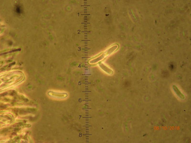
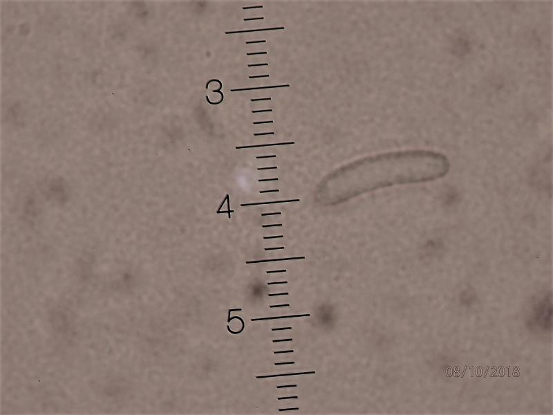
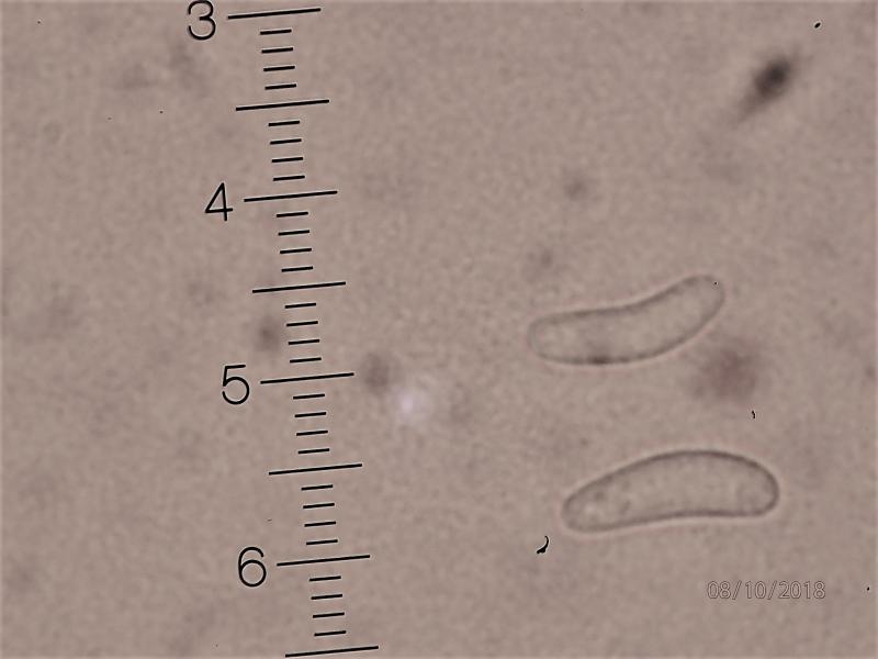
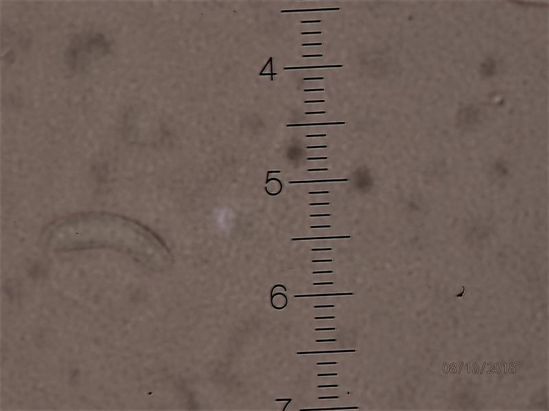
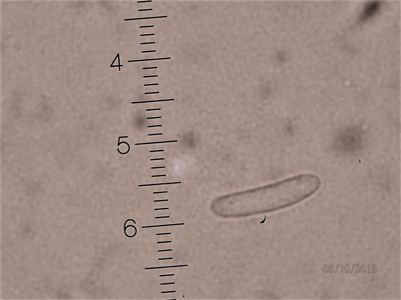
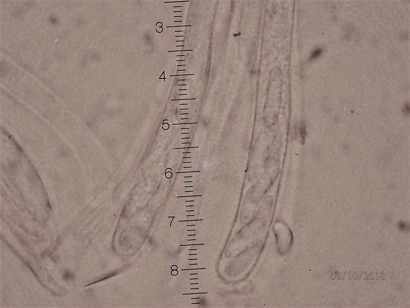
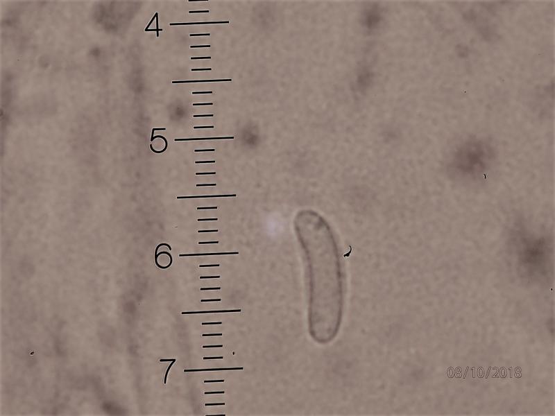
 I came across these dense mats of apothecia seated on a dark, but whitish hoary subiculum, growing on the bark of a huge, fallen deciduous tree (most likely Salix caprea or Popolus tremulae), in a nature reserve i Southern Norway. I guess this is an Encoelia, but it doesn't seem fit any of the species known from Norway. It comes closest to E. pruinosa. However, the apothecia are only 1-2 mm in diam. and the paraphyses ar not noticeably widened at the tips. The spores are curved, 11x3 microns, apprarently with some sort of thickening or body at each end, becoming evident during focussing. The attached micrographs are taken at 1000x oil immersion, phase contrast, in water. I have not yet used other media. Does anyone know this species?
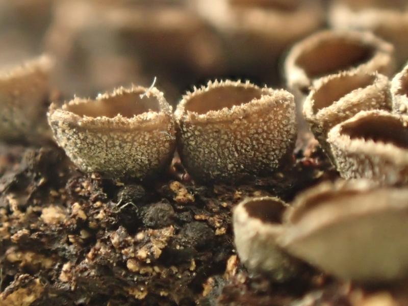
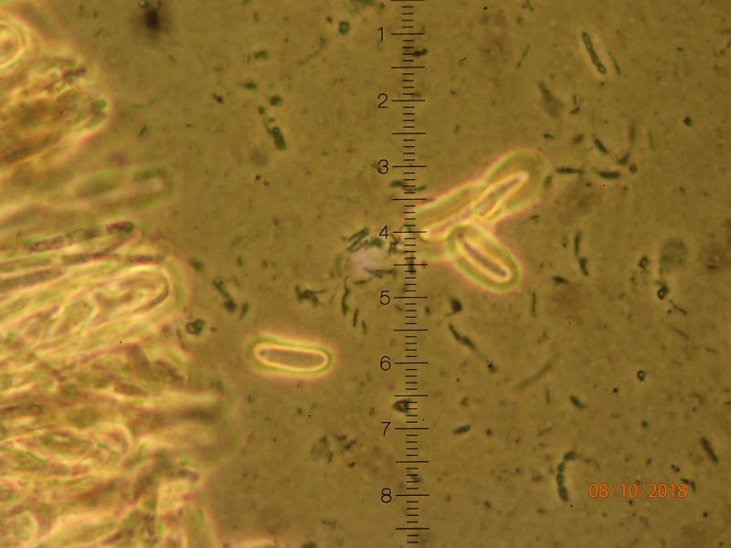
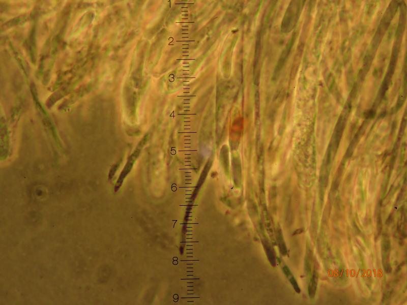

Indeed, Encoelia pruinosa )or now Sclerencoelia pruinosa) would also be my first option.But it should occur on living trunks. There should be lots of crystals outside, which is probable because of the pruinose surface.
Spore size could be larger, but apo size seems to be variable.
To clarify whether it is Salix or Populus needs to look at a radial or tangential section for homo/heterocellular radial rays.
Zotto
Hello Edvin,
very interesting fungus.
The photos are with phase contrast as you write. Why? I do not use it at all and would much prefer to see "normal" photos. I think I am not alonè with this (?).
Best regards, Lothar
There is enough contrast when mounting in water, especially when cells are alive. Look recent contribution by Elisabeth.
What you mean with polar thickenings of the spores, I assume you mean the light polar region seen especially in one spore. I am unable to say in photos with phasecontrast, but I could see clearer without that device, maybe polar oil drops.
I don't understand the grainy background but otherwise the photos are splendid.
So from the scale I get 11.5-13.5 x 2.6-3.3 µm which would fit well with other collections.
Would it be possible to share some apothecia for research?
This found is very interesting for me because from Europe I have seen only specimens collected on 19 century in Norway. It is important to undestand if Sclerencoelia pruinosa in Northern America we have studied with molecular methods, and current yours European specimens turn into the same species. In N-Am pruinosa is parasitic and it is possible that it is introduced from Europe where it does not destroy its native host. I would like to see, if there is stromatic tissue under apothecia - it was clearly developed in historical specimens and deviating character comparing with Sclerencoelia fascicularis.
Please, could you send a piece of your specimen to:
Kadri Pärtel
Chair of Mycology
Department of Botany
Institute of Ecology and Earth Sciences
University of Tartu
Ravila 14a
50411 Tartu
Estonia
kadri.partel@ut.ee
With best wishes from Estonia and thanking in advance,
Kadri