
12-04-2026 17:56
 Hardware Tony
Found on dead stems in February earlier this year
17-04-2026 19:16
 Enrique Rubio
Hi to everybodyI would appreciate any assistance r
14-04-2026 05:32
Ethan CrensonHi all, A few weeks back a friend pointed out som
17-04-2026 15:14
 Bruno Coué
Bonjour.Récoltes du 16/04/2026, sur feuilles mort
12-04-2026 15:52
Gernot FriebesHi,I'm looking for help with this anamorph collect
14-04-2026 21:52
Gernot FriebesHi,found on dead leaves of Carex elata. Conidia: 4
16-04-2026 22:09
Buckwheat PeteHello, I'd like to ask about this older specimen:
15-04-2026 19:33
Fátima Durán ManzanequeHi!! I need help, I found this Ascomycete but I d
14-04-2026 20:31
Gernot FriebesHi,can this be Psilachnum lateritioalbum on Phragm
12-04-2026 12:22
 William Slosse
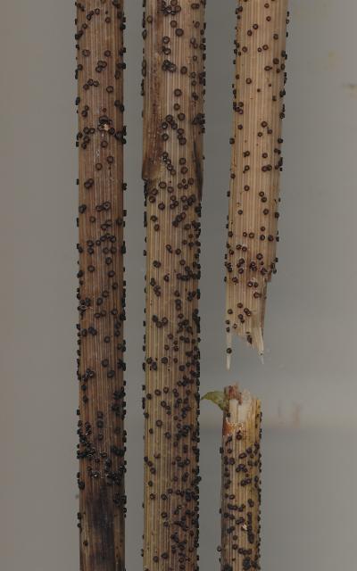
In a dune grassland in Oostduinkerke (Belgium), on
 Hello,
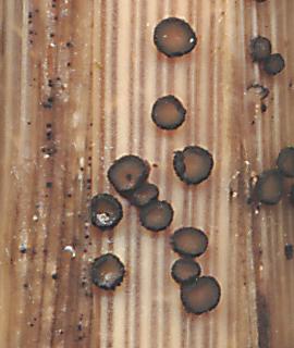
This collection appears to be the same species as one presented by Chris Yeates five years ago (http://www.ascofrance.fr/search_forum/24157) and tentatively identified as Micropeziza karstenii. Mine was growing on dead culms of Phalaris arundinacea completely submerged in water. Many of the ascomata were immature, so perhaps the ascospores would not be discharged until the water level went down and the ascomata were exposed to the air.
I have not yet been able to find any records of this species from North America and would like to be more certain of the identification before adding the record to our biological survey. Any comments would be appreciated.
Dave
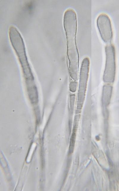
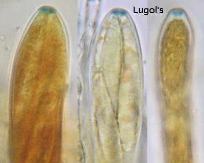

this requires a living turgescent ascus in order to see if the spores are septate already inside the living asci. Judging from the appearance of the spores I suspect so, which would mean that it can hardly be a Micropeziza. Members of that genus I only know as having at maturity non-septate spores, i.e., spores are ejected when non-septate. Moreover, M. karstenii/poae/cornea has a high lipid content, unlike yours.
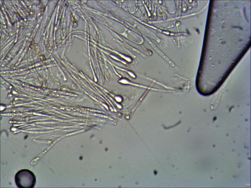
In Micropeziza the paraphyses are apically often swollen, and the VBs inside are more or less restricted to this inflated part. In your species the VBs are very elongate and reach far down. The spores are also distinctly larger than in that species.
What genus this belongs I cannot tell. Niptera could be a better alternative. You should test KOH whether it causes a yellow reaction at the moment when it comes in contact with the VBs. Niptera often shows this reaction.
Zotto
Thank you for your valued observations. I was afraid the identification was too easy! The fungus is very abundant in it's habitat so I will work further on getting a name for it.
Dave

david, did you restudy the specimen, particularly regarding a possible KOH-reaction?
Zotto
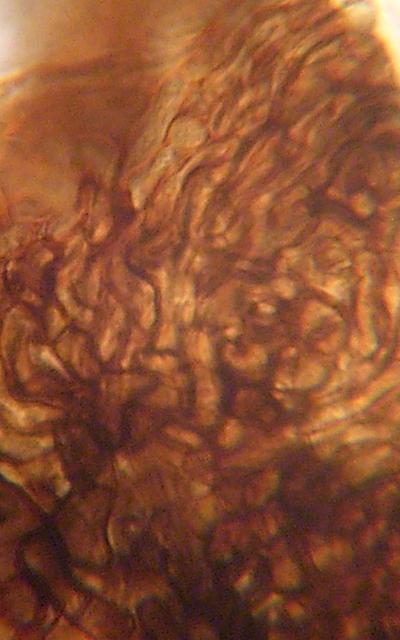
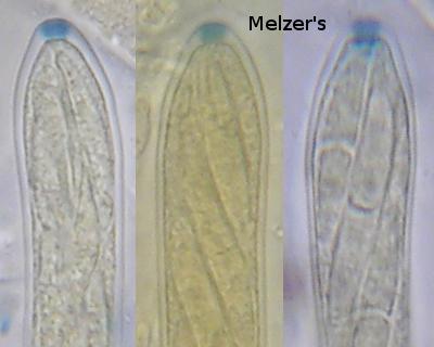
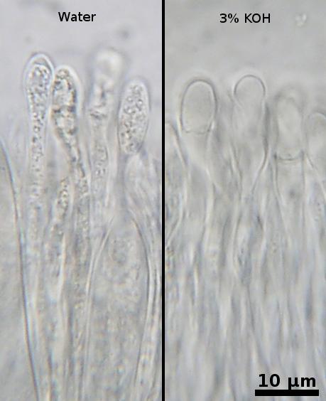
I tried applying 3% KOH to a water mount but did not see any fleeting yellow colour. The specimen is now dried and the paraphyses contain only a finely granular content, quite different from the fresh condition. The KOH instantly clears the cell, but without any colour changes.
The fungus is interesting enough that I am planning to revisit the locality and collect more, hopefully in a more mature condition. Unfortunately this requires a 2-hour car trip from home followed by two more hours in a canoe. Only another mycologist could appreciate such an obsession!
Dave
The photos aren't very good, but do show that amount of swelling. There is some obscure yellow colour in the KOH mount, but I don't think this is the reaction your are describing Zotto.
If the newly collected material is more mature perhaps we can get a culture and some useful sequences from it.
Dave
Another difference is the high lipid content in excelsior vs. a very low content in David's fungus.
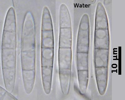
When you go to Dennis 1978 the spores of N. excelsior measure 50-80 x 3-4 µm.
From David's scale I measure 28-33 x 4.7-5.2 µm. David, did you also measure the spores?
Species Fungorum does not accept Niptera but Belonium, an antiquated opinion. This index is not at all up to date in every case, although it is continuously improved, as far as I know by one person concerning all fungi (!).
An unpublished ITS sequence of M. excelsior exists which yields Mollisia minutella as closest match (94%). But I must admit that no true Niptera (s.str.) sequence exists in GenBank.
Zotto
By the way, your photos are splendid!!