
05-05-2026 22:40
Gernot FriebesHi,I believe this is a Plagiostoma growing on a Sa
04-05-2026 18:13
 Stephen Martin Mifsud
ID request for what seems to be a true aquatic fun
04-05-2026 16:39
Stephen Martin Mifsud
ID request: This specimen was collected in Malta o
28-07-2011 18:31
 Alex Akulov
Dear FriendsToday I made the pdf file of Velenovsk
28-04-2026 20:07
 Lothar Krieglsteiner
... on twig in the air at standing Ceratonia siliq
04-05-2026 09:50
 Castillo Joseba
Me mandan el material seco de Galicia,(España) re
02-05-2026 12:42
Alain BRISSARDBonjour à tousJeuidi 30 avril dernier on m'a remi
02-05-2026 13:06
Pauline. PennaBonjour Please can someone help me with this id
01-05-2026 22:45
 Thierry Blondelle
Bonjour à tous, Une récolte sur bouse séchée d
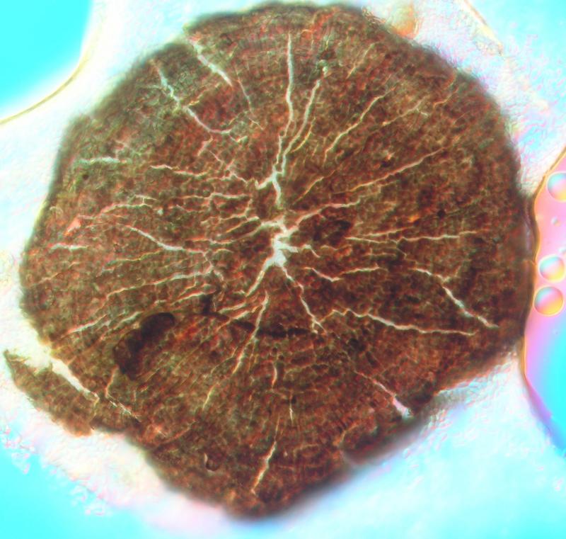
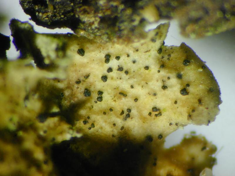
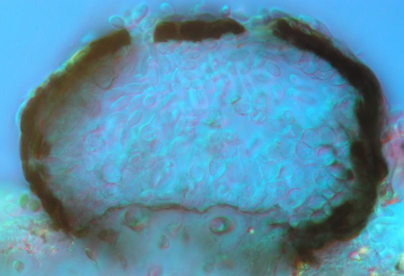
 Hello,
I found a strange (to me) coelomycete growing as well on fallen needles of Picea abies as on Cladonia digitata growing at the foot of the tree.
Description: Conidiomata solitary, sessile, on the squamules and podetia of Cladonia digitata, cushion-like to suborbicular, black, 100–150 µm in diam., opening by radially splitting of the wall (habitually like the peristome of bryophytes); wall brown, composed of one layer of pseudoparenchymatous cells, 1–2 µm thick, cells in surface view more or less in radial lines, rectangular, c. 3–4 × 4–4.5 µm; pycnidial cavity first filled with thin-walled, hyaline, isodiametric cells, 3–4 µm diam., later these cells are dissollving, some of them turning to broadly ampulliform conidiogenous cells, 4–6 × 3.5–5 µm; conidia non-septate, ellipsoid, hyaline, both ends rounded, with one minute guttule near each end, (3.5–)4.1–5.3(–6.0) × (2.0–)2.1–2.7(–3.0) µm, l/b = (1.3–)1.7–2.3(–2.8) (n = 20).
Strange to me is the splitting of the pycnidial wall and the conidiogenous cells developing inside the lumen and not on the inner side of the wall. Fist idea was Rhizospaera, but this has other pycnidia. Actinothyrium has fitting pycnidia, but here the conidia are filiform. Can anyone help?

Hi Wolfgang,
Strange to find these thyriothecia on needdles as on Cladonia. On needles grow Microthyriaceae. On Cladonia we can observe some Lichenopeltella. ¨Pehaps you have the asexual morph of one of them, and the best thing to do is probably to wait for the sexual morph.
Alain
I examined it carefully, it is really the same taxon on the needles as on Cladonia. Microthyriaceae is a good hint, but I have no idea how asexual state of this family look like. Lichenopeltella I think I can exclude because of the missing basal plate and the thickenings around the ostiole.
Wolfgang