23-02-2026 11:22
Thomas Læssøehttps://svampe.databasen.org/observations/10584971

29-11-2024 21:47
Yanick BOULANGERBonjourJ'avais un deuxième échantillon moins mat

07-02-2023 22:28
Ethan CrensonHello friends, On Sunday, in the southern part of

19-02-2026 17:49
Salvador Emilio JoseHola buenas tardes!! Necesito ayuda para la ident

19-02-2026 13:50
Margot en Geert VullingsWe found this collection on deciduous wood on 7-2-

16-02-2026 21:25
 Andreas Millinger
Good evening,failed to find an idea for this fungu
Mould which easily break when mounted in water or Stains
Stephen Martin Mifsud,
02-12-2017 08:36
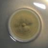
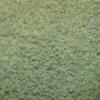
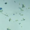
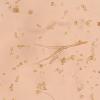
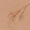
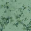
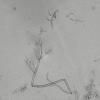
 Hi, I got this mould as a contaminant while studying another microfungus inoculated from a decaying log. On various media (PDA, SDA, Czapek) it forms an olive green growth (3cm / 14C / 7 days) with a yellowish tinge. First indication is that of a Penicillium sp. but under the microscope I could not make heads and tails. The prime character I can describe is that the phialides and hypha of the conidiogenesis apparatus breaksing up into constituent pieces giving this polymorphic observation of spores, hyphae and septate(?) phialides of various shapes and sizes. These septate hyphae have tiny projections indicating that they used to bear spores and hence part of the fruiting organ.
 Rarely I see spores in short chains but often they are very hygrophobic and entrapped in tiny bubbles hindering the view of the conidiaphore+apparatus. Is there a specific mould with this character - breaking out easily? I strongly doubt it is a Penicillium as I managed to see a fruiting part with budding spore.
Angel Pintos,
02-12-2017 09:32

Re : Mould which easily break when mounted in water or Stains
Looks like Cladosporium sp.
regards
Angel
regards
Angel
Joey JTan,
02-12-2017 17:03
Re : Mould which easily break when mounted in water or Stains
Yes this is a Cladosporium sp. The conidial chains easily fragment and can be seen best by making a tape mount. Gently press a small piece of transparent tape against the colony and mount it in water (place it on a drop of water, don't just stick it to the slide itself). You should see the chains still connected. The larger, 2-celled, shield-like ramoconidia are indicative of Cladosporium and give rise to branching conidia chains.
Stephen Martin Mifsud,
07-12-2017 23:29

Re : Mould which easily break when mounted in water or Stains
Hi thank you for your replies. It took time to come back but I did not forget you. The tape method did a lovely job. I cut a small squarish piece of tape using tweezers and clean scissors, laid the sticky surface on the outer part of the colony, gave a very gentle press (like a touch rather than a press), placed the sticky surface of a drop of water on a mounting slide and mounted under a microscope. It was interesting to observe that this species had clamp junctions.
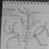
Now, I could see the conidiogenesis structure very well and I drew a quick illustration :-)
I don't think I dare to follow the Cladosporium key of a meticulous monograph to reach species level!
Now, I could see the conidiogenesis structure very well and I drew a quick illustration :-)
I don't think I dare to follow the Cladosporium key of a meticulous monograph to reach species level!

