
06-05-2026 11:25
 Castillo Joseba
Me mandan el material seco de Galicia (España) re
28-04-2026 20:07
 Lothar Krieglsteiner
... on twig in the air at standing Ceratonia siliq
05-05-2026 22:40
Gernot FriebesHi,I believe this is a Plagiostoma growing on a Sa
04-05-2026 18:13
 Stephen Martin Mifsud
ID request for what seems to be a true aquatic fun
04-05-2026 16:39
Stephen Martin Mifsud
ID request: This specimen was collected in Malta o
28-07-2011 18:31
 Alex Akulov
Dear FriendsToday I made the pdf file of Velenovsk
04-05-2026 09:50
Castillo Joseba
Me mandan el material seco de Galicia,(España) re
02-05-2026 12:42
Alain BRISSARDBonjour à tousJeuidi 30 avril dernier on m'a remi
02-05-2026 13:06
Pauline. PennaBonjour Please can someone help me with this id
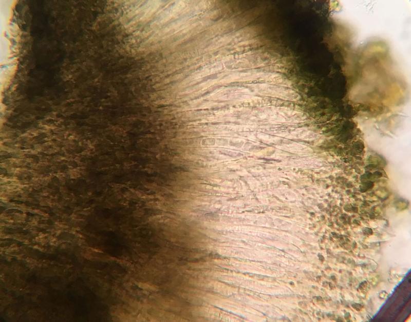
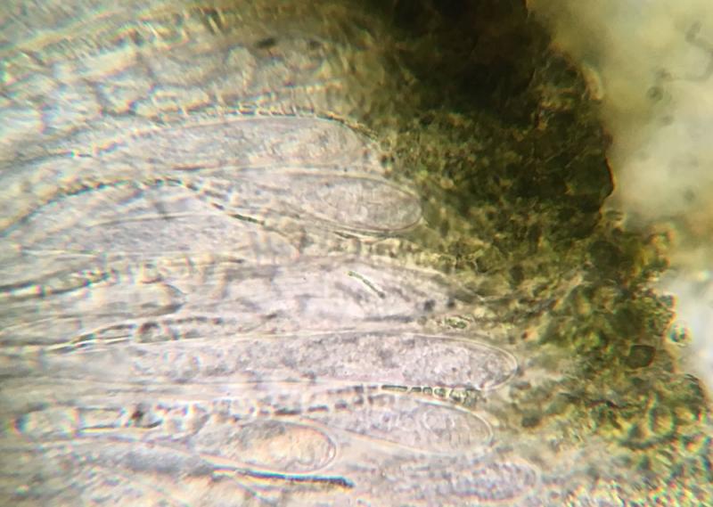
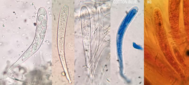
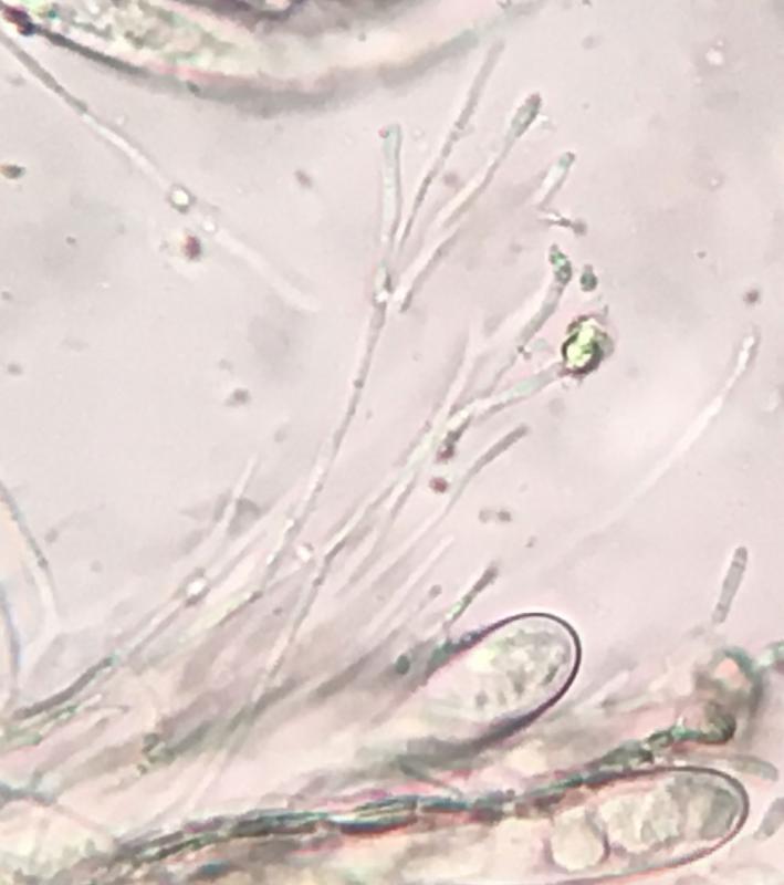
resembles Patellariales on resupinate toothed basidiomycete
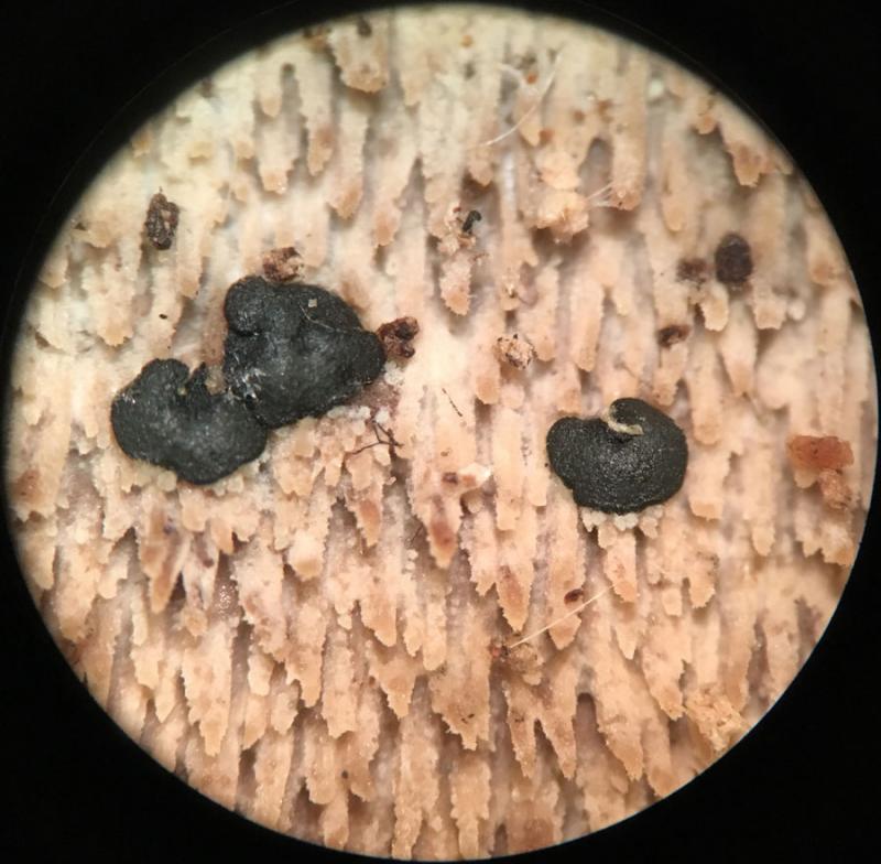
Ethan Crenson,
10-11-2017 06:45
Viktorie Halasu,
10-11-2017 10:26

Re : resembles Patellariales on resupinate toothed basidiomycete
Hello,
I think one of those asci might belong to and intrahymenial parasite (some very simple one, like Helicogonium). The lumpy spores remind me on packs of secondary spores in Tympanis, but I'm not sure, which one is host and which is parasite. Both kinds of asci have the same IKI- reaction?
Viktorie
I think one of those asci might belong to and intrahymenial parasite (some very simple one, like Helicogonium). The lumpy spores remind me on packs of secondary spores in Tympanis, but I'm not sure, which one is host and which is parasite. Both kinds of asci have the same IKI- reaction?
Viktorie
Hans-Otto Baral,
10-11-2017 10:32

Re : resembles Patellariales on resupinate toothed basidiomycete
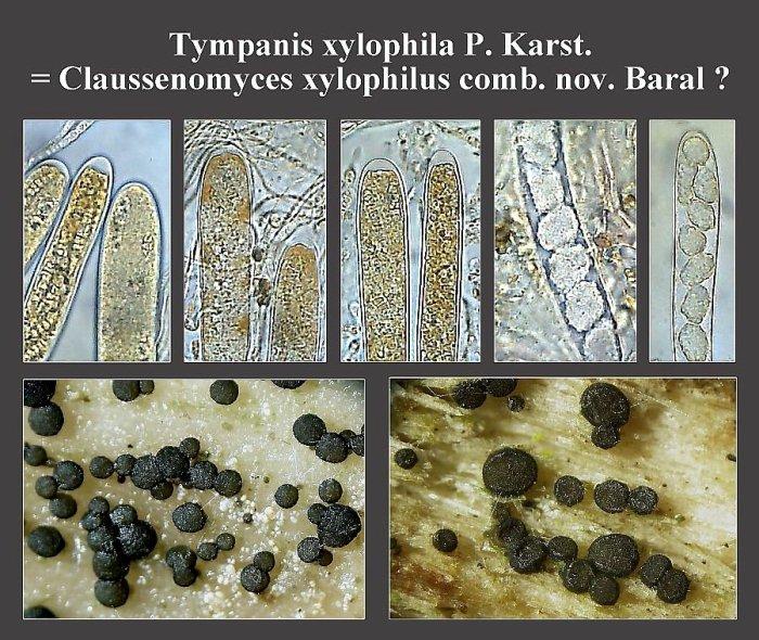
The similarity with Helicogonium is actually strong, but this here is not parasitised, it is a member of Claussenomyces, perhaps C. atrovirens. Actually I consider both genera to belong in the same order Phacidiales.
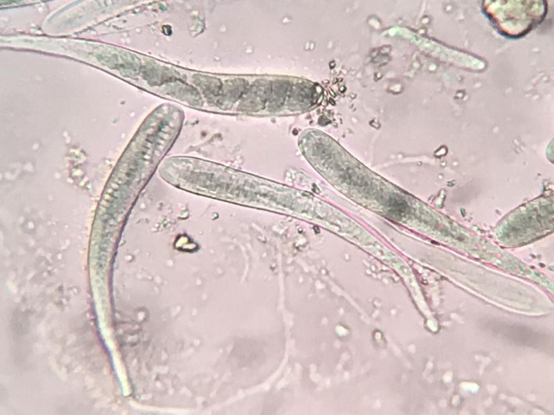
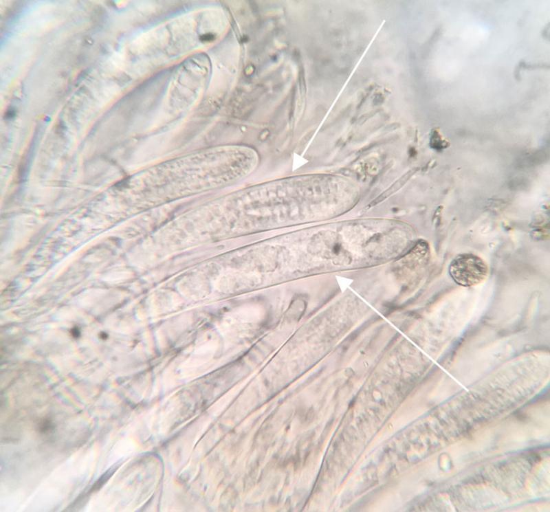
What we see in the asci is the septate primary ascospores and the many conidia that they form directly from the spore wall. These remain grouped around the primary spores and form eight "balls" that are ejected as entities. But when you kill the asci the balls disintegrate, and this is the currently figures state of a dysfunctional ascus, where the conidia fill the entire ascus lumen.
What we see in the asci is the septate primary ascospores and the many conidia that they form directly from the spore wall. These remain grouped around the primary spores and form eight "balls" that are ejected as entities. But when you kill the asci the balls disintegrate, and this is the currently figures state of a dysfunctional ascus, where the conidia fill the entire ascus lumen.
Peter Püwert,
10-11-2017 11:09

Re : resembles Patellariales on resupinate toothed basidiomycete
Hi all,
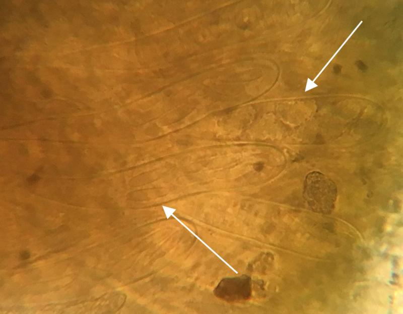
we have also already observed this occurrence. Here an old and change-dignified collage.
Greetings Peter.
we have also already observed this occurrence. Here an old and change-dignified collage.
Greetings Peter.
Hans-Otto Baral,
10-11-2017 11:43
Re : resembles Patellariales on resupinate toothed basidiomycete
The generic name Claussenomyces will probably be restricted in the future to the type species C. jahnianus, which is morphologically very different from the atrovirens/xylophila group. Which name will be used instead is still open.
Ethan Crenson,
10-11-2017 15:01
Re : resembles Patellariales on resupinate toothed basidiomycete
Thanks everyone! I assume that the growth on top of the rusupinate tooth is a coincidence and not parasitism or some other relationship?
Hans-Otto Baral,
10-11-2017 15:59
Re : resembles Patellariales on resupinate toothed basidiomycete
It is actually not often that these fungi grow directly on other fungi, but some association, e.g. with Dacrymyces, occurs in this group.
Ethan Crenson,
10-11-2017 16:43
Re : resembles Patellariales on resupinate toothed basidiomycete
I have just read the paper by Korf and Abawi "On Holwaya, Crinula, Claussenomyces and Corynella" which contains a key to 4 species of Claussenomyces including C. atrovirens. The defining features in the key seem to be apothecia size, ascospore size & septations and the presence or absence of an ionomidotic reaction. Does testing the ionomidotic reaction simply require applying 5% KOH aqueous solution to crushed apothecia and checking for brown pigments? I would like to try this, but I don't have many apothecia to work with, so I thought I'd get some advice before diving in. Thanks again!
Quijada Luis,
10-11-2017 16:45
Re : resembles Patellariales on resupinate toothed basidiomycete
Hi All,
dear Ethan I think that the correct id for your fungus is C. hydnicola that usually grown on this type of fungi, the primary ascospores only have transversal septa and they are shorter than C. dacrymycetoideus. So if you have a fresh sample I would be very happy if you send me part of it. Right now I am working in a project at Harvard University with the family Tympanidaceae and Claussenomyces is one of the genus included.
Best wishes,
Luis
P.S.: please write me an email to lquijull@gmail.com
Hans-Otto Baral,
10-11-2017 17:18
Re : resembles Patellariales on resupinate toothed basidiomycete
The ionomidotic reaction I do not remember exactly where it occurs. maybe in C. jahnianus. This C. atrovirens group shows dissolution of olive pigment but not by staining the medium around.
To test it, simply add KOH to a water mount and view. You can also put an apo in a drop of KOH and view it under the bino at the very moment.
But in this case I would spare the apos for Luis. Yes, C. dacrymycetoideus is more likely since the spores lack longitudinal septa.
To test it, simply add KOH to a water mount and view. You can also put an apo in a drop of KOH and view it under the bino at the very moment.
But in this case I would spare the apos for Luis. Yes, C. dacrymycetoideus is more likely since the spores lack longitudinal septa.