
11-05-2026 12:32
 Bernard CLESSE
Pourriez-vous m'aider à identifier cette héloti
13-05-2026 15:26
 François Freléchoux
Bonjour,Voici une récolte faite il y a quelques j
12-05-2026 15:41
 Nicolas VAN VOOREN
Dear Ascolovers, especially interested in Pezizale
13-05-2026 12:05
 Thierry Blondelle
Bonjour à tous,J'aimerais avoir confirmation de c
10-05-2026 23:17
 Andreas Gminder
Hello,today we found in a moist steep decidous for
28-04-2026 20:07
 Lothar Krieglsteiner
... on twig in the air at standing Ceratonia siliq
27-04-2026 20:52
Lothar Krieglsteiner
Found on hanging tiwg of Olea europaea in dried-ou
11-05-2026 20:22
Lothar Krieglsteiner
on attached twig of standing Ficus caricaquite uns
29-04-2026 10:44
Lothar Krieglsteiner
growing at moist, drying-out soil at the side of a
Hymenoscyphus on fern rhizome
Edvin Johannesen,
20-09-2017 11:27
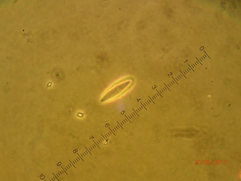
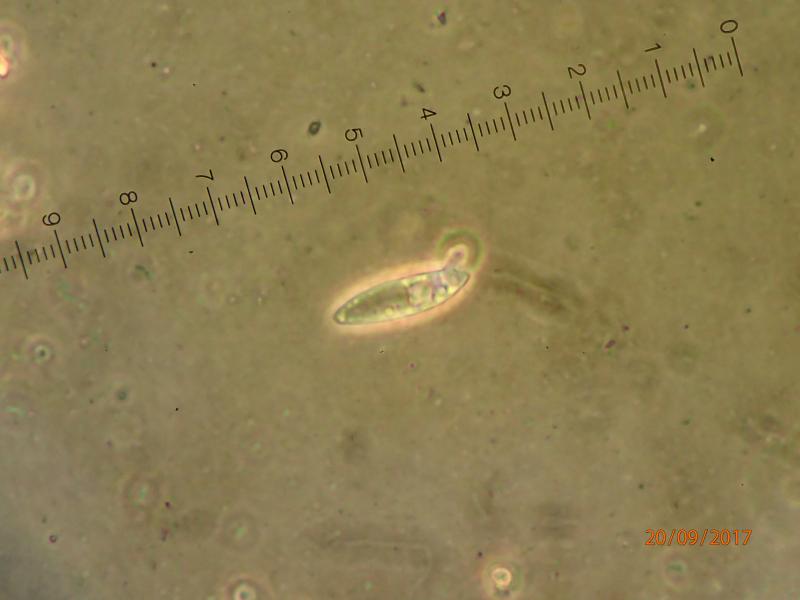
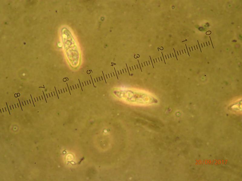
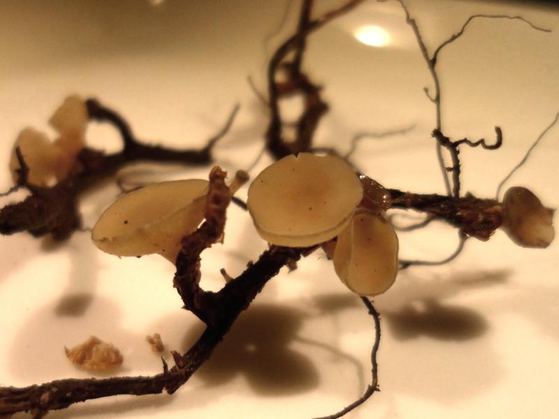
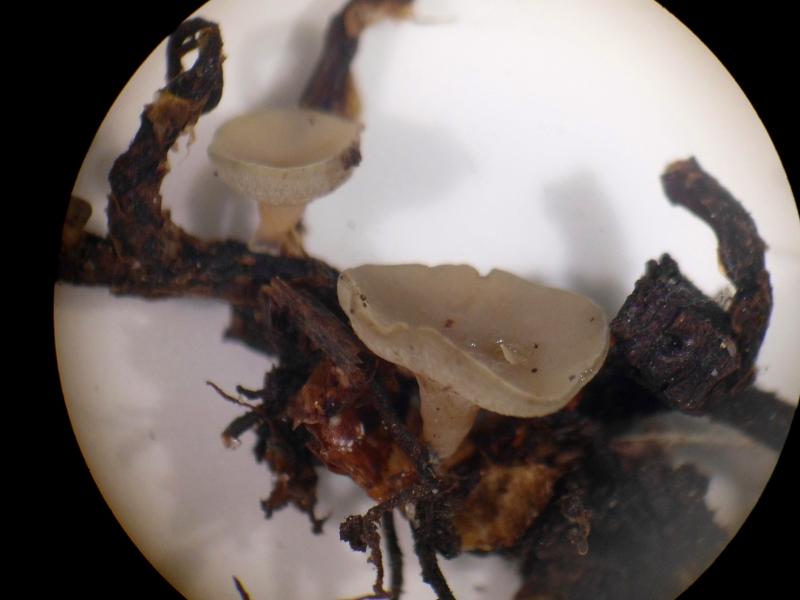
 This Hymenoscyphus (?) was found on Gymnocarpium dryopteris rhizomes in Central Norway. The spores measure approx. 14-16 (-20) x 5 microns and are elllipsoid to slightly inequilateral/navicular and often somewhat pointed at one end. A substantial portion of the spores are germinating with a hypha arising from one end of the spore. Spores are becoming one-septate at maturity. Excipulum consists of a pseudoparenchyma - large spherical/semiprismatic cells up to 30 microns.
I don't find any Hymenoscyphus with this combination of host/substrate and spore measures. Can anyone help? Am I in the wrong genus?
Hans-Otto Baral,
20-09-2017 12:00

Re : Hymenoscyphus on fern rhizome
Hmm, this seems to be a very interesting collection, but it would require a lot of further data to find out what it could be. How large are the apos?
The species could belong in the relationship of Cudoniella, but to verify this I should see the living paraphyses (guttulate?). Did you test the ascus apex with iodine? The shape of the amyloid ring would be important.
typical Hymenoscyphus species have prismatic cells or even t. porrecta.
I am unaware of such a species on fern rhizomes.
Zotto
Edvin Johannesen,
20-09-2017 12:40
Re : Hymenoscyphus on fern rhizome
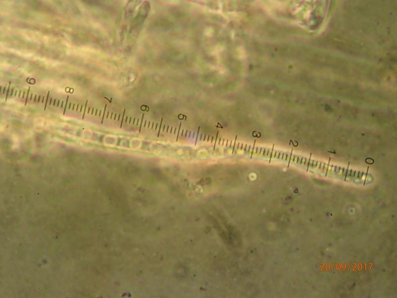
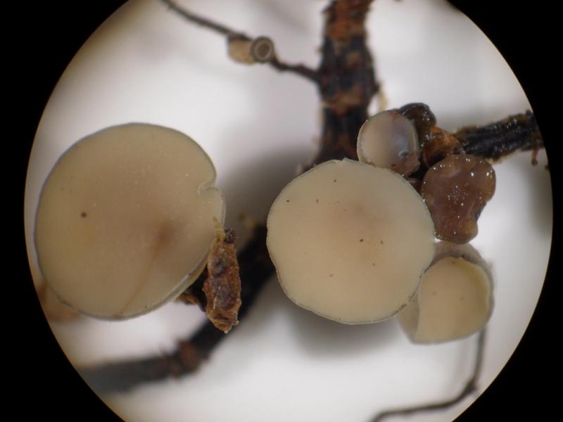
The apos are 4-9 mm in diameter, short-stalked. I am not able to see any ascus apex/pore staining with iodine (Meltzer's) (I tested the Meltzer on a Peziza yesterday).
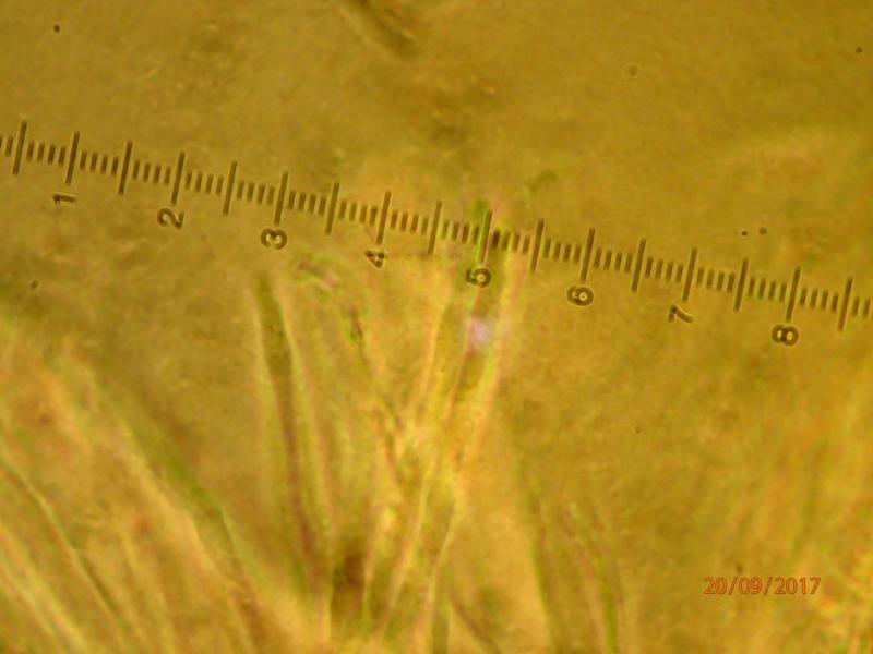
Ascus pore:
Ascus pore:

Edvin Johannesen,
20-09-2017 12:41
Re : Hymenoscyphus on fern rhizome
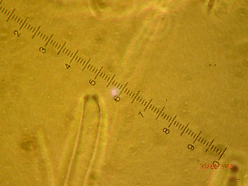
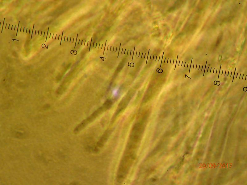
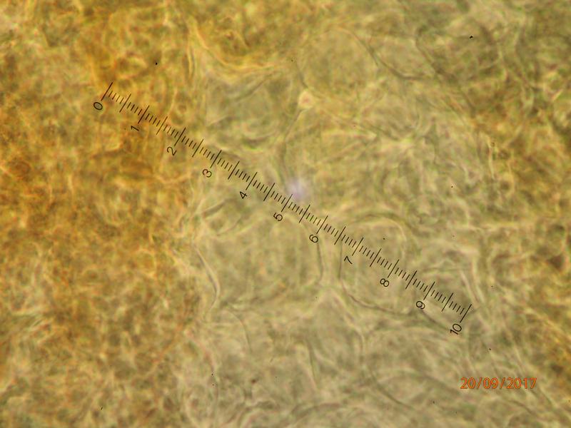
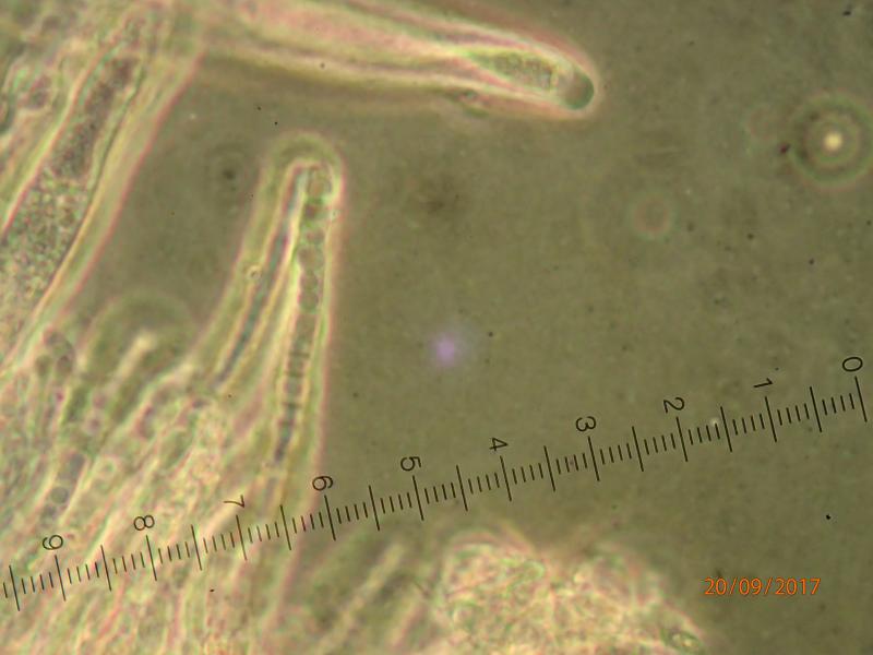
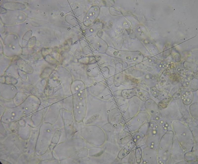
Paraphyses:
Edvin Johannesen,
20-09-2017 12:41
Re : Hymenoscyphus on fern rhizome
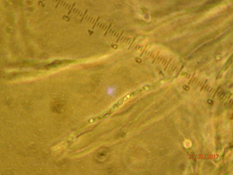
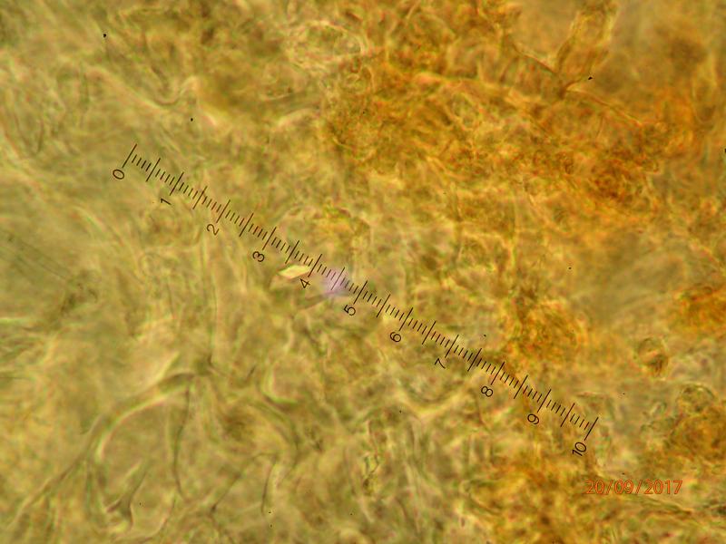
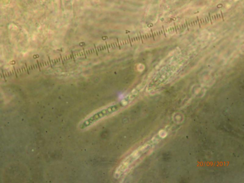
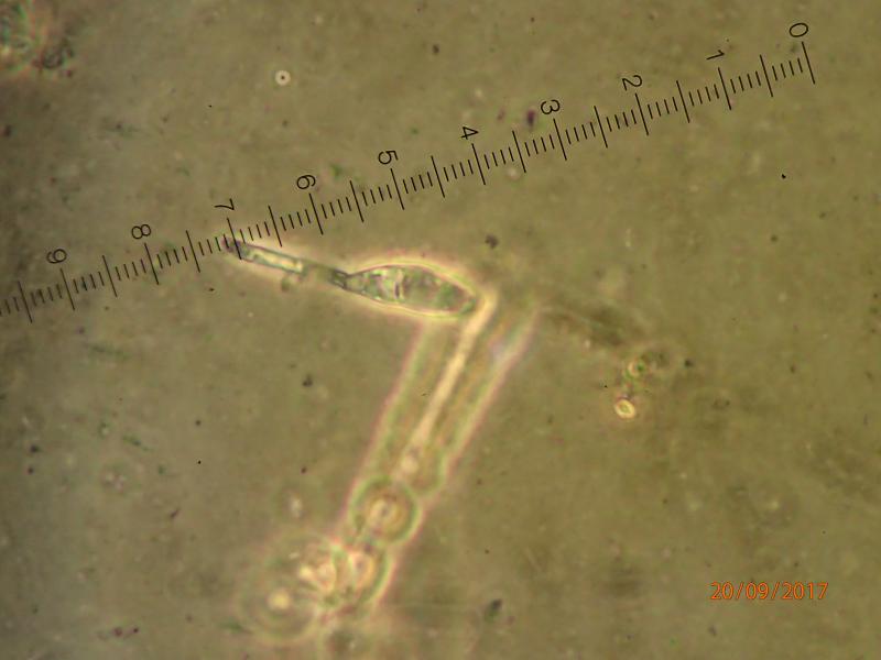
Germinating spore:
Edvin Johannesen,
20-09-2017 12:42
Re : Hymenoscyphus on fern rhizome
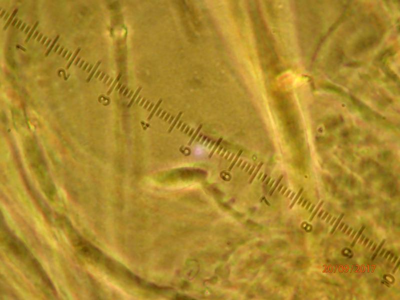
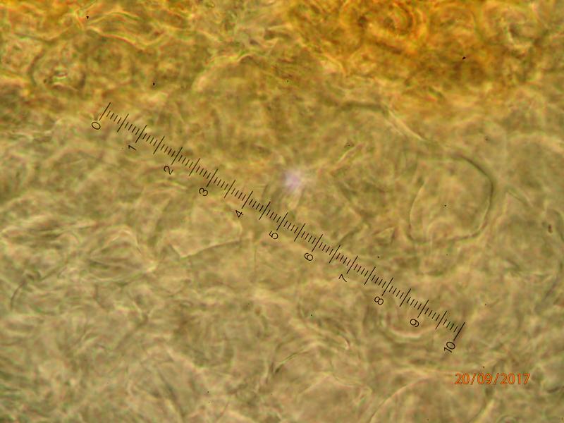
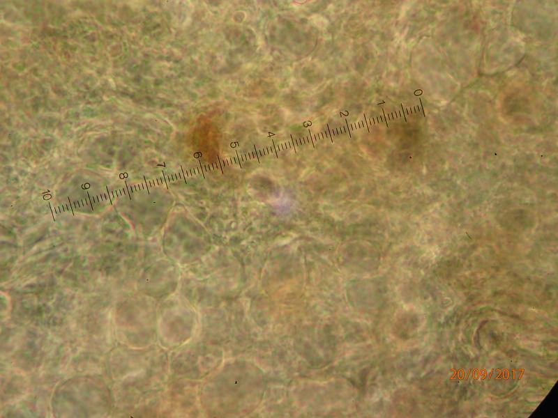
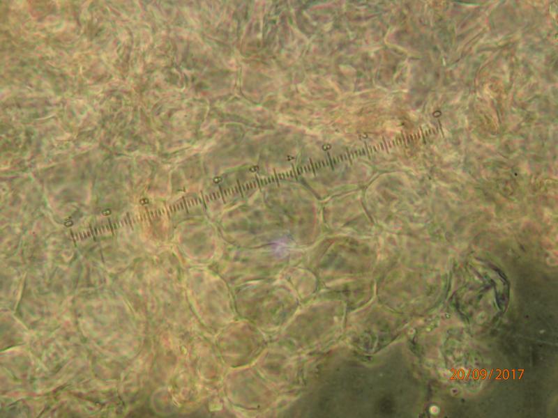
Excipulum:
Hans-Otto Baral,
20-09-2017 13:13
Re : Hymenoscyphus on fern rhizome
I am not sure if you mounted the fresh specimen and in what medium. The elements are all dead, which makes it very complicated.
With that size I would consider Cudoniella tenuispora but the large size and the angular excipulum rule out that option.
To show the variation of spore size would also be helpful, and please in water in order to see the contents.
Sorry for being so demanding.
With that size I would consider Cudoniella tenuispora but the large size and the angular excipulum rule out that option.
To show the variation of spore size would also be helpful, and please in water in order to see the contents.
Sorry for being so demanding.
Edvin Johannesen,
20-09-2017 13:32
Re : Hymenoscyphus on fern rhizome
Sorry. I will try to mount in water. More to come.
Edvin Johannesen,
20-09-2017 14:04
Re : Hymenoscyphus on fern rhizome
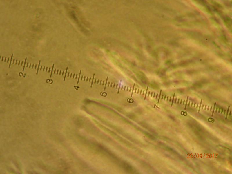
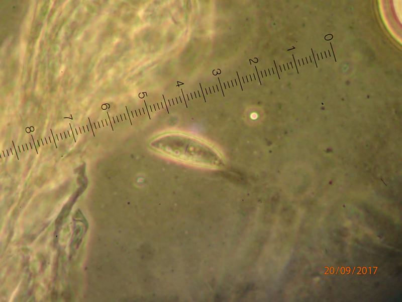
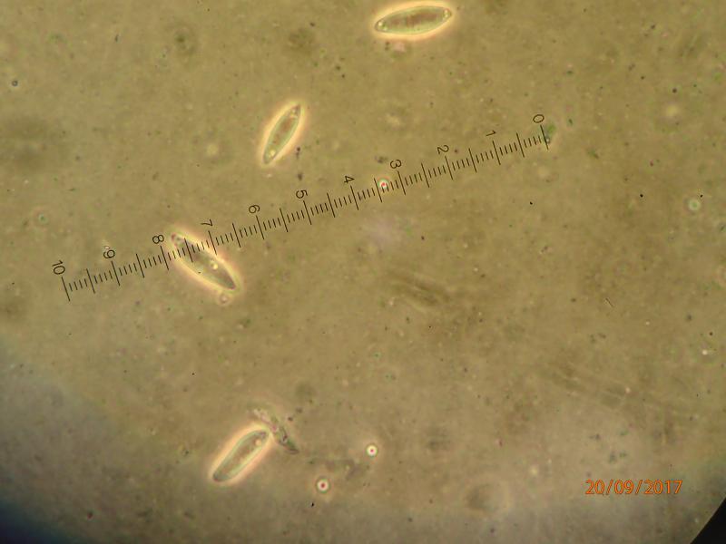
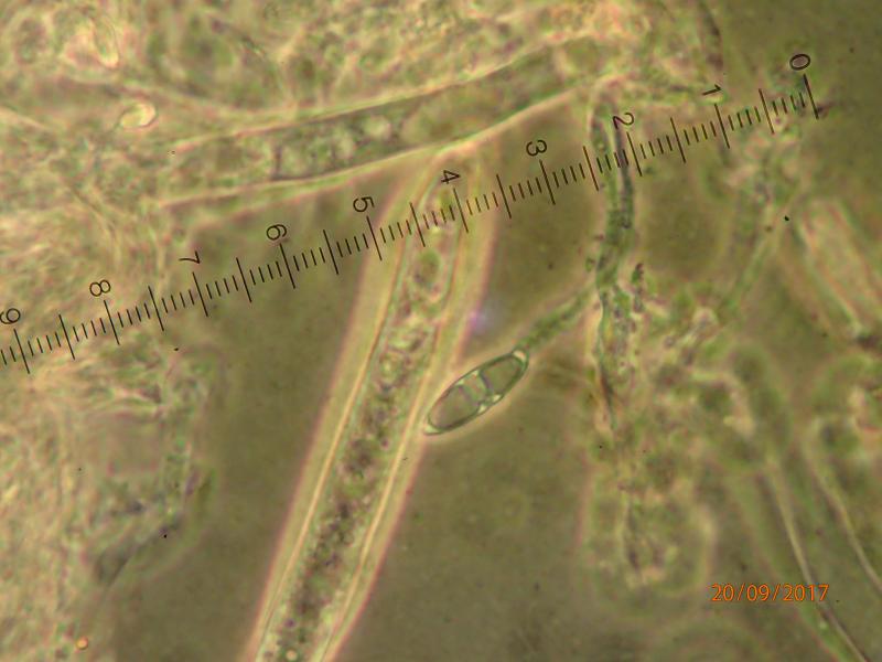
Mounted in water, 1000x, Phase contrast.
Edvin Johannesen,
20-09-2017 14:05
Re : Hymenoscyphus on fern rhizome
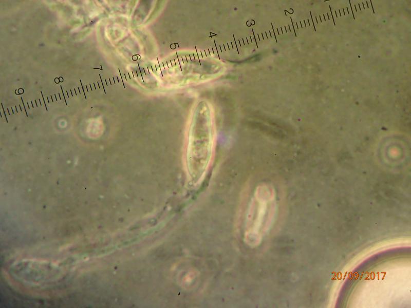
And more:
Edvin Johannesen,
20-09-2017 14:06
Re : Hymenoscyphus on fern rhizome
And finally (sorry if the quality is still bad):
Edvin Johannesen,
20-09-2017 14:07
Re : Hymenoscyphus on fern rhizome
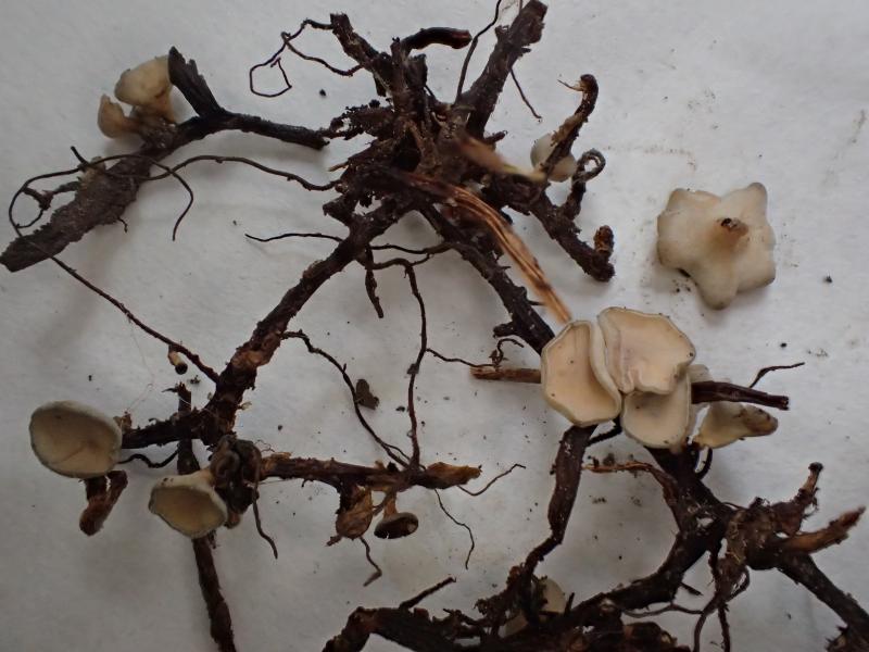
Images of fresh specimens from the collector. The mounts were made from semi-dried specimens, still soft.
Hans-Otto Baral,
20-09-2017 17:10
Re : Hymenoscyphus on fern rhizome
Thanks, yes, these are living spores and they contain a few small oil drops at the ends. The phase contrast that you use is difficult for me to interpret. For the spores it seems o.k., but difficult to tell the refraction index (strongly or weakly refractive).
In the paraphyses I see some large round drops but I am not sure are they refractive (I assumne so) or not. Under bright field this would have been clear.
Spore size in C. tenuispora is 12-16 x 4-5.5 µm, so I think this would fit. The septum is very probably a matter of the overmature spores.
The only thing that makes me wonder is the angular texture. That of C. tenuispora is an intricate prismatica, and the cortical cells also contain refractive drops and form hairs, here in surface view.
Edvin Johannesen,
21-09-2017 12:12
Re : Hymenoscyphus on fern rhizome
OK, thanks. I shall have to check the cortical cells.