11-05-2026 12:32
 Bernard CLESSE
Pourriez-vous m'aider à identifier cette héloti
13-05-2026 15:26
 François Freléchoux
Bonjour,Voici une récolte faite il y a quelques j
12-05-2026 15:41
 Nicolas VAN VOOREN
Dear Ascolovers, especially interested in Pezizale
13-05-2026 12:05
 Thierry Blondelle
Bonjour à tous,J'aimerais avoir confirmation de c
10-05-2026 23:17
 Andreas Gminder
Hello,today we found in a moist steep decidous for
28-04-2026 20:07
 Lothar Krieglsteiner
... on twig in the air at standing Ceratonia siliq
27-04-2026 20:52
Lothar Krieglsteiner
Found on hanging tiwg of Olea europaea in dried-ou
11-05-2026 20:22
Lothar Krieglsteiner
on attached twig of standing Ficus caricaquite uns
29-04-2026 10:44
Lothar Krieglsteiner
growing at moist, drying-out soil at the side of a
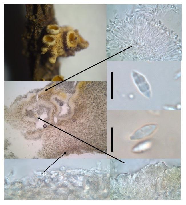
Stromatic cupulate coelomycete from Belize
Joanne Taylor,
07-09-2017 00:57
An interesting stromatic cupulate coelomycete was collected in Belize. it was growing on a seedling tree, on the stem and whether it was pathogenic or not, was unlcear. The description is below. It is very distinctive so surely must be described?
Creamy white stroma on host tissue (approximately 5mm), unclear whether it is bursting out of cuticle or superficial. Forming several cupulate conidiomata, often with stromatic short fat finger like projections.
In section hymenium can be convoluted and folded while appearing flat from above, orange coloured. The excipulum consists of textura intricata cells which are hyaline, thick walled with a narrow lumen, have crystalline encrustations and vary little from the stromatic tissue except in the upper margin where they are appear in a clear matrix.
The conidiogenous cells line the hymenium and are densely packed in some sort of matrix and will not separate even after treatment with 10% KOH. Conidiophores cylindrical/filiform, branched? or not, producing cylindrical filiform conidiogenous cells with small collarettes and are possibly percurrent with annelations.
Conidia are hyaline, fusiform with a truncate base and an apiculate apex, three septate with a germslit particularly visible in KOH (9-10 x 3 um).